Abstract
Background: High competitive level judo practice from a very
young age, may lead to an increasing risk of injury. The aim of this
study is to identify injury risk factors in a young high-level judo
population to develop future specific strategies for injury prevention.
Methods: An individual questionnaire was applied by physicians
to cadet and junior athletes that competed in international judo
tournaments in 2014. Statistical inferential analysis was performed to
identify the major injury risk factors and injury patterns in these
ages.
Results: 212 athletes from 13 different countries (53%
portuguese) answered the questionnaire. Male predominance was found, and
a mean age of 16,8 ±1,5 years with 9,7±3,1years of judo practice. We
found 347 injuries, with 83% of the athletes reporting at least one
injury. The majority of injuries occurred to the limbs (85%) during
training period (71%), in the throwing phase (87,6%), as a consequence
of direct contact (72%) and with articular involvement (62%). Sprain was
the most common type (36%) and shoulder the most specific segment
affected (25%) with an occurrence twice as high during defense movements
[p=0,018]. Injury occurrence was associated with training load
(p=0,001), whilst the number of injuries with the age [p=0,005], need to
lose weight (p=0,007) and training load (0,001).
Conclusions: Increasing age, need to lose weight and training
load were identified as injury risk factors for these ages. We also
verified that shoulder injuries are significantly more frequent during
defense movements, being these ones that led to more severe injuries.
Keywords: Judo; Sports injury; Trauma; Young athletes; Martial arts
Introduction
Judo is a combat sport often related to injuries since it is
associated with a significant direct contact [1,2]. Although injuries
occur, judo is considered one of the most safest sports activity due to
low injury rates [3], being in fact considered by UNESCO as the best
sport for children and young people between 4-21 years old. Nowadays,
because of the early competitive practice, specialization and intensive
training load among young athletes, we are seeing different injury
patterns and severity in these ages [2,4,5]. The potential vulnerability
of these athletes due to the physical and physiological processes of
growth exposed to high levels of activity, is a reason of concern [4,6]
which must stimulate the development of this specific knowledge, in
particular the analysis of risk factors in order to provide preventive
injury programs and educative measures. With this study, we intend to
reinforce the epidemiologic knowledge specific to judo in these
particular ages, and, by knowing the risk factors and more common injury
features and patterns, use them to help.com developing the strategies
to control and prevent injuries.
Methods
This was a retrospective cohort study, in which an individual
questionnaire was applied to cadet and junior athletes that participate
in international competitions in 2014, in Portugal (Cadet European Judo
Cup and Junior European Judo Cup, both in Coimbra city). The
questionnaire was written in 4 languages (portuguese, english, french
and Spanish) and it was designed and applied by a group of physicians,
focusing on the athlete's intrinsic characteristics, training method and
practice level, and, when it exists, sports injury and its treatment.
We defined "mild injury" - without any stop of the activity; "moderate
injury"-stop for one training session or competition; "severe
injury"-stop for more than one training session or competition [7-9].
The study was approved by the Ethics Comitee of the Faculty of Medicine
of the University of Coimbra, and all of the athletes included in this
study had to sign the informed consent form. Statistical analysis was
performed using the IBM SPSS Statistics 21.0 program (p<0.05).
Results
Demographic data
Some of the preliminary descriptive epidemiological data were
published previously in an early phase of the study (Table1 & 2)
[10]. Based on the information obtained from the questionnaires, we
analyzed the answers of 212 young high competitive judo athletes, from
13 different countries (52% Portuguese). Of these, 118 were cadet and 94
junior with a mean age of 16.8±1.5 years and male predominance (68.4%).
The more common weight categories were -60, -66e -73Kg in male gender
and -57, -63 and -70Kg in female gender. We found that 205 athletes
compete in categories with weight limit and that 127 (62%) have a real
weight higher than that of their category. Most of the athletes were
black belt (55.6%) and assume a right-handed dominance grip position
(73.1%) while performing judo techniques. We verified that athletes with
higher rank were the ones who did more judo trainings sessions per week
(Rho=0.254, p<0.001), and hours per day [Rho=0.313, p<0.001] and
per week (Rho=0.338, p<0.001). The average number of years of judo
practice was 9.7±3, 1 (1-16), the number of judo training sessions per
week 5.5±1.9 (2-11) and hours per day 2.57±0.9 (1-6). All athletes
reported having warm up sessions prior to judo training, averaging
23.3min±8.8 [10-60], while 12.7% answered that they do not usually
stretch after activity. Around 65% of the athletes do gym sessions and
strength workouts averaging 2.83 sessions per week. Out of these, 24.1%
perform 4 or more gym sessions per week. We found a statistically
significant correlation between the age of the athlete and hours of
training per day (Rho=0.244, p<0.001) and per week (Rho=0.280,
p<0.001). We also verified that the athletes who need to lose weight
to compete, were the ones who had more judo trainings per week, judo
training hours per day and per week and a higher number of gym
sessions/strength workout per week (Table 3). Regarding athletes that do
gym sessions, we found a statistically significant correlation with
age, need to lose weight and judo training load [number of trainings per
week, training hours per day and per week]. Comparing portuguese
athletes with foreigners regarding the frequency of gym sessions, we
found that foreigner athletes do 3.9 times more this type of workout
than the portuguese (x2= 19.578, p<0.001; OR=3.861 [IC95%:
2.088-7.142]) and that the judo training sessions are on average 14
minutes longer than the portuguese ones [p=0.038].
Table 1: Demographic Data: athlete intrinsic characteristics, training method and practice level
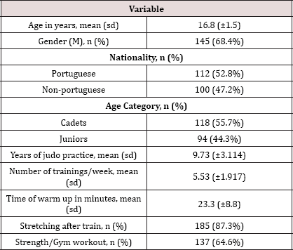 M: male. sd: standard deviation
Table 2: Frequency, type, distribution and severity of sports injuries in high-competitive level judoka
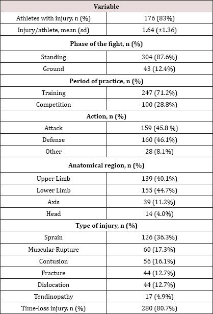 sd: standard deviation
Table 3: Statistically significant correlation with the athletes who need to lose weight
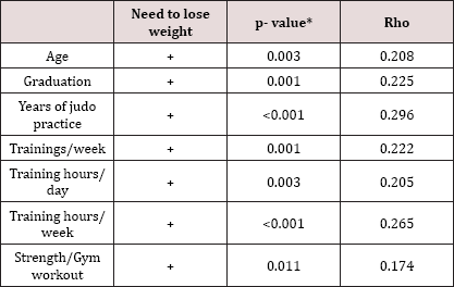
*Spearman's correlation
Frequency of injuries
We found 347 injuries in 83% of the athletes, while 17% reported no
injuries due to judo practice. The average number of injuries per
athlete was 1.64±1.36 (Table 2). We verified a statistically significant
correlation between the presence of injury and the number of judo
trainings per week [p=0.005] and of judo training weekly hours
[p=0.014]. Regarding the number of injuries, this was statistically
significant correlated with age, need to lose weight, number of
trainings per week and daily and weekly judo training hours (Table 4).
We found no statistically correlation between the occurrence of injury
and rank, gender, grip dominance, warm up, stretching, gym sessions or
years of practice. We have noticed a tendency for injuries in those who
do gym sessions but with no statistical significance. We also did not
find any statistically significant correlation between the weight
category and the number of injuries, frequence by specific anatomical
segment and type of injury or its severity. We observed that the
presence of injury was related with the age category (x2=8.593, p=0.003)
with a probability of injury 3.34 times higher in junior athletes when
compared with cadets (OR=3,344 [IC95%: 1,445-7,743]).
Table 4: Statistically significant correlation with the number of injuries.
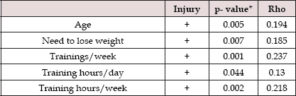 *Spearman's correlation
Action and phase of combat
Most of the injuries (71.2%) occurred during training sessions (62%
in the pre-competitive phase] [p<0.001 [IC95%: 0.661-0.759]) and
throwing phase (87.6%) (p<0.001, [IC95%: 0.837-0. 909]). The
occurrence of injury was similar for both defense (45.8%) and attack
(46.1%) movements, and 8,1% of the injuries occurred without any
interference of the opponent. However, we observed that the occurrence
of shoulder injuries was 1.8 times higher during defense movements
(x2=5.629, p=0.018; OR=1.838 IC95%: 1.108-3.050). We verified that of
all injuries that occurred during groundwork phase, most (55.8%)
occurred during a defense movement of the athlete. We also found that
the upper limb injuries were more frequent during groundwork phase (55,
8%) while those in the lower limbs were during standing phase (46, 7%)
(Table 5). Even tough we found groundwork injuries more common during
trainings regarding its absolute number, these injuries were 2.4 times
higher in proportion during competition (x2=7.490, p<0.006;OR=2.435
[IC95%: 1.269-4.670]).
Table 5: Statistically significant differences between
anatomical region, phase of combat, injury type, injury mechanism and
injury severity.
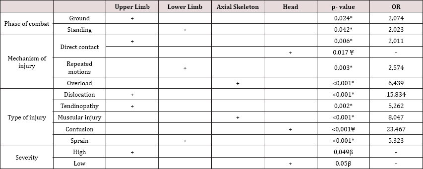 ** Chi-square; ¥ Fisher's Test; ß Mann-Whitney's Test
Injury location
Regarding injury location, we found a statistically significant
higher number of limb injuries [84,8%], with 40,1% in the upper limb and
44,7% in the lower limb. Out of the rest of injuries reported, 11,2%
occur in the axial skeleton and 4% in the head (Table 2). Analyzing all
injuries by specific anatomical segment we verified that shoulder was
the most commonly involved [n=87, 25.1%] followed by the knee (n=68,
19.6%), ankle (n=37, 10.7%) and spine (n=31, 8.9%). About half of the
injuries occurred on the right side of the body, 40% on the left one and
10% on the median region. We verified with statistically significant
difference that most of the injuries had an articular involvement
(61.7%],[p<0.001, [IC95%: 0.563-0.668]).
Mechanism and types of injury
The most common mechanism of injury identified was direct contact
[n=249, 71.8%], followed by dynamic overload (n=50, 14.4%) and cyclic
overuse (n=48, 13.8%). On the other side, the most common types of
injury identified were sprain [36.3%], muscular rupture (17.3%) and
contusion [16.1%] (Table 2). Fracture and dislocation occurred with the
same frequency (12.7%) and were the ones that had with higher injury
severity. Tendinopathy was found in 4.9% of the cases. Regarding the
type of injuries, we found with statistical significance, that
dislocation and tendinopathy were more common in the upper limb, sprain
in the lower limb, muscular rupture in the axial skeleton and contusion
in the head (Table 5). We also verified that the probability of muscular
rupture occurrence was 2 times higher than any of the other types of
injury during competition (x2=5.838, p=0.016;0R=2.020 [IC95%:1.
134-3.597]). Regarding the mechanism of injury, we found, with
statistically significant difference, that the occurrence of contusion
was 2.4 times more common than any of the other types of injury during
training [x2=5.289, p=0.021;OR=2.376 (IC95%: 1.1175.055)]. We also
observed with statistically significant differences that upper limb
injuries are more frequent as consequence of direct contact and that
those of the axis and lower limb as consequence of dynamic overload
(Table 5). We noticed that the mechanism of injury was statistically
correlated with the type of injury (x2=139,283, p<0,001). We verified
that muscular rupture was related with dynamic overload, tendinopathy
with cyclic overuse, dislocation, fracture, contusion and sprain with
direct contact.
Injury severity
Regarding the impact and severity of the injuries, we verified that
81% resulted in cessation of the activity. We also noticed with
statistically significant difference, a higher proportion of severe
injuries during defense movements and of mild injuries on solo movements
(no movement/action from the opponent] [H1=27.109,p=0.014) (Table 6).
In 18% of the injuries no treatment was needed, while 72% needed some
kind of treatment. About 72% of the treated injuries required elastic
contension, functional bandage or immobilization. We also noticed that
56.2% of the injuries required physiotherapy and 6.6% required surgery
mainly because of knee pathology (52.2%)-meniscal tear or anterior
cruciate ligament rupture. We did not find any statistically significant
correlation between injury severity and its occurrence during training
or competition, phase of combat, sports season, mechanism of injury or
rank. The most severe injuries were joint dislocation and bone fracture
and the less severe was muscular rupture (Table 6). We found that the
most severe ones were statistically correlated with the number of years
of judo practice of the athletes, and we also verified that athletes
that perform more judo trainings per week, tend to have more severe
injuries, but without statistical significance (r=0.098, p=0.0690).
Regarding the severity of the injury, this was found to be correlated
with the type of treatment [p=0.007], with some of the most severe ones
leading to surgical procedures [p=0.045] and mild injuries to no
procedure at all [p=0.003]. Analyzing injury severity between portuguese
and foreign athletes we found no differences between both groups.
Regarding the anatomical segment, we verified that the most severe
injuries occur in the upper limb and the less severe in the head (Table
5).
Table 6: Statistically significant differences between injury severity, action, injury type and years of judo practice.
 * Kruskal-Wallis; β Pearson's correlation
Discussion
Demographic data, injury incidence patterns and risk factors
Based on the results collected in our study, we obtained a diverse
sample of athletes from different european countries, with a normal
distribution regarding age category and nationality [portuguesese versus
foreigners], with a predominance for central weight categories similar
to the european trend. Considering the need for high technical
requisits, specialization and load training intensity in such a young
age, these athletes are more likely to develop injuries in their sports
activity [2,4,5,11].With respect to the frequency of injuries, we
verified that 83% of the athletes had at least one injury, a number that
seems clearly underestimated: for example, we didn't find any ear
injuries, skin scratches or epistaxis reported in our study, and we
noticed a low number of small articular injuries although they are
common in judo practice [3,12-15]. The main reason for that is probably
related with the socio-psychological profile of judo formers, having
higher pain thresholds in relation with the high training intensity. In
addition, this also happens due to the demanding competitive calendar
and athletes' will of the athletes to pursue their objectives, which
makes them train in suboptimal conditions, sometimes hiding injuries,
without medical support, reducing quality, intensity and training
performance, ultimately leading to chronic injuries or re-injuries
[16-19].
We verified that 72% of all injuries occur during training period, an
outcome that is also described in the literature [16,20]. This is
possibly related to poor training strategy programs with excessive
intensity and training load in young ages, which predisposes to a high
musculoskeletal fragility [13,21,22]. Another reason for this fact is
that many athletes undergo strenuous strength workout programs while
training hard before competition, all the while maintaining hydric and
caloric restriction diets for several days [12,23-27]. This attitude
might lead to situations of severe dehydration fluid and electrolyte
imbalance which can potentially influence both the physical and
psychological performance of the athletes [12,2325,28-30]. It is also
important to highlight the weight differences between partners during
practices [8], which become even more important in the pre-competitive
period, during which most of the athletes are already on a diet at these
ages. Because of this, it is important to evaluate some risk/benefits
for the athletes, when considering the dynamic balance of upgrading
skills by fighting with a heavier and more experienced partner and the
potential risk of injury because of it before a competition.
Similar to other studies, we found that most ofthe injuries (74%)
occurred during the throwing phase [31]. This is mainly explained
because most of the action during a fight occurs in the standing phase,
with associated explosive attacking throws and defense resisting
movements counteracting the action [32-34]. verified that a greater
injury severity was related to defense movements, which is consistent
with the literature [31]. We also found that the occurrence of
groundwork injuries was 2.4 times higher in competition than in
training, which is probably related with higher fight intensity in this
phase of combat during competition [31].
In line with the literature, we observed a total of 85% of limb
injuries [13,35], 72% of the injuries due to a direct contact mechanism
[35-37] and that 62% of all injuries were articular. This articular
involvement is mainly explained by the direct contact mechanism but also
because most of the action occurs by working in closed kinetic chains,
which, because of the strong musculoskeletal structure of the athletes,
causes movement fulcrums such as the joints to become more prone to
injury [13,16,21,35,38] .Similar to other studies, shoulder was the
anatomical segment most commonly affected [19,39] and we observed that
its shoulder injury occurrence was 1.8 times higher during defense
movements, mainly caused by the resistance of the defender in an attempt
not to fall on his back [32-34] .
In our study we verified an 81% of time-loss injuries, result that,
as previously stated, seems clearly related to an underestimation of
mild injuries by the athletes, that remember more accurately severe
injuries, like dislocation or fracture, because of the obvious
repercussion in their sports activity and overall daily activity [40].
Despite this limitation in data quality, we can assume that the
information collected is very accurate for severe injuries related to
judo practice in young athletes. In 72% of the cases, athletes used some
kind of elastic or functional bandaging or immobilization, usual
practice in judo high level athletes, that do it often in a daily basis
practice [41].
Limitations
This study provide.com information based in a retrospective analysis
of the past injuries of the athletes. Thus, the quality and objective
description of the injuries by the athletes, and their treatment, was
not the most accurate. The young age of our sample, as well as the
subjective impact and significance of the injury reported are
athlete-dependent and also contribute to a higher reporting of severe
injuries, gives the idea of a larger number of time-loss-injuries while
many mild injuries are underestimated or not even reported at all. At
the same time, this allows.com a very precise and objective description
of the most severe injuries of the athletes.
Conclusion
Judo is commonly associated with injuries, risk that based in our
study is greater in athletes that need to lose weight and who are
exposed to a more frequent and intensive training loads. In fact, in our
high competitive population cohort, we found that 83% of the athletes
reported at least one injury, most of them in the limbs, during the
training period, in the standing phase of the combat, as a consequence
of direct contact and leading to time-loss injuries. Shoulder was found
to be the most common specific anatomical segment injured and its
involvement was also significantly higher when associated with defense
movements. We observed that lower limb injuries were more common during
throwing phase and upper limb injuries during groundwork phase. The most
common type of injury was sprain, with the most severe injuries being
associated with defense movements. Most of the injuries required some
kind of functional bandage/elastic taping and surgery was performed in
7% of all cases mainly due to meniscal tears and ACL rupture, leading to
arthroscopic partial meniscectomy and/or ACL reconstructions. As a
final consideration, we emphasize that this study provides and
reinforces present epidemiologic data and knowledge regarding judo
injuries in such a young high competitive level population. Based in our
results and better knowledge of risk factors and common injury features
and patterns, we will now use them to develop strategies to control and
prevent injuries to this particularly young population.
For more
Lupine Publishers Open Access Journals Please visit our website:
https://lupinepublishers.us/
For more
Open Access Orthopedics and Sports medicine articles Please Click Here:
https://lupinepublishers.com/orthopedics-sportsmedicine-journal/
To Know More About
Open Access Publishers Please Click on
Lupine Publishers