Monday, 22 August 2022

Lupine Publishers| Investigation Of Surgical Nurses Knowledge and Practice Levels for Multimodal Analgesia

 Lupine Publishers| Journal of Medical Science 


Abstract

Objective: This was a descriptive study with the aim of determining knowledge and practice levels of nurses working in surgical clinics for multimodal analgesia and its administration.

Materials and Methods: The study was conducted with 132 nurses working in the surgical clinics of three training and research hospitals. The data were collected between April and June of 2018 using the “Knowledge and Application Questionnaire on Multimodal Analgesia” developed by the researchers. The data were analyzed using number, percentage, and the Chi-square test.

Results: The mean age of the nurses who participated in the study was 37.5 years. Further, 47.7% had been working for 11– 20 years, and 60.6% had a bachelor’s degree. It was determined that 59.1% of the nurses did not receive any pain training, and 93.2% did not receive any education about multimodal analgesia. In the Knowledge and Application Questionnaire on Multimodal Analgesia, they obtained a score of 9.83 ± 2.38 points, which showed that the nurses’ knowledge and practice score for multimodal analgesia was moderate.

Conclusion: In this study, it was observed that the nurses were in need of knowledge about multimodal analgesia management and non-pharmacological interventions. It seems important that hospital administrators should initiate in-service training programs, including effective pain management and nursing interventions.

Keywords: Nurse; multimodal analgesia; surgery

Introduction

Pain is a symptom that is felt in different parts of the body for various reasons, leading to physiological and psychological reactions and causing the individual to seek help. The perception of pain is affected by many emotional and behavioral factors, such as environment, gender, culture, education, and experiences of the individual [1]. One of the most important causes of pain that can be caused by many factors, such as accidents, traumas, and acute and chronic diseases, throughout the life is surgical interventions. Pain after surgical interventions is an acute experience that begins with tissue destruction following surgical trauma and progressively decreases and ends with tissue healing. Postoperative pain is the most important factor in the development of complications, such as pneumonia, thrombosis and ileus; conversely, long and severe pain with ineffective pain management leads to prolonged hospital stay and increased opioid use [2-4]. In the management of postoperative pain, adequate analgesia may not be achieved with a single agent [5].

Recently, multimodal analgesia method has been used for postoperative pain management [5-6].In multimodal analgesia, a lower dose of analgesic is used due to the additive and synergistic effects of drugs; therefore, fewer side effects occur and more effective analgesia is provided. For this purpose, non-steroidal anti-inflammatory drugs, which are combined with opioids for postoperative analgesia in major surgeries, are widely used [7,8]. Although nurses have an important role in the evaluation and management of pain, Senyuz and Kosasli reported that pain is evaluated incorrectly and treated insufficiently because nurses lack knowledge in the assessment and pharmacological and nonpharmacological treatment of pain [9]. In the pain management process, because pain is a person-specific symptom, nurses should have sufficient knowledge for taking patients’ history, making continuous observation, and determining an appropriate pain assessment method. In order to evaluate effectiveness of pain management, it is necessary to use a scale and check whether the intervention is effective on the patient’s pain [10].

For effective assessment and control of pain and prevention of pain-related complications, nurses’ knowledge on multimodal analgesia management and application in multidisciplinary teams was considered to be important, and the study was planned to determine the knowledge and practice levels for multimodal analgesia and its administration of nurses working in surgical clinics.

Objective

The study was conducted as a descriptive study in order to determine the knowledge and practice levels of nurses working in surgical clinics for multimodal analgesia and its administration.

Materials and Methods

Population Sample

The initial study population comprised 190 nurses working in general surgery, neurosurgery, urology, otolaryngology, orthopedics and traumatology, and cardiovascular surgery clinics of three training and research hospitals. The study was completed with 132 nurses who agreed to participate in the research. The data were collected between April and June 2018.

Data Collection Tools

In this study, a literature review was conducted and the “Knowledge and Application Questionnaire on Multimodal Analgesia” developed by the researchers [11-15]. was utilized. This form consists of two parts. In the first part, there are sociodemographic questions, including descriptive characteristics of nurses, and in the second part, there are questions that determine the level of knowledge about multimodal analgesia and its administration. A total score of knowledge and practice was calculated by giving a score of 1 for every correct answer for the questions in the questionnaire, and a score of 0 point was awarded for every wrong answer. Accordingly, a score of ≤7.45 points was determined as a low knowledge and practice score, a score between 7.46 and12.21 points as medium, and a score of ≥12.22 as high. The low and high score ranges were determined by subtracting the standard deviation number of 2.38 from 9.83, which is the average knowledge and practice number, and adding the standard deviation number to this number. The values between the low and high score ranges were considered to be the medium knowledge and practice scores.

Data Collection

After the approval of the ethics committee and permissions of the institution were obtained, the nurses were informed about the study, and their written consent was obtained. The data were collected by conducting face-to-face interviews at a convenient time during the working hours of the nurses. The interviews lasted for approximately15–20 minutes.

Data Evaluation

In the analysis of the obtained data, frequency distributions and Chi-square statistical methods were used with the program of IBM SPSS Statistics 15.0 (IBM Corp. Released 2012. IBM SPSS Statistics for Windows, Version 15.0. Armonk, NY: IBM Corp.). A P value <0.05 was considered statistically significant.

Limitations of the Study

The study aimed to reach the entire population, but it was completed with 132 nurses who agreed to participate in the research. This was a limitation of the study.

Ethical Dimension of the Study

The study was conducted in compliance with the approval of Yıldırım Beyazıt University, Social Sciences and Humanities Ethics Committee, Ankara (23/02/2018-10) and the application permissions obtained from Atatürk Training and Research Hospital (13/04/2018-E.4834), Gulhane Training and Research Hospital (12/04/2018-E.952), and Yenimahalle Training and Research Hospital (09/04/2018-E.3902).

Results

The mean age of the nurses participating in the study was 37.5 years. Further, 94.7% were female, 68.9% were married, 47.7% had worked for 11-20 years, and 60.6% had a bachelor’s degree. Additionally, 25% of the nurses were working in general surgery, 24.2% in brain surgery, 16.7% in orthopedics, and 12.1% in urology clinics. The socio-demographic characteristics of nurses are given in (Table 1). It was determined that 59.1% of the nurses who participated in the study did not receive pain education, and 93.2% did not receive any training on multimodal analgesia (Chart 1). In the examination of the answers given by nurses on the knowledge and application about multimodal analgesia, it was determined that 94.7% gave the correct answer for the statement “Multimodal analgesia method is the use of multiple drugs instead of a single drug”; 83.3% for the statement “In multimodal analgesia, it is recommended to use drugs that affect different parts of pain pathways together in selection of drugs”; and 82.6% for the statement “Multimodal analgesia provides sufficient analgesia with drugs with additive or synergistic effects.” It was determined that 69.7% of the nurses gave the wrong answer for the statement “Multimodal analgesia was initiated to be administrated to increase the effect of opioid drugs”; 59.1% for the statement “A different scale is used in the assessment of pain of patients in multimodal analgesia”; 58.3% for the statement “Non-pharmacological treatments are also performed in multimodal analgesia” (Table 2).

When the answers given to the “Knowledge and Application Questionnaire on Multimodal Analgesia” were evaluated out of 16 points, nurses obtained 9.83±2.38 points. Therefore, nurses’ knowledge and practice score on multimodal analgesia was moderate. In terms of knowledge and practice levels on multimodal analgesia, it was found that 15.9% (n=21) of the nurses had low score, 75.8% (n=100) had moderate score, and 8.3% (n=11) had high scores (Chart 2). In our study, some individual and occupational characteristics (level of education, clinics they worked, and working year), which were considered to have an effect on the total score averages of knowledge and practice levels of the nurses on multimodal analgesia, were examined. In the statistical analysis results, when mean scores of the nurses’ knowledge and practice levels about multimodal analgesia were compared to their professional years and the clinics they worked, no significant relationship was found (p>0.05).

Chart 2: Distribution of nurses’ scores in terms of their knowledge and practice levels on multimodal.

(SCORE: LOW SCORE–MEDIUM SCORE–HIGH SCORE)


Discussion

In pains requiring severe and moderate opioid use, multimodal analgesia have advantages, such as preventing prolonged abuse of opioids, providing effective analgesia with drug use at low doses, providing early organ function recovery, early recovery, and an early discharge from the hospital [16-17]. Nurses have an important place in multimodal analgesia because they play an effective role in the assessment of pain, the implementation of treatment, followup of patients, monitoring the effectiveness of the practice, and providing training [18]. In a study conducted by Erden et al., it was reported that 70.2% of the nurses did not receive pain training and that hospitals ignored training on pain. In our study, it was determined that 59.1% of the nurses did not receive any training on the subject of pain in activities, such as hospital orientation training or seminars, congresses, symposiums, 93.2% did not receive any education about multimodal analgesia, and the ratio of nurses who received training was low.

It is recommended that executive nurses should plan in-service trainings and provide in-clinic training for nurses regarding pain and multimodal analgesia. Multimodal analgesia is an analgesia that aims to affect different pain pathways by using two or more analgesia and analgesia techniques and aims to use less analgesia with the synergic effect of analgesia [19-20]. In our study, it was found that most of the nurses gave the correct answers for the statements that were asked to determine the nurses’ knowledge status about the definition of multimodal analgesia and mechanism of action, such as “Multimodal analgesia method is the use of multiple drugs instead of a single drug” (94.7%), “In multimodal analgesia, it is recommended to use drugs that affect different parts of pain pathways together in selection of drugs” (83.3%), and “Multimodal analgesia provides sufficient analgesia with drugs with additive or synergistic effects”(82.6%). It is considered that nurses working in surgery have sufficient information about the definition of multimodal analgesia.

In the study by Ozveren et al., it was reported that nurses’ knowledge level about non-pharmacological methods is not at a desired level. In a study by Bicek, it was found that nurses lack of knowledge about pharmacological methods. Similarly, De Rond et al. reported that nurses had no awareness of non-pharmacological treatment of pain. In our study, it was found that most of the nurses gave wrong answers for the statements that were asked to determine the nurses’ knowledge status about the practice of multimodal analgesia, such as “A different scale is used in the assessment of pain of patients in multimodal analgesia” and “Nonpharmacological treatments are also performed in multimodal analgesia.” The results of our study are in parallel with the literature. It was determined that the nurses’ knowledge of the administration of multimodal analgesia was not sufficient. Erden et al. reported that the knowledge and behavior score of the nurses about pain was moderate (10.76±1.98), and they did not have sufficient knowledge about pain physiology and the pharmacological management of pain.

In the study by Lebovits et al., the nurses’ level of knowledge about pain was found to be low. In our study, the answers given to the “Knowledge and Application Questionnaire on Multimodal Analgesia” were evaluated out of 16 points, and the nurses obtained 9.83±2.38 points. Based on this result, the nurses were found to have a medium level of knowledge and application about multimodal analgesia. Nurses have a significant role in pain control because they spend longer time with their patients during the postoperative care and treatment process [18,21]. Therefore, it is indispensable for the success of pain treatment for the nurses to be knowledgeable and experienced about pain [21-22]. The lack of sufficient knowledge and practice on this subject may result in not providing effective analgesia, and it may prevent patients from having a comfortable postoperative period. There are many complications in which pain may not be relieved.

Due to pain, anxiety, inability to move, and deep breathing, especially after abdominal and thoracic surgery, and inability to effectively cough leads to hypoxia, atelectasis, pneumonia, and lung infection [23-25]. For this reason, it is recommended that nurse executives should provide regular training programs, periodically including effective pain management and nursing interventions.

Conclusion

In this study, it was revealed that nurses are in need of knowledge about multimodal analgesia management and non-pharmacological interventions [26-29]. It seems important for hospital managers to initiate in-service training programs, including effective pain management and nursing interventions.

Read More About Lupine Publishers Journal of Medical Sciences Please Click on below Link:

https://lupine-publishers-medical-sciences.blogspot.com/

Saturday, 20 August 2022

Lupine Publishers | Calcium Supplementation: A Review of Oral Calcium Intake on Human Health

 Lupine Publishers | Journal of Oncology


Abstract

Adequate calcium intake is essential for the maintenance of bone health and the preservation of bone mineral density. The lay person believes that calcium is always good for health. Successful marketing and various clinical practice guidelines have made prescribing calcium supplements a billion dollar market in recent years. The aim of this study was to scan the literature and find out whether calcium supplements should be prescribed to all patients with fractures so as to improve their bone healing or should it be restricted to elderly post- menopausal females who have osteoporotic bones. The inference drawn was that dietary calcium is easier to absorb and may have beneficial effects as compared to calcium supplements and it should be limited to the total daily intake to 1000–1200 mg optimally from dietary sources. Calcium supplementation does not significantly reduce fracture risk in postmenopausal women but it reduces the risk of osteoporosis. It increases the risk of urolithiasis. A favourable role of calcium has been seen in postmenopausal women, elderly population, children and adolescents. Calcium is a double-edged sword, which may be both potentially crucial and perilous and hence should be prescribed with caution.

Keywords: calcium; Osteoporosis; Dementia; Kidney stones; Constipation

Introduction

A frequently asked question by a healthy, young lady presenting with a limb fracture being managed conservatively with immobilisation in a plaster is “Should I take Calcium supplementation so that my fracture heals well?.” Adequate calcium intake is essential for the maintenance of bone health during growing phases [1] and the preservation of bone mineral density in elderly individuals [2]. Calcium supplementation is a widespread practice in different age-groups and has been promoted widely to improve bone density [3]. The lay person believes that calcium is always good for health due to successful marketing and various clinical practice guidelines and prescribing calcium supplements has become a billion dollar market in recent years and has been taken by millions of both men and women, children, adults, and the elderly wishing to improve their skeletal health.

Aim

The aim of this review article was to find out whether calcium supplements should be prescribed to all patients with fractures so as to improve their bone healing or should it be restricted to elderly post- menopausal females who have osteoporotic bones.

Discussion

A. Introduction: Osteoporosis is one of the leading causes of disability in the elderly. Because calcium deficiency contributes to osteoporosis, daily dietary calcium intake of 1,000–1,200 mg is recommended [4]. Such a large calcium intake through diet alone can be difficult; therefore, calcium supplements are widely used [4]. Epidemiological studies have demonstrated that a significant number of population throughout the globe fail to achieve the recommended daily calcium intake [5]. In addition to its pivotal role in bone metabolism, the potential role of calcium in non-skeletal tissues has also been investigated, particularly in elderly people [6].

B. Role of Calcium: Calcium is an important and integrative component of human body with 99% of the body’s calcium being contained within the skeleton [7]. Calcium homeostasis plays a major role in maintaining human life activities, such as maintenance of the skeleton, regulation of hormonal secretion, transmission of nerve impulses, and vascular activities [8]. The homeostasis of calcium is mainly maintained by both parathyroid hormone (PTH) and calcitonin [9].

Benefits of Calcium Supplementation

A. Osteoporosis: Osteoporosis is a skeletal disorder associated with aging and characterized by compromised bone strength due to reduced bone mass and reduced bone quality leading to increased bone fragility thereby predisposing a person to increased risk of fracture, notably at the vertebrae, hip, and forearm. It is suggested that Vitamin D and calcium supplementation, either in the form of calcium supplements or dietary calcium, plays a positive role in prevention od osteoporosis in people of different ages and genders. Calcium supplementation plays a protective role for bone health, improving bone mass density (BMD) and decreasing morbidity of osteoporosis and osteoporotic fractures in different genders and age-groups [10].

B. Postmenopausal women: Bone remodelling is accelerated in the peri-menopausal and postmenopausal periods and is characterized by a decrease in estrogen production and an increase in resorption of calcium from bone resulting in a marked decrease in bone density. Calcium supplementation may be recommended in postmenopausal women with a history of osteoporotic fractures, a diagnosis of osteoporosis, vitamin D deficiency, or a high risk for osteoporosis (eg, primary ovarian insufficiency) [11] but are not of any proven benefit in primary prevention of fractures in community dwelling asymptomatic premenopausal women [12].

C. Pregnant and lactating women: Calcium supplementation in pregnant or lactating women for the skeletal health of foetus and mother is of doubtful benefit. In a study in pregnant women in Gambia, West Africa, with low calcium intakes, calcium supplement resulted in significantly lower bone mineral content, bone area, and BMD at the hip throughout 12-month lactation. The women also had greater decreases in bone mineral during lactation at the lumbar spine and distal radius and had biochemical changes consistent with greater bone mineral mobilization [13]. In pregnant and lactating women with low calcium intakes, calcium supplementation is recommended [14].

D. Children: For growing children, bone modeling (i.e., formation over resorption) is the predominant skeletal process which requires mineralization; hence, calcium requirements are increased, particularly during neonatal and pubertal growth spurts. For healthy children, there has been no recommendation for routine calcium supplementation, however children with a high risk of osteoporosis (eg, celiac disease, inflammatory bowel disease, or congenital bone disorder) or low calcium intake may benefit from calcium supplementation. In a study conducted in rural Gambian children accustomed to a low-calcium diet, calcium supplementation resulted in higher bone mineral content and BMD [15].

E. Cardiovascular System: A study conducted among an elderly Chinese population indicated that dietary calcium intake could reduce the risk of death from all causes and cardiovascular diseases [16]. Another study conducted among postmenopausal women revealed that high intake of dietary and supplemental calcium were associated with a decrease in the mortality of ischemic heart disease [17].

F. Gastrointestinal tract: A randomized, double-blind trial performed on people with a history of colorectal adenomas, receiving either 1,200 mg/d elemental calcium or placebo has revealed a significant reduction in the risk of recurrent colorectal adenomas with calcium supplementation6. Calcium carbonate is more often associated with gastrointestinal side effects, including constipation, flatulence, and bloating [18]. The explanation behind those findings is the ability of calcium to combine with bile acids in the intestines, reducing the rectal epithelial proliferation rate [19].

Adverse effects of Calcium Supplementation

A. Cardiovascular System: It is estimated by a metaanalysis (including eleven randomized controlled trials) that calcium supplements have up to 30% increase risk for myocardial infarction [20]. The explanation behind these findings is that calcium supplements potentially contribute to elevated serum calcium levels and possibly accelerated cardiovascular calcification [21].

B. Gastrointestinal Diseases: Calcium supplements may increase the incidence of constipation, severe diarrhoea, and abdominal pain [22].

C. Renal Stones: A major concern regarding the safety of calcium has been the occurrence of kidney stones. An abnormality of urinary super-saturation is one of the main factors for the formation of kidney stones23. Individuals who consumed any amount of supplemental calcium had an increased risk for kidney stones compared with individuals who did not consume supplemental calcium [24].

D. Age Related Macular Degeneration: A recent crosssectional study found that >800 mg/d of calcium consumption in people aged >67 years may increase the risk of age related macular degeneration compared with those who do not take calcium [25].

E. Dementia and Alzheimer’s Disease: Some trials have reported an association between calcium supplementation and increased risk for vascular events [26]. Vascular risk factors are related to vascular dementia and Alzheimer disease [27]. Calcium supplementation might have direct toxic effects on vulnerable neurons, because the increased calcium levels may amplify ischemic cell death and worsen the outcome after cerebrovascular events [19]. calcium influx and intracellular calcium overload have a crucial role in apoptosis and necrosis [28]. The mechanism of calcium supplements in the pathogenesis of dementia could be the steep increase in serum calcium levels caused by the supplements [20]. Calcium plays a central role in the mechanisms of cell death. In necrosis, the transmembrane influx of calcium ions activates proteases that are responsible for degrading critical proteins and disrupting membrane function [29].

F. Dietary Calcium: Dairy products in the form of milk, yogurt, and cheese are rich sources of calcium. About 72 percent of calcium comes from milk, cheese and yogurt and from foods to which dairy products have been added as in pizza, lasagna, dairy desserts. The remaining calcium comes from vegetables (7 percent); grains (5 percent); legumes (4 percent); fruit (3 percent); meat, poultry, and fish (3 percent); eggs (2 percent); and miscellaneous foods (3 percent) [30].

G. Difference between Dietary Calcium and Supplementary Calcium: The difference between dietary calcium and calcium intake by supplements could be explained by variations in corresponding changes in serum calcium concentration. Dietary intake does not increase the serum calcium levels to the same extent as supplements20. In addition, dietary calcium intake involves simultaneous intake of all other components of calciumcontaining foods and much lower dosages of calcium than calcium supplements.

Conclusion

Dietary calcium is easier to absorb and may have beneficial effects as compared to calcium supplements. Calcium from food sources has not been linked to morbidity, so limiting dietary consumption would not be advised. The recommendation should be to limit the total daily intake to 1000–1200 mg, optimally from dietary sources. Different dosages of calcium may induce different results. A favourable role of calcium has been seen in postmenopausal women, elderly population, children and adolescents. Calcium prevents osteoporosis but at the same time, increases the occurrence of cardiovascular diseases and risk of urolithiasis. Calcium supplementation does not significantly reduce fracture risk in postmenopausal women but it reduces the risk of osteoporosis. Calcium is a double-edged sword, which may be both potentially crucial and perilous and hence should be prescribed with caution. The issue of safety in people receiving calcium has long been a matter of debate. There is an abundance of data for beneficial effects and side effects in the literature on the role of calcium. All taken together, it is important to balance the advantages and disadvantages of calcium supplementation on human health [31].

Read More About Lupine Publishers Journal of Oncology Please Click on below Link:
https://lupine-publishers-cancer-journal.blogspot.com/

Friday, 19 August 2022

Lupine Publishers | Foraminifera Distribution in Itapitangui River Mangrove, Cananeia (Sao Paulo, Brazil) and its Environmental Relations

 Lupine Publishers | Journal of Oceanography and Petrochemical Sciences


Abstract

Investigation of foraminifera assemblage through thirtyt-wo surface samples along the transect in Itapitangui river mangrove was done in july / 89. Foraminiferal fauna composed mainly of Ammotium salsum, Arenoparrella Mexicana, Haplophragmoides wilberti, Miliammina fusca and Trochammina inflata occur on mangrove area of Cananeia-Iguape estuary-lagoon system at bottom salinity range from 6 to 30, bottom temperature range from18 -9°C and organic carbon percentage values range from 0.47 to 5.7. The low diversity pattern of foraminiferal population inhabiting the mangrove area of Itapitangui River and the lack of foraminiferal zonation is similar to that encountered in mangroves from all over the world.

In July 1989, a survey of foraminifera zoning was carried out by means of thirty-two surface samples along a transect in the Itapitangui river mangrove, Cananéia-Iguape estuarine-lagoon system. The foraminifera fauna found is mainly composed of Ammotium salsum, Arenoparrella mexicana, Haplophragmoides wilberti, Miliammina fusca and Trochammina inflata. This fauna has been found in muddy and sandy sediments where the bottom salinity ranges from 6 to 30, the bottom temperature ranges from 18oC to 19oC, and the organic carbon content ranges from 0.47% to 5.70%. The diversity pattern of the foraminifera population inhabiting the Itapitangui river mangrove area and the absence of zonation of foraminifera species are similar to those found in the mangroves worldwide.

Keywords: Association of foraminifera; Mangroves; Salinity; Foraminifera zonation

Introduction

Lagoons and estuaries situated between sea and continental domains are under the influence of hydrological and chemical fields’ variability. Those environments contain ecosystems that are very rich and diverse, where sea and freshwater species can reproduce and develop, maintaining the ecological balance of coastal regions. The Cananeia-Iguape estuary-lagoon system is one of the largest complex environments from south of Brazil. Benthic foraminifera fauna is used as biological indicators because these assemblies receive influences from several environmental factors such as salinity, temperature, substratum type, organic carbon, pH, Eh and ebb energy. Environmental variability control foraminifera assemblages delimiting particular microhabitats and establishing ecologically different environments. From these environments, the estuaries have characteristics of complex systems, where various environments parameters are responsible for the change of faunal composition in small geographic areas [1]. A remarkable characteristic resulting from the foraminifera assemblages is a transition towards the sea from agglutinated to a calcareous fauna. This faunal change could be gradual, but generally is a sudden fact. The characteristics of stressing conditions in estuaries environments, such as daily ebb oscillations that provokes abrupt changes in salinity allow only a few species, highly adapted, can tolerate these changes. Meanwhile, the species that are able to tolerate such changes and dominate the environment will get abundance food and low competition among species, dominating with high reproduction [2-4].

The foraminifera fauna distribution of estuaries has been widely studied and developed by the micropaleontology group from the Oceanographic Institute of Sao Paulo, through master’s degree dissertations, PhD thesis and papers resulting from international cooperation projects. Among these works, we can summarize: [5-10,11,12]. Our main objective is to add to the knowledge of the ecology of benthic foraminifera species from Itapitangui river mangrove in the Cananeia-Iguape estuary-lagoon system. This area remains well preserved from anthropic action, and it is believed that the respective fauna is subjected only to the natural stress typical from transitional environment. Resulting data from well preserved natural mangrove environments and its foraminifera fauna are valuable to compare with other mangroves from all over the world forming a basis to study the distribution of foraminifera fauna [13].

Study Area

The Cananeia-Iguape estuary-lagoon system is located south of Sao Paulo state and it is a low-energy environment formed by a complex group of islands. In the north part of it is delimited by the estuary of Ribeira de Iguape river, and in the south by Trapande Bay, where many rivers flow into the system the estuary-lagoon system is separated from adjacent ocean by 70km long barrier island (Island) Long). The Itapitangui river is located in the southeast part of Cananeia city (Figure 1). The Itapitangui river mangrove fringes are colonized by Laguncularia racemosa and Rhizophora Mangle. It can also be found isolated species of Avicennia shaueriana and fields of Spartina sp. The mangrove vegetation has an area of 2,000ha as interpreted in aerophotogrametrics by [14]. This area is microtidal with mean tidal range about 1.5m. The sampling area is well sheltered from severe wave activity and is subjected to periodic flooding as a result of fluctuations of the adjacent water body. Bottom sediments are mostly mud and sand. Rainfall, local tributaries rivers and tidal channels control area discharge.

Materials and Methods

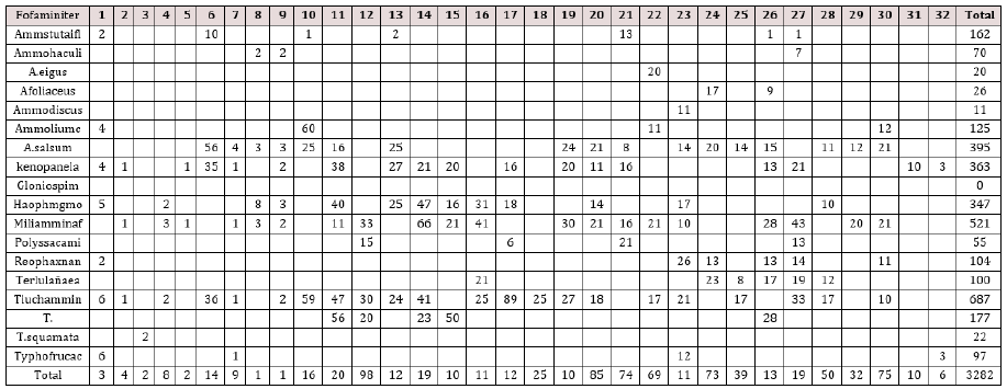
Thirty-two surface samples (10cm3 area, 1 cm deep) were collected along a transect across the Itapitangui river mangrove in July / 89 (Figure 1). The first sample was taken on the channel, and the subsequent ones toward mangrove vegetation at every 50m. Samples were taken from sediment retained around supporting roots from Rizophora Mangle and Laguncularia Racemosa. To collect the foraminiferal samples, it was used a small, serrated corer, as described by [15]. This corer cuts the mangrove material, mainly roots, without compressing anything. Samples containing sediment were fixed with 5% buffered formalin, stained with rose Bengal, washed over at 0.063-micron sieve and dried. In order to separate foraminifera from sediment each sample were floated in carbon tetrachloride and examined for foraminiferal specimens. All specimens of foraminifera were identified and counted. Sediment samples for organic carbon analysis were also collected and performed the Gaudette [16-20] method. Salinity measurements were obtained from interstitial water in bottom sediment. These values were determined using Goldberg 10.433 portable refractometer. The temperature was obtained with manual thermometer. Species diversity index of Shannon-Wiener function H and Simpson Dominance were calculated for each station. The physical and chemical data (salinity and amount of organic carbon) in sedimentation were correlated with foraminifera data (diversity and dominance) through “nonparametric” correlation method of Spearman, accepting as the criterion for recognition, significant differences <0.05. The number of individuals was evaluated to group species and stations. The matrix was logarithmized (log x+1) and submitted to Bray Curtis index. A few samples and rare species were not taken into account for the matrix data. Samples and species that were removed from the calculation were samples 3, 18 and 31, and the foraminifera species Ammobaculites exigus, Ammodiscus sp., Glomospira compressa and Trochammina squamata. The grouping was obtained by minimum variation. The final calculation and dendogram were done by Fitopac program.

Figure 1: Map of study area and location of sample sites.

Lupinepublishers-openaccess-ocean-journal

Results

In the 32 samples collected along the Itapitangui river mangrove transect, was found a total abundance of 3282 individuals, belonging to 18 foraminifera species (Table 1). The foraminiferal fauna from this area is dominated by agglutinated species belonging to Textulariina sub order. Ammotium salsum, Arenoparrella mexicana, Haplophragmoides wilberti, Miliammina fusca and Trochammina inflata are the dominant foraminiferal assemblages found in the mangrove-studied area. The diversity and dominance species index, salinity, and organic carbon values found in Itapitangui mangrove are shown in (Table 2). Minimum diversity values were in sample 29 (0.662) and maximum diversity values were in sample 26 (2.011). The minimum dominance values were in sample 26 (0.137) and the maximum diversity values were in sample 29 (0.516). Bottom salinity values range from 6 to 30. The highest values occur near Itapitangui Sea at the tidal channel, and the lowest value in the upper mangrove region. Bottom water temperature values remaining between 18 and 19 °C. Organic carbon percentage values range from 0.47 and 5.7.

Table 1: The DR lesion types descriptions.

lupinepublishers-openaccess-ocean-journal

Table 2: The DR lesion types descriptions.

lupinepublishers-openaccess-ocean-journal

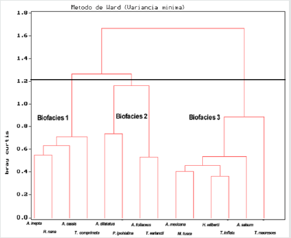
Figure 2: Dendrogram showing faunal similarities of fourteen species.

Lupinepublishers-openaccess-ocean-journal

The cluster analysis, performed on the 32 samples containing 18 species, defined 3 biofacies (Figure 2). The found species of biofacies 1 are Ammoastuta inepta, Reophax nana, Ammotium cassis and Typhotrocha comprimata. Biofacies 2 presents Ammobaculites dilatatus, A. foliaceus, Polysaccamine hypohyaline and Textularia earlandi. Biofacies 3 consists of Arenoparrella mexicana, Milammina fusca, Haplophragmoides wilberti, Ammotium salsum, Trochamina inflata and T. macrescens. The biofacies 3 is characterized by the dominance of true mangrove foraminifera fauna. These findings suggest that foraminiferal zonation is unclear and change in foraminiferal assemblage may not be directly associated with mangroves. On the other hand, mangrove foraminiferal assemblage is rather controlled by others environmental factors such as salinity, temperature and substrate organic content [21]. The physical chemical data (bottom salinity and organic carbon percentage) were correlated with biological data (diversity and dominance). The results obtained in this study have not revealed any clear relations between the environmental parameters and foraminiferal fauna.

Discussion and Conclusion

The mangrove foraminiferal assemblage found in this study presents a low faunistic diversity, probably due to environmental changes such as daily salinity and temperature variations. The foraminifer’s patchy distribution contributes to recognizing several faunal provinces suggesting different microenvironments along the samples collected from the mangrove transect. The foraminiferal fauna identified in Itapitangui river mangrove consists of all agglutinated species, characteristics from mixohaline environments. It was not found any calcareous species from ocean adjacent areas. This result may suggest that there is a minimum ocean influence, through ebb flows, capable of transporting foraminifer’s tests. The high organic carbon percentage present in the transect mangrove, may be responsible for the development of true mangrove fauna entirely agglutinated. Besides that, bottom salinity seems to be another influential factor upon foraminiferal distribution in Itapitangui mangrove. Many authors are in agreement with that, and the importance of this attribute is well discussed [13,16]. The occurrence of agglutinated foraminifera is also probably due to freshwater discharge in the system. According to [17], the impact of fresh water, draining into the estuarine environments, promotes a reduction in calcareous species and increase in agglutinated individuals’ number and diversity. Diversity pattern expected for mangrove area is always lower than coastal regions, due to freshwater dilution from rivers. In hyposaline waters, the number of species varies about 10 species, mostly agglutinated species. In the other hand, in regions where discharge of fresh water is less significant, this number increases over 25, and calcareous shapes predominate. The low number of foraminifera species found in study area is a good confinement indicator of marine influence in mangrove area. Many authors have obtained reductions in the number of species when sampling stations are far from the adjacent ocean region [3,7]. Dominance and diversity results obtained from the foraminiferal fauna, seem to be at random, not showing any kind of distribution pattern. The salinity role upon foraminifera functioning system is getting started.

It is known, however, that effects from reduction of salt in waters may have a great influence on habitat restriction of mixohaline species. In the same way, the lack of inorganic salts availabilty in fresh water is an extreme limiting factor, mainly to the calcareous species, because calcareous species takes from the chemical elements necessary to construct its tests. The assemblage found in this study is considered a true mangrove fauna by the dominance of Mexican, Arenoparrella Milammina fusca, Haplophragmoides wilberti, Ammotium salsum and Trochamina inflata. These species dominate mangroves assemblages in Sumatra. The pattern found in the Itapitangui mangrove indicates that zonation does not seem to exist in this particular environment and foraminiferal distribution shows a uniform assemblage pattern along the study area. Usually, benthic foraminifera can be used as indicator of freshwater influence and gradient of marine influence in mangrove area, but in the present study salinity variation might have not been noted in the area and has not provided any control in foraminifera assemblage able to delimit ecologically different environments.

Read More About Lupine Publishers Journal of Oceanography Please Click on Below Link:
https://oceanography-lupine-publishers.blogspot.com/

Thursday, 18 August 2022

Lupine Publishers | Recent data on the initial Acheulian occupation of the South Caucasus in the Early Pleistocene

 Lupine Publishers | Journal of Anthropological and Archaeological Sciences


Abstract

For a long time all the data indicated that in the Early Pleistocene the South Caucasus was briefly inhabited by Oldowan people (Dmanisi site, ~1,8 MYR), whereas the Acheulian tool-makers appeared in the region only in the Middle Pleistocene. However, studies conducted recently in Armenia, South Caucasus revealed there two Early Pleistocene sites with Early Acheulian industries including around 4000 artifacts. On the basis of U-PB dating and paleomagnetic readings the key Karakhach site was attributed to the Olduvai subchron (1.95-1.78 MYR). The second site named Muradovo has no absolute dates but must be synchronous to Karakhach because of their similarities in both deposits and lithic industry. To judge by scarce paleoecological evidence these sites were occupied in conditions of subtropical climate and periodical volcanic ashfall. Almost all the Early Acheulian artifacts were made of natural slab-like pieces of local rhyodacite. Cores and flakes are rather rare. The large tools are dominated by pics, choppers and crude handaxes. There are also certain special tool types that along with very old age of the considered Early Acheulian industries suggest their local an autonomous appearance.

Keywords:South Caucasus; Early Pleistocene; Early Acheulian occupation

Introduction

Before the beginning of the present century there was only a single evidence of human occupation of the Caucasus in the Early Pleistocene. It was the Dmanisi site, Georgia (South Caucasus) (Figure 1) where deposits dated around 1.8 MYR contained a typical Oldowan industry (small core-choppers and flakes) in association with remains of early hominids [1-3]. Acheulian artifacts found in the region were collected primarily in the numerous surface occurrences and dominated by flat and carefully flaked handaxes indicating Late Acheulian age [4-6]. A majority of these localities were discovered in the volcanic uplands of the South Caucasus (Armenia, southern districts of Georgia) where typical Late Acheulian handaxes are accompanied by the Levallois products. Judging by both absolute dates and bio-stratigraphic data for few stratified sites (Azykh, Kudaro I и III, Cona, Nor Gekhi 1) the Late Acheulian industries existed in the different parts of the Caucasus in the interval around 600-300 KYR [7-10], i.e. in the Middle Pleistocene. Hence, all the research results suggested an enormous chronological break between the Oldowan and Acheulian occupations of the region [6].
However, there were also some reasons to doubt in so late appearance of the Acheulian tool-makers in the Caucasus in comparison with other regions. Today it is known that the earliest Acheulian industries in Africa originated in the East African rift zone just after 1.8 MYR [11-15]. These lithic industries with complicated technologies and diverse large tools (crude handaxes, pics, cleavers, choppers, large scrapers etc.) developed in conditions of semi-open volcanic landscape and on the base of local lava rocks. The first manifestation of the Early Acheulian in the Near East was revealed in the Ubeidiya site (Israel) dated to 1.4-1.6 MYR [16, 17] and located in the Syrian-Arabic, or Levantine rift zone continuing the East African rift system. The Early Pleistocene ecosystems within these areas were analogous and favorable for the Acheulian tool-makers [18]. Since the considered tectonic structures extend further to the south of the Caucasus on may expect to find there a similar palaeoecological situation [6]. Then, it is impossible to agree with N. Rolland that the Armenian high plateau situated between the Near East and the Caucasus was a physical barrier for inter-regional migrations of Early Paleolithic populations [19]. In any times humans were able to overcome this upland thanks to long river valleys and intermountain depressions cutting the high relief in sub-meridional direction [20]. During the Early Pleistocene there was a less elevated relief that facilitated moving of early humans within and across the upland [6, 21].

The northern volcanic areas of the Armenian plateau occupy the south of the Caucasus isthmus. During the Early Pleistocene in the Southern Caucasus there were diversified environments with small-scale topographic complexity (cliffs, lava flaws, natural traps etc.), available water supplies (lakes, springs) and varied food resources obtained from mosaic landscapes [6, 22]. These conditions are believed to be very suitable for inhabitance of Early Paleolithic humans. Moreover, the palaeoenvironmental setting of the Dmanisi site located in the South Caucasus shows striking similarities to those of East Africa and the Middle East such as volcanic landscape with small lacustrine basins and the combination of open and wood ecotones [23]. To judge by analysis of both pollen and fauna composition of the Dmanisi site, around 1.8 MYR in the South Caucasus there was a semi-dry and warm climate similar to the present-day climate of the Mediterranean region [1, 24]. Then, the southern areas of the Caucasus must be especially attractive for Acheulian people thanks to abundance of various volcanic rocks of high knappable qualities which were available as large-sized pieces. It was the ideal raw material for the production of various large tools (handaxes, cleavers, pics etc.) and large flake blanks characteristic for the Acheulian techno-complex [6]. Hence, there are certain prerequisites for an earlier appearance of the Acheulian in the South Caucasus. To verify this speculation, it was necessary to search there for the Early Pleistocene tool-bearing deposits. Accordingly, the specially-purposed studies have been arranged in the beginning of this century in the territory of Armenia.

Materials and Methods

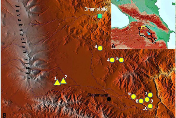
In 2003 the joint Russian-Armenian expedition launched studies for Lower Paleolithic and especially Acheulian traces in the north of the Armenian volcanic upland (South Caucasus, Armenia), in the Lori intermountain depression [6, 25, 26]. This area (Figure 1) was believed to be prospective because of proximity to the Dmanisi site and abundance of lava rocks of the Early Pleistocene age [27, 28] which may be used for manufacturing Acheulian large tools. The lavas as well as tuffs and ashes erupted by the local Javakhetian ridge in the interval of 2.65-1.7 MYR occur in most sediment sections of the area and provided an opportunity for U-Pb absolute dating of the Acheulian artifacts found in these deposits [29]. The dating results were validated with a series of paleomagnetic readings [28]. While no paleontological remains preserved in the acidic volcanogenic sediments, some palaeoecological data were obtained by an analysis of paleosoils [30] and phytoliths, i.e. petrified plant remains. The tools were described in accordance with the classification systems designed for the Early Acheulian industries of Africa and the Near East [31, 32].

Figure 1.

Lupinepublishers-anthropological-and-archaeological-sciences

Studies of the reference Early Acheulian sites

Figure 2:

Lupinepublishers-anthropological-and-archaeological-sciences

Figure 3:

Lupinepublishers-anthropological-and-archaeological-sciences

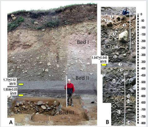
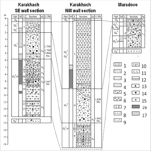
The studied area is the Lori intermountain depression (1400 – 1800 m a.s.l.) located near to the northern border of Armenia (Figure 1). A bottom of the depression is filled by the Gelasian basalts and basaltic andesites with K-Ar dates from 2.51+0.12 to 2.08+0.10 MYR [27, 28]. The lavas came from the volcanic Javakhetian ridge, which flanks the depression in the west. Nearly to this ridge two sites with the very early Acheulian-bearing deposits have been discovered (Figure 1, 2). The key site was revealed in the Karakhach quarry (N41°04ʹ25.64ʺ; E44°07ʹ14.25ʺ; H=1800 m) located at the foot of the Javakhetian ridge [26, 33]. At Karakhach the sediment sequence is observed in two walls of the quarry and in several test pits. The sediments may be subdivided into three principal units designated as the Beds I-III [28]. In the larger north-western wall the sediment sequence is as follows (from the top to downwards (Figure 3):

I. Non-stratified unit composed of badly rounded pebbles and boulders of different volcanic rocks that could be sedimented by a mud-rock flood; up to 9 m. The lower 2.5 m part of the unit has the reverse and lower has normal magnetic polarity
a) White ash with the normal magnetic polarity; up to 0.4 m.
b) Gravel-sand lens with dispersed pebbles; up to 0.8 m. The upper part of the lens shows the normal polarity and the lower part shows the reverse one.
II. Poorly stratified unit of dacite agglomerate tuff, possibly redeposited in the upper part; up to 5 m. The tuff consists of pumice-like clasts of the pebble and rarer boulder size cemented by ach. The SIMS U-Pb dates are 1.944+0.046 MYR 0.5 m above the visible bottom of the quarry and 1.826+ 0.02 MYR in the top of the tuff [28, 29].
III. Lens-type layers of boulders and finer gravel with loam, sand matrix and interbeds of dacitic ash; 3.5 m (visible in the test pit 1).

In the south-eastern wall of the quarry the bed I is reduced up to 1.5 m. The lower part of the bed has the reverse magnetic polarity. The layers Ia и Ib are absent. The layer II (tuff) is 3.1–3.2- m thick and shows the reverse polarity. There are the SIMS U-Pb dates: 1.75+0.02 Ma in 1 m from the bottom and 1.804+0.03 Ma directly above the bottom of the tuff. Additionally, the SIMS U-Pb date 1.799+0.044 Ma was made for the middle part of the Bed II somewhat beyond. The lower levels of the Bed II show the reverse magnetic polarity [28, 29].

The Bed III in the south-eastern part of the quarry was partly excavated in the test pit 2. The observable section 8.2 m thick consists of 14 layers:

a) 1-2: Brownish loams; 0.15–0.5 m. The layer 2 contains large boulders. Redeposited paleosoils [30] with phytoliths.

b) 3-6: Alternation of lens-type layers of boulders, pebbles and finer gravel with loam and sand matrix; 2.7–3 m. All these layers show normal magnetic polarity.
c) 7-9: Ash with the erosional top; ~0.7 m. There is the lens of gravel and pebbles III.8 in 0.1–0.4 m from the top. The layer is characterized by normal magnetic polarity The SIMS U-Pb date is 1.947+0.045 Ma (28, 29).
d) 10: Boulders and pebbles with greyish sandy loam matrix and the ash lens in the middle level; 2.2 m. The loam under the ash has the normal magnetic polarity.
e) 11: Yellow-brownish loam; 0,15 m, paleosoil [30] with phytolyths.
f) 12-14: Layers of boulders and finer gravel with loam and sand matrix; 2 m. Layer 13 includes paleosoil [30].

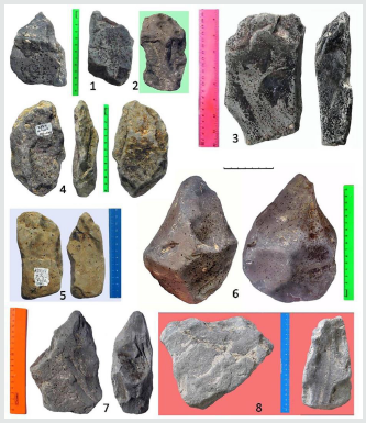
Acheulian artefacts were extracted from the Beds II and III [26, 33]. A majority of finds was made in the Bed III. The test pit 2 near the SE wall yielded 2963 lithic artifacts (Figure 4) extracted from the layers III.2-4 and III.6-14. The richest collections were obtained from the layers 3, 4, 6 and 10. The presence of such large tools as crude handaxes, pics and choppers is indicative of the Early Acheulian industries. Several large flakes marking the Acheulian technologies have been found too. Smaller tools include pointed tools, side scrapers and end scrapers, chisel-ended tools and other variable implements. As most tools were fashioned on natural slab-like pieced of rhyodacite as both flakes and cores are quite rare. Similar technological and morphological features of lithic artifacts in all the layers suggest a single Early Acheulian industry. Analogous artifacts (N=136) were also found in the test pit 1 exposing the Bed III near the NW wall. The lower levels of the Bed II limitedly excavated in the south of the quarry yielded 340 artifacts of andesite. There were several choppers and picks resembling those from unit III, but no handaxes. The Bed II industry is also distinguished for a larger portion of knapping by-products (flakes, chunks, cores) and the predominance of small-sized tools (end scrapers, side scrapers and pointed pieces). The Early Acheulian industry of the unit II may be a successor of that from the unit III. Certain differences may reflect adaptation to raw material of lower quality or/and functional features of the excavated areas.

Additionally, the Early Acheulian industry analogous to the Bed III industry of Karakhach have been found in the Muradovo site (1649 m a.s.l.) located 3.5 km eastwards, on the terrace of a small stream running from the Javakhetian ridge (Figure 1). At Muradovo the sediment sequence has been excavated up to the depth around 7 m from the terrace surface [25, 26, 33]. In the top there are thin horizons of the Holocene soil (layers 1-2) with clearly redeposited Late Acheulian pieces originating probably from eroded Middle Pleistocene sediments. The layer 3 is a yellow-brownish paleosoil yielding a more archaic Acheulian industry with single pics. The lower layers 4-5 and 7-9 may be described as sandy loams rich in boulders, pebbles and gravel, which resemble those of the Bed III at Karakhach. The layer 6 consists of volcanic ash but it has not been dated yet. To judge by increased parameters of magnetic susceptibility and high content of inorganic phosphorus the layers 4-9 were also deposited in conditions of more or less volcanic activity [34].

Figure 4:

Lupinepublishers-anthropological-and-archaeological-sciences

For today the layers4-9 of Muradovjo have yielded about 900 artifacts made of natural slab-like pieces of rhyodacite and andesite. Like in the Bed III of Karakhach large tools in the Early Acheulian industry of Muradovo are dominated by pics, choppers and crude handaxes [26, 33]. In both sites there are also some special tool types such as elongated bar-shaped chisels, fan-shaped choppers and short pics. Besides the Karakhach and Muradovo sites isolated finds of the typical Early Acheulian tools were made in several surface localities in the foothills of the Somkhetian ridge flanking the Lori depression in the east.

Interpreting the research results

The Karakhach quarry sediments lean against the slope of trachydacites with K-Ar date of 1.96+0.08 MYR, which probably form the rock bed of the section. Taking into account the series of absolute dates and paleomagnetic data the Bed III with numerous Early Acheulian artifacts is attributed to the Olduvai subchron with a normal polarity (1.95–1.77 MYR). The overlying Bed II (tuff) with rare Early Acheulian pieces showing primarily reverse polarity deposited in the beginning of the Upper Matuyama, i.e. a little earlier than 1.77 MYR [28]. Most Acheulian layers of the Bed III contain boulders, pebbles and gravel resulted from water activities. However, poor sorting and unequal rounding of the rock fragments indicate rather temporal streams as well as slope washing including local mud flows of low power. The Early Acheulian artifacts have been were found in two test pits at the distance about 100 m and most pieces were slightly rounded. They are unlikely to occur in situ but the displacement was hardly distant. One may suppose that Early Acheulian people lived there not in a certain camp but rather in the particular habitat with small and periodical watercourses that rounded and removed the discarded artifacts within the place before became completely buried. At the same time, both paleosoils and ash lenses preserved within the Karakhach sediment sequence suggest that there were also some periods without considerable water activity. Compared to Karakhach the Muradovo site is situated 3.5 km further away from the Javakhetian ridge and has no absolute dates yet. However, the lower pebble and gravel deposits of Muradovo are very similar to those of Karakhach excluding a presence of interbedded paleosoils [28] and contain an analogous Early Acheulian industry [26, 33] that indicate the same or, at least, somewhat younger age within the Early Pleistocene.
Up to the present certain data on climate and lahdscape were obtained only for the Karakhach site. To judge by characteristics of three paleosoils within the Bed III they formed in sub-tropical conditions [30]. The numerous fytoliths extracted from these paleosoils show the predominance of subtropical plants (A.A. Golyeva, pers. com.). The same climate and vegetation have been interpreted from pollen data for the coeval Oldowan site at Dmanisi [1, 24] situated not more than 25 km north from Karakhach. Given more or less amounts of ash in the Bed III at Karakhach and layers 4-9 at Muradovo, in the period of their formation (subchron Olduvai and possibly somewhat later) the local Early Acheulian people lived in conditions of periodical ashfalls. The Bed III at Karakhach is covered with thick tuff deposits consisting of ashes and pyroclasts (Bed II) where Acheulian pieces were met only in the lower horizons. So, just after the subchron Olduvai an increased volcanic activity gradually led to termination of the Early Acheulian occupation in the Karakhach habitat and probably in its surroundings too.

Concluding Remarks

The reliable results of studies conducted recently in Armenia had revealed that in the South Caucasus there was no chronological hiatus between the Oldowan and the Acheulian occupations. The Acheulian tool-makers first appeared in the region Southern Caucasus in the very Early Pleistocene and inhabited there during the Olduvai subchron (1.95–1.77 MYR) together with the Oldowan people. Then, it should be pointed out that the considered industries have even an older age then the Early Acheulian in East Africa and differ from the latter in both technologies and tool types. In most Early Acheulian industries of East Africa large tools were fashioned mainly on large flakes and for this reason were dominated by handaxes and cleavers. This technological variant of Acheulian was designated by G. Sharon as the Large Flake Based (LFB) Acheulian [35]. In contrast, in the Early Acheulian industries of the South Caucasus a majority of tools was made of natural slablake pieces. In this Slab-like Blank (SB) Acheulian large tools are dominated by pics combined with choppers and crude handaxes without any cleavers. Some special tool types such as elongated chisel-ended tools, choppers of sub-triangular and sub-rectangular form and some others were defined too [33, 36]. Both very early dates and particular features of the considered industries suggest a possibility of autonomous appearance of the Acheulian in the South Caucasus. Then the local Early Acheulian is continued in local Middle Acheulian [33]. It should be noted that another Early Acheulian industry of similar age (1.95–1.78 MYR) was discovered in the Taman peninsula, North-Western Caucasus [37], where might be one more center for development of Acheulian. This evidence perfectly fit with the concept stating that the Acheulian techno-complex independently originated in multiple places of the Old World [38].

Acknowledgments

The author is grateful to the former director of the Armenian- Russian archaeological expedition S. Aslanyan and its present director A. Petrosyan for the possibility to take part in field work in Armenia. Then many thanks are expressed to numerous archaeologists and specialists in natural sciences who provided successful digging the considered sites as well as collecting and interpreting various data. The studies presented in the paper were funded by the Russian Foundation for Basic Research, project 18- 00-00592.

Read More About Lupine Publishers Journal of Anthropology and Archaeology Please Click on Below Link:
https://journalofanthropologicalsciences.blogspot.com/

Monday, 15 August 2022

Lupine Publishers| Headache and Eye-A Mutual Relationship

 Lupine Publishers| Journal of Ophthalmology


Opinion

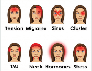
Headache is probably the commonest neurological complaint worldwide. According to the World Health Organization (WHO), 1.7-4% of the adult population of the world have headaches on 15 or more days every month. Sometimes these symptoms are misunderstood and confused with Migraine Attacks or with normal tension headaches. It is also one of the commonest problems because of which people come for a glass/eye check-up. Very often the belief is that a change of eye power is the reason for headaches. Students who constantly keep reading books or keep doing their academic exercises have more chances to suffer from eye related problems like Eye strain, Fatigue, Eye pain, Headache etc. When a patient comes to you with complaints like headache after some continuous reading it is important to be able to distinguish if it is related to the eyes or not. Headache can be felt in a variety of places on the head, including the temples, sinuses, forehead and back of the head. Eye pain associated with a headache can be caused by a few different types of headaches, as well as other seemingly unrelated conditions. But regardless of the cause, this type of pain can be mild, moderate, or even unbearable

Figure.

Lupinepublishers-openaccess-ophthalmology-journal

Figure.

Lupinepublishers-openaccess-ophthalmology-journal

Relation to the Eyes

Ask These Questions to your Patient if they have Complaints Related to Headache (Table 1)

There are many diseases related to the eyes which can cause headache. But there are also some types of muscle imbalances which commonly occurs in young age. Let us discuss about the common causes of headache related to eye which occurs mainly in young adults.

Table 1:

Lupinepublishers-openaccess-ophthalmology-journal

Uncorrected/Under corrected/Over corrected Refractive Error: The prevalence of Refractive error in young adults has increased in past few years [1], it is extremely important to perform the visual acuity test and retinoscopy before going to any other conclusion for the cause. As we all know, the visual acuity of the patient might be 20/20 even when there is manifest hyperopia or small amount of astigmatism is present. All young patient mandatorily undergo cycloplegic refraction regardless of their refractive status or visual acuity. Cycloplegic refraction gives the accurate amount of refractive status of the eye. Be it emmetropia or any types of ammetropia [1].

Convergence Insufficiency (CI): It is one of the most common cause of headache in young adults. It is an eye problem where eyes tend to drift outwards when reading or doing any close work. If this drifting happens it leads to double vision. To prevent the double vision, eyes must work extra hard to turn the eyes back in and this leads to all kinds of asthenopic symptoms like headache, eye pain etc.

Figure.

Lupinepublishers-openaccess-ophthalmology-journal

• Treatment: One of the common treatments of CI is pencil push-up exercise and cat card exercise (Figure 1)

Figure 1.

Lupinepublishers-openaccess-ophthalmology-journal

Accommodative Spasm (AS): It is another common cause of headache in young adults. Due to continuous viewing/reading near objects, especially associated with stressful situations, the ciliary muscles remain in the condition of contraction. This causes headache and transient blurry vision for distance due to pseudomyopia [2]. Advising the patient to avoid near work for some time and do some stress relieving exercises can help.

Treatment: There are various exercises & therapy treatments for the AS. Better consult an orthoptist/vision therapist’s clinic for the treatments of these kind of problems.

Accommodative Insufficiency (AI): It is characterized by an inability to focus or sustain focus at near, demonstrated clinically by an insufficient amplitude of accommodation based on age. Children with poor general health are more prone to this condition.

Treatments: Hart Chart (Figure 2) will help and even cat card (Figure 1).

Read More about Lupine Publishers Journal of Ophthalmology Please Click on below Link: https://ophthalmology-lupine-publishers.blogspot.com/

Lupine Publishers| Non-Alcoholic Fatty Liver Disease and its Interplay with Various Metabolic Disorders

 Lupine Publishers| Journal of Gastroenterology and Hepatology


Abstract

Nonalcoholic fatty liver disease (NAFLD) is described as exposition of multiplex liver metabolic disturbance interconnected with obesity. NAFLD is depicted by steatosis, excessive accumulation of fats in liver, due to triglycerides export and oxidation of fatty acid from plasma and de novo synthesis. Hepatic steatosis can therefore be explained as biochemical outcome of inconsistency between interfused mechanisms of lipid biotransformation. This condition is allied to a range of various modifications in lipoproteins, fatty acids, and glucose metabolisms in organism. So, above metabolic disfunctions are suspected to be the origin of possibility for adverse cardiometabolic risk agents related to NAFLD, like dyslipidemia, Type 2 diabetes mellitus (T2DM), and insulin resistance. Reactive oxygen species (ROS) generation participates as known inducer of inflammation and oxidative stress, that exacerbate this disease. These disorders are hallmarks that worsen NAFLD complications, so far participate in developing advanced stages of NAFLD and incline the body to CVD and T2D. The reciprocal risks exist among these diseases. Given the sharp growing prevalence and persistence of NAFLD, and its complexity that provoke additional metabolic syndrome, this review discusses various mechanisms of developing NAFLD, interaction with other associated hallmarks, aiming to clarify beneficial mechanisms for improvement.

Keywords: Nonalcoholic fatty liver disease; oxidative stress; Type 2 diabetes; noncoding RNAs; cardiovascular disease

Introduction

The liver is known as metabolically complex organ due to its multiple biological activities, involving detoxification of various endogenous metabolites and exogenous toxic substances, formation of biochemicals needed for degradation and the synthesis of protein. The liver also serves an exigent task in metabolisms like lipid homeostasis and glycogen storage regulation [1]. Increased activity of mitochondria consequent to the fatty acids hyper-afflux from oxidation of fatty acid, produce free radicals that generate liver oxidative stress [2]. Abnormal function of liver may engender several metabolic impairments, such as NAFLD (Marrero et al., 2002). NAFLD is explained as the grouping of excess fat into liver cells, that does not result from alcohol consumption. The existence of fats in the liver is normal; however, if the concentration of fats exceeds 5%-10% of the weight of liver, then it is called a fatty liver (steatosis). Thus, NAFLD is explained as the amassing of liver fat of >5% of liver weight with <10g of daily alcohol consumption (Bayne, 2010). Besides these, generation of NAFLD was discovered to be linked with insulin resistance, the latter is known also as a critical risk factor for developing type 2 diabetes (T2D) [3]. NAFLD comprise a large spectrum of manifestations extending from simple steatosis, continuing to nonalcoholic steatohepatitis (NASH) and cirrhosis. Furthest, NAFLD correlates with significant higher risk of developing hepatocellular carcinoma (HCC) [4]. The worse hallmark of this disease is the significant interdependence with various attributes of metabolic syndrome (MetS), such (T2DM), obesity or dyslipidemia [5]. MetS is hallmarked by the aggregation of several impairments involving elevated blood pressure, obesity, dyslipidemia, insulin resistance and proinflammatory activities [6]. Moreover, studies by different researchers elucidated NAFLD as the leading inducer of liver diseases and the main reason for impaired liver function worldwide; also considered as an inherent part of MetS (hypertension, hyperglycemia, central obesity and dyslipidemia) [7,8]. The evolution of MetS has been coincided with a growth in liver disorders including NAFLD, and was revealed to correspond with disorders, like cardiovascular disease (CVD). Particularly, NAFLD has been taken as hepatic exposition of MetS [9].
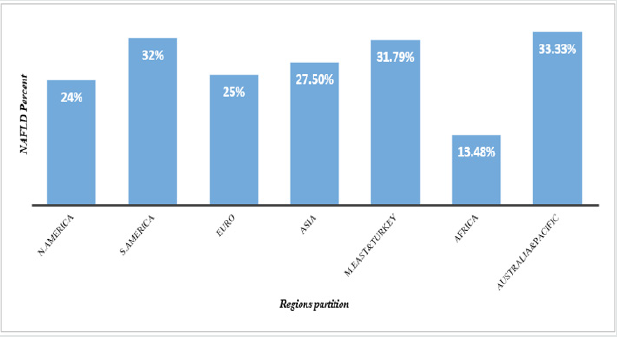
The NAFLD prevalence increases quickly around the world and noted as a camouflaged epidemic. The Universal estimation of NAFLD prevalence in general population has overpassed 25% [10], See Figure 1. In developing countries such as, China; and India, the number of NAFLD patients has been augmenting sharply over the last decades [11]. However, no medications presently approved for NAFLD. The primary therapeutic intervention in NAFLD, same as in other MetS is gained from lifestyle improvement, promoting equitable low-energy diet, together with promoting physical activity. So, these are prime remunerative approaches for this condition [12,13]. Lifestyle moderation has improved the metabolism syndrome features; however, more effort needs to be made in addressing various MetS components [14]. Thus, to provide better understanding that will help different players involved in management of MetS disorders. This review discusses various mechanisms of developing NAFLD and its interplay with other metabolic disorders, while clarifying beneficial mechanisms for improvement.

Figure 1: NAFLD Prevalence in global remarkable regions.
N. America; North America, S. America; South America, Euro; Europe, M. East; Middle East.

Lupinepublishers-openaccess-gastroenterology-hepatology-journal

Prevalence of NAFLD

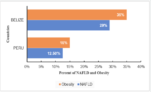
Figure 2: Influence of obesity on NAFLD.

Lupinepublishers-openaccess-gastroenterology-hepatology-journal

The NAFLD prevalence increases quickly in every region in the world as a masked epidemic. The worldly estimation of NAFLD prevalence in general population has surpassed 25% [10] (Figure 2).
In region of South and Central America, NAFLD prevalence varies depending on obesity rate, where Belize has the highest obesity prevalence of 35% with NAFLD 29% and Peru has the lowest obesity prevalence of 15% with NAFLD 12.5% [10]. The Occurrence of obesity aggravate NAFLD prevalence. In addition, NAFLD is sharply elevating worldwide, coincident with the augmented prevalence of obesity. Currently NAFLD has become the major chronic liver disease; NAFLD prevalence in adult population of developed countries is approximately 30% [9]. NAFLD has also become a considerable liver disease in children in response to the elevation of childhood obesity prevalence. Obesity may initiate production of excess ROS and systemic oxidative stress [15] which result in protein and lipid oxidation [16] (Figure 3).

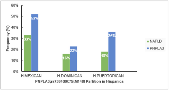
Figure 3: Influence of Genetic PNPLA3 on NAFLD in USA.
H. Puerto Rican; Hispanic from Puerto Rican origin, H. Dominican; Hispanic from Dominican origin, H. Mexican; Hispanic from Mexican origin.

Lupinepublishers-openaccess-gastroenterology-hepatology-journal

The study from sub-region of USA that compared NAFLD in Hispanic from different origins, showed the greatest NAFLD prevalence in Hispanics from Mexican origin (33%) than those from Caribbean origin (Puerto Rica (18%), Dominic Republic (16%)), (P<0.01). The greater NAFLD prevalence remained in Hispanics of Mexicans than Dominican or Puerto Rican origin, even after regulating other factors that discovered to contribute in NAFLD; such as insulin resistance, levels of triglyceride and C-reactive protein, hypertension, serum level of high-density lipoprotein, waist circumference, body mass index (BMI), sex and age [17]. Prevalence of NAFLD might be explained by elevated polymorphism prevalence in the gene encoding patatin-like phospholipase domain-containing 3 (PNPLA3; rs738409 C/G, M148I); especially in Hispanics where it accounts (49%), compared to Non-Hispanics whites (23%) and African-American (17%) [18,19]. Moreover, PNPLA3; rs738409 counts 52% in Hispanics Mexican, 23% in Dominicans [20] (Eric et al., 2019) and 36% in Hispanics Puerto Rican [21]. According to a genome-wide association study [19] elucidated that a non-synonymous sequence variation (rs738409) in PNPLA3 that replaces methionine for isoleucine at residue 148 (I148M) correlates with disproportion in liver lipid profile and the possibility of generating NAFLD. Thus, higher PNPLA3; rs738409 C/G, M148I in Hispanics suggested to take part in fueling NAFLD high prevalence [22] (Figure 4).

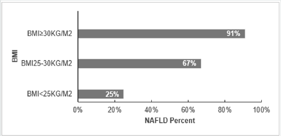
Figure 4: NAFLD Prevalence with BMI in Europe.

Lupinepublishers-openaccess-gastroenterology-hepatology-journal

BMI correlates with NAFLD, and it has been revealed as a factor that elevates the probability of emerging NAFLD. In years, 1980-2008, the BMI-mean augmented worldwide by 0.4 kg/ m2 for males and by 0.5 kg/ m2 for females per decade. This coincides with obesity prevalence that increased simultaneously from 4.8 to 9.8% for men and from 7.9 to 13.8% for Women [23]. This factor was investigated in the region of European Union and it’s clear that similarly to other regions, in Europe the higher risk of acquiring NAFLD is elevated coincidently with the increases of BMI, 25% (BMI<25kg/ m2), 67% (BMI 25-30 kg/ m2) and 91% (BMI ≥30 kg/ m2) [24]. Together with other risk factors (obesity or T2DM) which more likely associate to sedentary life, are increasing in Europe fueling the NALFD prevalence and further the impairments such as cirrhosis and HCC [25].

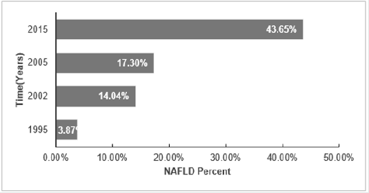
The NAFLD prevalence among adults of sub- region Shanghai was considered to show high growing trend of NAFLD with time, it’s obvious that NAFLD prevalence is elevating with time [26]. The research showed diversity of NAFLD prevalence in different subregions of Asian countries, in Taiwan (15%-27%), Korea (24%- 40%), and Japan (9%-18%) [27]. Additionally, prevalence in India has increased from 28% in 2015 to 31% in 2016 in the rural region of Haryana [28]. Timely elevation of Fatty Liver was also revealed in different sub-regions of China [29]. Shanghai with high rate in development that goes together with life behavioral changing; the incremental in NAFLD is seen to increase sharply in adults in recent years. Findings supported by [30-33], shown in Figure 5.

Figure 5: Timely prevalence of NAFLD in shanghai adult.

Lupinepublishers-openaccess-gastroenterology-hepatology-journal

NAFLD Pathogenesis

Numerous researches have reported that metabolic impairment or disturbance is the basic abnormality in NAFLD [34]. At the center of this discovered metabolic abnormality there is insulin resistance, that also known among the initiators of NAFLD. Thereafter, by provoking oxidative stress, fatty liver may evoke inflammation and hepatocyte injury which may cause the disease to tend to NASH and cirrhosis. The overload of triglycerides (TG) as fat droplets in hepatocytes cytoplasm was reported as the main event of NAFLD, which is a precondition for the succeeding NASH, as it was shown by liver biopsy that 5%-10% of hepatocytes have fat droplets [35]. Increased moving of both TG and free fatty acids (FFA) to the liver, reduced hepatic using of FFA, decrease in export of TG from liver, disturbed beta-oxidation of FFA in hepatocytes result in stockpiling of TG within hepatocytes cytoplasm [34,36,37]. Another main stimulus for liver de novo fatty acid synthesis, is the surplus carbohydrate from either dietary origins or hepatic de novo gluconeogenesis [36,37].
Obesity can be defined as a chronic low-grade inflammatory condition. There are cytokines related to obesity, such as interleukin-6 (IL-6), leptin, adiponectin, and tumor necrosis factoralpha (TNF-α) that play a remarkable role in NAFLD evolution. Research findings reported that adipose tissue may be the inducer of inflammatory mediators and adipokines, such as pro-inflammatory ones (IL-6, TNF-α, and leptin) and anti-inflammatory (adiponectin) effects [38]. Even though, these hormones and cytokines may ordinally work in balance, the homeostasis is disturbed in NASH that result in increased TNF-α and reduced adiponectin levels. Several mechanisms that likely involve in hepatocellular injury in setting of NAFLD, many of them produce the ROS. A liver with surplus fat can be more prone to stressors, such as adipokines, reactive ROS, and cytokines than normal liver [39,40].
In the study done by Yang and colleagues, obese mice with fatty liver cleared endotoxins less than nonobese controls [38]. Factors that perform key functions in the evolution of NASH from simple steatosis remain unclear [41-43]. Some known possible ways are oxidative stress through increasing ROS and reduced antioxidants, lipid peroxidation, reactive metabolites, 4-hydroxynonenal and malondialdehyde, adipose tissue products. Other discovered ways are Fas-ligand, transforming growth factors-β1, respiratory chain deficiency along with mitochondrial dysfunction, and intestinal microbiota by augmented intestinal permeability, elevated energy gaining from diet, intestinal leak, bacterial lipopolysaccharides (LPS), TNF-α, and endotoxins [44].

Hallmarks in NAFLD

NAFLD and oxidative stress

Oxidative stress increases the loss of structure and function of healthy cells, DNA and also damage of important macromolecules. These conditions are the main roots for chronic diseases such as cancer, stroke, cardiovascular impairment including diabetes [45]. Oxidative stress has been found to disrupt insulin signaling process, which result in insulin resistance in cell [46]. Yet, impaired insulin signaling mechanism remains unclear [47] (Rains & Jain, 2011). Besides, Elevation of oxidative stress is generally a considerable factor in degenerative diseases, including chronic fatigue neurodegenerative diseases. Moreover, oxidative stress can be suggested to mediates the transition from simple steatosis to steatohepatitis and, disruption of metabolic balance. Furthest, the above cited impairments are the crucial features that lead to steatohepatitis in lipid-laden hepatocytes [48]. Oxidative stress arises due to the lower antioxidant capacity and/or overproduction of the ROS [47]. Various mechanisms by which these two scenarios initiate oxidative stress in organism are described in the following paragraphs.

Several factors initiate oxidative stress in diabetes. The main origin of oxidative stress is mitochondria. Mitochondria use around 98% inhaled oxygen, from which 0.2-2% produce ROS [47,49]. A part of the used oxygen is reduced to water, and the left oxygen converted to oxygen free radicals through oxidative metabolism of mitochondria [50]. Oxygen is required for human life, even though, it may damage and kill the cells when it produces ROS [51]. The ROS are engendered by the reduction of molecular oxygen or from oxidation of water to give products such as superoxide anion, hydroxyl radical, hydrogen peroxide [47]. The hyperglycemia promotes low density lipoprotein (LDL) peroxidation and followed by the production of free radicals [52,53]. The other significant factor in the production of free radicals is glucose oxidation. In its enadiol form, glucose become oxidized in a transition-metal dependent reaction to give enadiol radical anion transformed into reactive radicals [54]. Other mechanism of oxidative stress in diabetes is the engendering of advanced glycated end products (AGEs) [55]. AGEs resulted from the covalent binding of the ketone or aldehyde groups of reducing sugars, in their way to free the amino groups of proteins [47].
Alternatively, Antioxidant may be defined as a substance that delay or inhibit the oxidation of substrate, this scenario comprises many mechanisms pointed out in oxidative stress production pathways. There exist various exogenous and endogenous components which can perform a considerable function in antioxidant defense and help in prevention of oxidative stress in organism including Catalase (CAT), glutathione peroxidase (GPx), superoxide dismutase (SOD) [56]. The alteration of endogenous antioxidant defense comes in case of hyperglycemia. In diabetes, both decreases and increases in the activities of major antioxidant enzymes like CAT, SOD, Glutathione reductase (GR), GPx have been noticed [57]. Studies have shown that in pancreatic islets 8-hydroxy-2-deoxyguanosine (8-OH-dG) and 4-hydroxynonenal (4- HNE) levels augmented which support that hyperglycemia might be a key inducer of oxidative stress in the β-cell and oxidative stress induced by glucose [58]. The β-cells of pancreatic islets are vulnerable to the genesis of ROS and the reduction of antioxidant enzymes activities [59]. In Goto-Kakizaki (GK)-rats, the levels of 8-OH-dG and 4-HNE were high in β-cells of pancreatic islets [60]. Many reports have elucidated that the increasing of peroxide content in tissues, plasma, and red blood cells of animals that have chemically induced diabetes [61,62].

Read More about Lupine Publishers Journal of Gastroenterology and Hepatology Please Click on below Link: https://currenttrendsingastroenterology.blogspot.com/