Showing posts with label Journal of Pediatric Dentistry. Show all posts

Wednesday, 20 September 2023

Lupine Publishers | Treatment of Infected Primary Teeth using Modified Antibiotic Paste

 Lupine Publishers | Journal of Interventions in Pediatric Dentistry


Abstract

Objectives: Treatment of pulpectomized primary molars with chronic infection using a mixture of three antibiotics: Metronidazole, Ciprofloxacin, and Doxycycline mixed with Macrogol or Propylene Glycol (modified 3MIX-MP) as an intracanal medicament before the complete cleaning and shaping and obturation.

Study design: A 7 years old child with infected primary molar came to our clinic for treatment. A detailed medical history and drug allergy were taken. Ciprofloxacin (500mg), Metronidazole (500mg) and Doxycycline (100mg) tablets divided in the proportion of 1:3:3 (one part of Ciprofloxacin, three parts of Metronidazole, and three parts of Doxycycline) and mixed with propylene glycol to form an ointment. Biomechanical preparation was done. The modified 3MIX-MP paste placed in the pulp chamber then temporary filling. The patient was recalled after 2 weeks. The tooth was obturated and restored then a stainless-steel crown placed. Then reevaluated at 3rd, 6th, and 12th months.

Results: Excellent clinical and radiographic success when compared to conventional pulpectomy and non-instrumentational lesion sterilization tissue repair therapy.

Conclusion: Treatment of Primary molar with modified 3MIX-MP, followed by instrumentation and obturation provided excellent clinical and radiographic success when compared to non-instrumentational lesion sterilization tissue repair therapy.

Keywords: Pulp infection; Pulpectomy; Modified antibiotic paste; Primary molars; Chronic, infected pulp; Modified 3 MIX-MP; Pulpectomy; Triple antibiotic paste; Primary teeth

Introduction

The first topical antibiotic introduced to endodontics was Grossman’s polyantibiotic paste in 1951, later many topical antibiotics have been introduced with varying combinations, few of those include Septomixine forte; PBSC (Combination of Penicillin, Bacitracin, Streptomycin and Caprylate sodium), and Clindamycin. However, none of these combinations has proven to be 100% successful in eliminating all the bacterial strains from the root canal system [1-5].

Materials and Methods

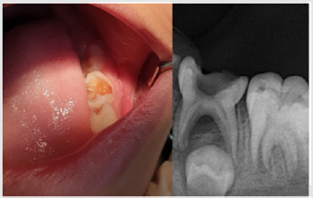
A child aged 7 years old with chronic infection related to the lower left primary molar came to our clinic for treatment of the infected molar (Figure 1). Treatment was explained to the parents and written informed consent was taken from parents before start of the study. A detailed medical history and previous illness with a history of drug allergy were taken from the parents, then the mentioned primary molar was diagnosed clinically, the molar was badly decayed with signs of chronic infection such as: gingival swelling and tenderness to percussion. A radiographic examination was done and a per radicular radiolucency was found, with no excessive root resorption. Commercially available chemotherapeutic agents such as Ciprofloxacin (500mg) (Omacip, NPI Pharma, Oman), Metronidazole (500mg) (Anazol, JPI, Saudi Arabia), and Doxycycline (100mg) (Tabocine, TPMC, Tabuk) tablets were obtained [6,7], then these tablets were crushed into fine powder using sterile porcelain mortar and pestle. These powdered drugs were transferred into three separate sterile glass containers, capped tightly and stored in the refrigerator until its use. Just before use, each powdered drug was divided in the proportion of 1:3:3 (one part of Ciprofloxacin, three parts of Metronidazole, and three parts of Doxycycline) and were mixed with propylene glycol and polyethylene glycol to form an ointment. Reddy GA et al. Trairatvorakul and Detsomboonrat, Jaya et al., Cruz et al. also followed the similar protocol of preparation of 3MIX antibiotic paste [8-11].

Figure 1: Preoperative illustration.
Lupinepublishers-openaccess-pediatric-dentistry-journal

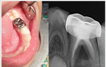
Figure 2: Postoperative illustration.
Lupinepublishers-openaccess-pediatric-dentistry-journal

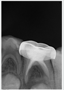
Preoperative clinical and radiographical signs and symptoms were recorded. The tooth was anesthetized using 2% Xylocaine with 1:80,000 adrenalin and isolated with rubber dam. Access opening was performed using round bur, Biomechanical preparation was done using k files from size 10–25. The root canals were chemically cleaned with 1% sodium hypochlorite solution and dried with paper points. The 3MIX-MP paste placed in the pulp chamber and pressed with dampened cotton pellet and temporized with Cavit. The patient was recalled after 2 weeks for evaluation. The tooth was obturated with reinforced zinc oxide eugenol (IRM, Dentsply) using lentulo spirals. Then restored with glass ionomer restorative material (Riva self-cure, SDI) and reinforced by placing stainless steel crowns (Figure 2). Further, the treated tooth was reevaluated both clinically and radiographically at 3rd, 6th, and 12th months intervals postoperatively (Figure 3). At the time of revisits, the tooth was examined clinically for any signs of failure that includes a report of spontaneous pain, presence of swelling, sinus tract and mobility. Radiographic evaluation was done to check the radiolucency and signs of resorption. The tooth was asymptomatic without pain, swelling, sinus tract and mobility also there was no increase in furcation radiolucency or development of root resorption which is abnormal for the age of the child.

Figure 3: 12 months Follow up.
Lupinepublishers-openaccess-pediatric-dentistry-journal

Results

Excellent clinical and radiographic success when compared to conventional pulpectomy and non-instrumentational lesion sterilization tissue repair therapy.

Discussion

This study was approved by “Research Ethics Committee, Taibah University, College of Dentistry, TU CD-REC”. The concept of Non-Instrumentation Endodontic Therapy introduced by Niigata university school of dentistry; Japan has gained reputation as it proved to attain 100% sterility in the root canal system [12- 15]. They recommended a technique similar to pulpotomy where debriding only the pulp chamber of chronically infected primary teeth and placing medicament (ciprofloxacin, metronidazole, and minocycline) near the root orifice without preparing the radicular portion. Cruz et al. suggested vehicles such as macrogol and propylene glycol (3MIX–MP) and demonstrated that these vehicles will carry the medicament deep into the dentinal tubules, thus aid in effective eradication of bacteria [11]. Metronidazole (Nitroimidazole compound) due to its wide spectrum of antibacterial action against anaerobes (Ingham et al. 1975) gained importance as the 1st choice drug for triple antibiotic paste preparation [16,17]. Metronidazole binds to the DNA and disrupts its helical structure and thus leads to rapid cell death. However, metronidazole even at higher concentrations could not eradicate all the bacteria thus indicating the necessity of some additional drugs to sterilize these lesions [15]. The two other antibacterial drugs, i.e. ciprofloxacin, and minocycline, in addition to metronidazole (3MIX) were added in an effort to eliminate all bacteria [8,10,15,18]. The 2nd choice of drug ciprofloxacin is a synthetic fluoroquinolone with rapid bactericidal action. It inhibits the enzyme DNA gyrase of bacteria. It exhibits very potent activity against Gram-negative bacteria but very limited activity against Gram-positive bacteria. Most of the anaerobic bacteria are resistant to ciprofloxacin. Hence, it is often combined with metronidazole in treating mixed infections. The 3rd choice of drug was minocycline. It is a semisynthetic derivative of tetracycline, primarily bacteriostatic, inhibiting protein synthesis by binding to 30S ribosomes in susceptible organisms and exhibits broad spectrum of activity against Gram-positive and Gramnegative microorganisms [3].

In our present study we replaced Minocycline with Doxycycline due to the difficulty in obtaining Minocycline, and before using the Doxycycline as a replacement we have done further searches for previous studies to ensure that both medications have the same effect and this replacement will not affect the efficacy of the mentioned mix. The already done studies concerning the difference between both Doxycycline and Minocycline revealed that still no statistically significant differences had been demonstrated in clinical trials when comparing Minocycline with Doxycycline, and investigators had concluded that both are equally effective. And they differ in their adverse event profile [19]. Considerably fewer adverse effects have been reported for Doxycycline than Minocycline; the adverse effects for Minocycline are 5 times more common than for Doxycycline [19]. We have followed the same protocol of Reddy GA et al. of extirpation of both necrotic coronal as well as all accessible radicular pulp tissue and then complete obturation, which is reported successful clinically over 16th month follow-up [9]. Although the previous studies have demonstrated that the LSTR (Lesion Sterilization Tissue Repair) technique as one of the successful techniques for management of chronically infected primary teeth, the controversies aroused about the duration of therapeutic activity of the medicament and leaving the infected material in the radicular region. So that the present study planned where in treated tooth were revisited after 2 weeks for medicament removal and obturation.

Conclusion

All the primary teeth with chronic infection which were treated using modified 3MIX-MP, followed by the instrumentation and obturation provided excellent clinical and radiographic success when compared to conventional pulpectomy and noninstrumentational lesion sterilization tissue repair therapy.

Read More About Lupine Publishers Journal of Pediatric Dentistry Click on the Below Link:

https://lupine-publishers-pediatric-dentistry.blogspot.com/


Wednesday, 26 July 2023

Lupine Publishers | Varied clinical and Oral Presentation of Beckwith – Wiedemann Syndrome - Report of a Case from Saudi Arabia

 Lupine Publishers | Journal of Pediatric Dentistry


Abstract

Beckwith – Wiedemann syndrome is congenital, genetic and epigenetic pathologies with low prevalence and diverse clinical presentations. It is characterized by triad of omphalocele, macroglossia and gigantism. This syndrome has been widely studied with a current emphasis on improvement of prenatal diagnostic techniques and a multidisciplinary approach towards treatment. We report a case of BWS from Saudi Arabia, with unique presentations and misleading history which delayed diagnosis, due to cultural and religion constraints.

Keywords:Congenital; Epigenetic; Genetic; Prenatal

Introduction

Genetic and epigenetic changes or a human genomic imprinting disorder is characterized by phenotypic variability which might shows its occurrence either as sporadic or inherited. The pathology presents wide range of effect on psychological and social wellbeing of patients and families. One such congenital, multigenic, multisystem human genomic imprinting disorder with complex molecular etiology and variable complex phenotype is Beckwith – Wiedemann Syndrome (BWS). Beckwith-Wiedemann Syndrome is most common overgrowth syndrome described by Beckwith in 1963 and Wiedemann in 1964 with similar findings. It is rare congenital deformity with low prevalence but at same time have high prevalence within genetic abnormalities of overgrowth [1]. The presentation of triad features of omphalocele (exomphalos), macroglossia and gigantism was described earlier as EMG syndrome which now is referred as Beckwith – Widemann Syndrome. The incidence of BWS reported is approximately 1:13700 births and the major cause is thought till date is genetic and epigenetic defects within the chromosome 11p15.5 regions [2].

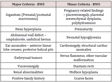
BWS presents wide array of clinical manifestations such as congenital abdominal wall defects as hernia (exomphalos), gigantism, macroglossia, nevus flammeus, ear pits/hearing loss, midface hypoplasia, cardiac anomalies, hemihypertrophy, genitourinary anomalies and musculoskeletal abnormalities. To standardize the diagnostic criteria various attempts have been made to classify the major and minor criteria. Elliot et al described the diagnosis of BWS with the presence of either three major features (abdominal wall defect, macroglossia, gigantism) or two major and three minor features (ear pits, nevus flammeus, hemi hyperplasia, nephromegaly, neonatal hypoglycemia) [3]. In spite of diverse clinical presentations of BWS, most of the cases do not show characteristic features at birth but develop later in life. Also, children with BWS have significantly increased risk of cancer during early childhood which need strict follow up and monitoring. Here, we present a case of BWS with unique dental and medical presentation and its differential diagnosis with literature review.

Case Report

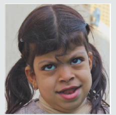
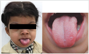
A 5-year-old female patient, accompanied by her mother, presented to the dental unit with complaint of decay tooth in upper front region of mouth. Extra oral examination revealed dysmorphic features, coarse facies and developmental problems (Figure 1). Intra oral examination of hard tissue showed high arched palate, decayed teeth in relation to 51, 52, 55, 61, 62, 74, 75, 84,85. Oral soft tissue examination revealed macroglossia, enlargement of fungiform papillae and mild loss of filiform papillae (Figure 2). Speech and feeding difficulty were noticed due to macroglossia. History revealed she is the youngest 7th child born out of consanguineous marriage in 30th week by cesarian section. She has a chronic history of constipation for 9 months of age. She passes hard stool once in every 8 to 10 days, by spending long time in washroom. It is associated with decrease in appetite and abdominal pain. She was given Movicol (half the adult dose) twice a day for constipation without any medical prescription. She was also tried with lactulose, glycerin suppository and mineral oil. Under medical supervision fleet enema and contrast enema were performed to relieve constipation and to rule out Hirschsprung disease.

Figure 1: Photograph showing dysmorphic features and hypertelorism.

Lupinepublishers-openaccess-pediatric-dentistry-journal

Figure 2: Macroglossia with enlarged fungiform papillae and loss of filiform papillae.

Lupinepublishers-openaccess-pediatric-dentistry-journal

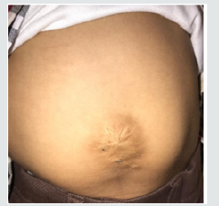
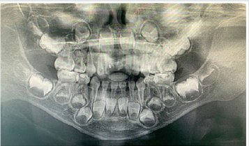
Other medical findings noticed omphalocele, ear pits, large child at 90th centiles, large rounded eyes with hypertelorism, abdominal soft lax, enlargement of kidney, distention of left renal pelvis with significantly distended urinary bladder, abnormal anatomy of the colon located in left abdomen and partial colonic non – rotation with no evidence of obstruction (Figure 3). Based on the clinical and past medical history a diagnosis of Beckwith – Wiedemann Syndrome (BWS) was made. Series of laboratory investigation were reviewed which presented negative urine examination, alpha – fetoprotein, karyotype, microarray and methylation analysis for BMS. Patient was advised for gene analysis and targeting testing for parents. The gene analysis of CDKN1C gene showed heterozygous alteration consistent with BWS but targeting gene tests were refused by parents. Panoramic radiograph was advised considering the patient chief complaint, which revealed multiple developing permanent tooth buds, protrusion of anterior teeth, open bite and increase in mandibular dimension (Figure 4). Under preventive measures the patient was treated for the decayed teeth and is under follow up from past 6 months.

Figure 3: Photograph showing abdominal wall defect with surgical scar.

Lupinepublishers-openaccess-pediatric-dentistry-journal

Figure 4: Panaromic radiograph showing multiple developing permanent tooth buds, open bite and increased mandibular dimension.

Lupinepublishers-openaccess-pediatric-dentistry-journal

Discussion

Diagnostic criteria for BWS is still a matter of research due to its varied clinical presentations and overlapping features with other various conditions. The presence of major and minor findings is generally helpful in establishing the clinical diagnosis (Table 1). The oral findings as mentioned in the literature and observed in our case has been tabulated in Table 2 [4,5]. The incidence of BWS is difficult to assess in Saudi Arabia, as most of the cases goes undiagnosed and unnoticed. Also attributed to its diverse clinical presentation and difficulty in diagnosing. In the present case, features of macroglossia, macrosomia, omphalocele, abdominal wall defect (treated immediately after birth and surgical scar observed clinically), Renal involvement, ear crease, high arched palate, open bite and increased mandibular dimension, leads to the diagnosis of BWS. Various molecular mechanisms and alterations have been involved in BWS such as abnormal methylation of H19DMR, loss of imprinting of IGF2, chromosomal rearrangements, loss of imprinting of LIT1, uniparental disomy of 11p15 and CDKN1C mutations [2]. The full gene analysis of CDKN1C gene profile were suggestive of BWS in our case and the alteration is thought to be located in the allele inherited from the mother. Parental testing was advised which was refused by the parents. There are various endocrine and overgrowth syndromes that was considered in the differential diagnosis. These included Simpson-Golabi-Behmel syndrome (mutation in X-linked gene, GPC3), Perlman syndrome (Increased risk of neonatal mortality), Costello syndrome (missense mutation in HRAS), Sotos syndrome (Mutation in NSD1) and Mucopolysaccharidosis type IV (lysosomal storage disorder) [6]. Oral findings like macroglossia of BWS needs differentiation from other lesions like lymphangioma, idiopathic muscular hypertrophy, hemangioma, rabdomyomas, amyloidosis, cretinism and acromegaly.

Table 1: Presenting major and minor features of BWS.

lupinepublishers-openaccess-pediatric-dentistry-journal

Table 2: Oral findings of BWS.

lupinepublishers-openaccess-pediatric-dentistry-journal

The overall risk of BWS for tumor development/malignancies is estimated to range from 4 – 21%. The tumors reported with BWS are mainly embryonal tumors such as Wilms tumor, hepatoblastoma, rabdomyosarcoma, adrenocortical carcinoma and neuroblastoma [7]. The prenatal diagnosis with current technology is increasing representing an important tool to determine some features of BWS before birth. In our case, parents were highly orthodox and refuse to share the detailed prenatal and ultrasonic reports. Few misguided information’s were given by mother which was later clarified with the reports from the subsequent medical hospitals. Patient’s parents were advised for periodic follow up with genetic counselling and the possibility of surgical interventions in the medical units, but they refused to follow and changed the hospitals every time. Hence, an effort was put forward to retrieve the information’s related to the patient while giving her the primary treatment for which she reported to our dental unit. This suggest the need of awareness required in the country like Saudi Arabia, where most of the cases goes unreported/unnoticed or parent’ consent not given or the cultural and religion barriers that prevent reporting such cases. Though the patient was treated with dental fillings, the follow up of the patients is been restricted by the family members.

Conclusion

Beckwith – Wiedemann Syndrome patients usually grow and do well despite being at increased risk of childhood cancer. Hence, strict follow up, awareness of parents and cancer screening is mandatory. Families, physicians and dentists should determine screening schedule including abdominal ultrasound in every three months, blood test to measure alpha-fetoprotein in every six weeks, dental check-up in every six months and other symptomatic treatment schedule as and when required.


Read More About Lupine Publishers Journal of Pediatric Dentistry Click on the Below Link:

https://lupine-publishers-pediatric-dentistry.blogspot.com/


Wednesday, 31 May 2023

Lupine Publishers | Paediatric Dentistry and Prevention from Teenage Pregnancy

 Lupine Publishers | Interventions in Pediatric Dentistry Open Access Journal


Abstract

Medical specialties have paid little attention to the teenage stage, including Dentistry. If to this we add the teen pregnancy trends due to economic, social or cultural circumstances, the results are young women with a high prevalence of caries, periodontal disease and early tooth loss.

Objective: To detect teenagers at risk through different educational institutions such as churches, health centers, sports clubs, etc.

Situation Analysis: The field study revealed a worrying number of adolescents who drop out of school or job due to pregnancy at a young age.

Intervention Strategies: Three different levels of prevention were stablished

a) Primary prevention: measures intended to prevent teenage pregnancy.

b) Secondary prevention: measures adopted when there was evidence of an ongoing pregnancy.

c) Tertiary prevention: measures taken to promote school reinsertion and reintegration into the labor market.

Results: Teachers, doctors, dentists, obstetricians, physical therapists, neonatologists and pediatricians worked in a multi and interdisciplinary way in order to educate and promote healthy living, and to avoid risk situations in this age group.

Keywords: Teenage pregnancy; Caries; Oral health; Education; Prevention

Abbreviations: UNFPA: United Nations Population Fund in Argentina; UNICEF: United Nations International Children’s Emergency Fund

Introduction

The topic to be developed is about an issue which has arisen in important sectors of young people from Chaco. Around sixteen million of young women between the ages of fifteen and nineteen give birth each year - roughly eleven percent of all births worldwide. Complications related to childbirth and pregnancy is the main cause of death among adolescent girls, especially in developing countries. In Latin America, ten percent of girls aged fifteen to nineteen are mothers. In Argentina, the number of teenage pregnancies has increased since 2001, representing a sixteen percent of pregnancies. The percentage recorded in some areas such as the Argentinian northeast and west was twenty five percent [1]. Teenage motherhood and fatherhood are more frequent among young, poor people who have a lower educational level. Eighty percent of teenagers who don’t have children attend school regularly, while twenty five percent of teenagers who have children don’t. The number of teenage mothers with incomplete primary education trebles the number of those with incomplete secondary education [2]. The national legislative framework contains rules as the Law 25.673 which ensures that young people have the right to access to sexual and reproductive health. The Law 26.150 states that individuals have the right to receive sexual integral education from elementary to superior level studies. The Laws 25.58 and 25.273 provide that the continued attendance at school of pregnant students is guaranteed. Carlos Dabalioni, Director of Children and Adolescents Department of La Plata City Hall, Buenos Aires, has stated that, although in some cases pregnancy is the result of misinformation, it goes beyond mere teaching teenagers how to take care of themselves; because the problem is, in many cases, the lack of social and family support. To many women, having a child is their only asset, the chance to have the family they didn’t have when they were younger, the only way to keep their partners or give the baby all they lacked. When that kind of support is missing, there is no point trying to teach young women how to take care of themselves. UNICEF’s Regional Office for Latin America and the Caribbean has claimed that “UNICEF is committed to focusing its efforts on the phases of adolescence as the opportunity to develop individual skills and abilities in favorable and safe surroundings, so as to enable the adolescent to contribute to and participate in the family, school, community and society” [3].

Adolescence Stages

Adolescence can be divided into three different stages, which entails different ways to deal with pregnancy:

Early adolescence (10 -13 years old):

a) Strong connection with the mother.

b) Denial of pregnancy.

c) Depression and social isolation caused by unplanned maternity.

d) The father is absent from the mother’s plans and decisions.

Middle adolescence (14-16 years old):

a) The mother sees the child as her possession and as an instrument to show independence from her parents.

b) Ambivalent attitude: blame and pride.

c) The father is given a more important role. He’s considered as a hope for the future.

Late adolescence (17-19 years old):

a) Adaptation to the reality impact.

b) Feelings of motherhood.

c) Search for affection, commitment, dedication from the baby’s father.

d) Mother’s desire to have a settled life with her partner.

It is important to highlight that a teenage pregnant won’t reach mental and emotional maturity earlier than expected. She will behave in accordance with the stage she is going through [4]. Consequences of an Unplanned Pregnancy

a) High risk of maternal mortality

b) Higher possibility of premature births

c) Risk of having a child with low birth weight

d) Difficulties in completing studies and having a life project.

At a global level, increased morbidity during teenage pregnancy is caused by:

a) Abortion

b) Anaemia

c) Urinary infection

d) Asymptomatic bacteriuria

e) Gestational hypertension

f) Preeclampsia – Eclampsia

g) Little weight gain

h) Maternal malnutrition

i) Haemorrhage associated with placental conditions

j) Preterm birth

k) Preterm rupture of the membranes

l) Cephalopelvic disproportion

m) Caesarean section

Levels of Prevention<./

Primary Prevention

The first level concerns the application of measures to prevent unplanned teenage pregnancy.

a) Information distribution about gradual and sequential reproductive physiology not only in school but also in all areas.

b) Appropriate use of mass media.

c) Fostering strong parents/school-children communication and collective reflection on adolescence issues.

d) Training of people who often deal with high-risk young people who quit school or job in order to help them reintegrate fully into society.

Secondary Prevention

The second level concerns the actions that should be taken if there is an existing pregnancy.

a) Activities to improve maternal health through the promotion of pre-natal and post- partum health care programmes for teenage mothers.

b) Assistance should be given to the teenage father, helping him to assume his social role.

c) Psychological support and information should be provided to young mothers who decide to place their children for adoption.

Tertiary Prevention

The third level concerns the monitoring of the mother/fatherchild bond and the support and fostering the parents’ reinsertion in the labor market. At an educational level, the emphasis is placed on the relevance of speaking about sexual and reproductive health with teenagers and their friends, parents, teachers and trustworthy adults, teaching teenagers how to resist social pressures and delay onset of sexual activity to prevent sexually transmitted diseases and unplanned pregnancies, teaching teenagers to support those who decide not to have sexual relations (they have to be prepared to say no and act firmly when faced with risk situations or threats), raising awareness about the importance of condom use during intercourse to ensure their protection, keeping reminding young people that they should avoid drinking alcohol or taking drugs when they are with their partners, so that they can make right and responsible decisions regarding sexuality and sexual behaviors, and promoting safe, healthy and responsible sexuality. Teenage pregnancy can be prevented, not cured. If an unplanned pregnancy happens, parents play a vital supportive role. They should teach their children to behave responsibly and confront life difficulties.

Caries Prevention during Pregnancy

It is well known that teeth and gums are affected during pregnancy since hormonal changes have a great impact on women’s gums. These may bleed spontaneously, be itemized and red, causing halitosis. There is a higher risk of tooth decay during pregnancy because of nausea, vomiting, reduced saliva pH and secretion, anxiety, and higher consumption of sweets. Caries can be prevented by adopting a good oral hygiene (for at least 2 minutes), brushing the teeth three times or more per day, consuming calcium-rich foods (such as milk, yogurt, cheese), proteins (meat, eggs), vitamins and minerals (fruits, vegetables, cereals, beans), avoiding sugary foods and drinks, and visiting the dentist once each trimester during pregnancy. Babies are born free from bacteria that cause tooth decay. Bacteria are spread through saliva when the mother kisses the baby in the mouth, or cleans the bottle or the pacifier, also when the baby’s first teeth appear. Babies shouldn’t sleep with the bottle in the mouth. The sugar contained in milk together with the bacteria produce an acid that can eat through the teeth, leading to dental enamel damage. To eliminate or reduce caries risk factors in the baby is necessary to use a mouthwash-soaked gauze to clean inside the baby’s mouth after breastfeeding or drinking from a baby bottle, brushing their teeth from the first moment they appear and visiting the dentist with the baby so they can monitor your child’s oral health from birth and every six months.

Materials and Methods

A Mother-Child Programmed was implemented in health centers, with the multidisciplinary professionals’ participation. Dental care and prevention were taught through games, as well as pre- birth gym. Efforts were made to empower individuals and government agencies, civil associations, academic institutions and the private sector.

Results and Discussion

The present research is based on data field extracted from the UNFPA, an international cooperation organism for development formed in 1969. It has been running in Argentina since 2003, promoting women, men and children’s rights to enjoy a healthy life and equality of opportunities [5]. In 2018, the UNFPA struggled to achieve 3 transforming, ambitious goals which promise to change every man, woman and child’s life: to put an end to the family planning unsatisfied need, to the gender violence [6] and to the preventable maternal death [7].

Conclusion

Early pregnancy and motherhood are strictly linked to human right issues. A pregnant child is pushed to drop out school. In all regions of the world, poor children with lack of education and living in rural areas are at risk of getting pregnant. Pregnancy can have devastating effects on the young mother’s health. Many teenagers are not physically prepared to get pregnant or deliver; therefore, they are more vulnerable to complications. Besides, teenage pregnancy has tremendous costs in girls’ education and incomes potential. In Argentina, efforts are being made to prevent teenage pregnancy, trying to change factors such as inequality of gender, poverty, sexual violence and coercion. Such an approach must include provision of suitable, integral sexual education for every young man and woman, as well as investment in girls’ education and measures to guarantee the access to information about sexual and reproductive health and services to facilitate young people’s life choices.

Read more about Lupine Publishers Journal of Pediatric Dentistry Please click on the below link:
https://lupine-publishers-pediatric-dentistry.blogspot.com/

Friday, 27 November 2020

Lupine Publishers | Isolation and Characterization of Candida Species from Dental Caries in Deciduous Teeth

 Lupine Publishers | Journal of Pediatric Dentistry


Abstract

The present study showed the presence of Candida tropicalis as a mayor fungus isolated of dental caries in deciduous teeth.

Keywords: Dental Caries, C Tropicalis

Introduction

Candida species is the most frequent fungus found in the oral cavity [1]. This microorganism provokes a pathology known as candidiasis in many forms [2], however, this yeast can be found in dental decay lesions, gingival and periodontal disease [3]. Candida albicans is the most frequent species of microorganism in all these lesions [4], however, other Candida species as Candida tropicalis, C glabrata, C. Krusei, C. guillermondii are less present in oral cavity [5]. Dental caries, is the most frequent lesions over world and its etiology is eminently microbial, being the Streptococcus mutans who produce the teeth demineralization and destruction [6]. The main of this study is to isolate and characterize the Candida species from dental caries in deciduous teeth.

Materials and Methods

Fifty children, female and male, from pediatric dentistry of Universidad Andina del Cusco, between 4 and 6 years old with dental caries are selected. Before remove and rehabilitate the dental caries, with a dental spoon excavator it was collected a caries sample and stored in 0.9% NaCl [7]. After that, the samples were sonicated and 100 ul aliquot was placed in CHROM Agar Candida medium (CHRO Magar, Paris, France) and were incubated for 48 hours at 37°C [7]. It followed the CHRO Magar Candida manual instructions to determine the presence of Candida species.

Results

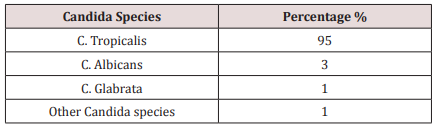
The Candida species most present in the dental caries in deciduous teeth were the C. tropicalis. Other species of Candida are found in less percentages (Table 1).

Table 1.

lupinepublishers-openaccess-journal-pediatric-dentistry

Discussion

Candida species is the most fungus found in oral cavity being the C Albicans the most pathological yeast of the Candida species [4]. This microorganism was found in many oral lesions as candidiasis, dental caries, gingival and periodontal disease [2,3]. Other Candida non albicans are founded in less frequency. However, C. tropicalis and C. glabrata has been described as emerging pathogens in recent years [8]. In the present study, C. tropicalis was presented in the most cases of dental caries in deciduous teeth, being this data corroborated with other studies who the main pathogen is the C. tropicalis. Most studies, in fact, found that the C. albicans as the mayor pathogen isolated from dental caries [6,9,10]. This difference of data can be explained by the geographical location of patients where Candida species can be found in amounts depending on the geographical area. In this study, other Candida species, can be found in less amounts. Despite limitations, the data obtained in the present study demonstrated the high rate of C. Tropicalis in dental caries in deciduous teeth, however, has not been determined which factor is involved in the pathogenesis of dental caries produced by C. tropicalis. It is also important study the oral microbiome in dental caries to dilucidated the role of Candida species, mainly C. tropicalis, in the development of dental caries in deciduous teeth.

Conclusion

Candida tropicalis is the most fungi founded in dental caries lesion in deciduous teeth in child between 4 and 6 years old.

Read More Lupine Publishers Pediatric Dentistry Journal Articles:
https://lupine-publishers-pediatric-dentistry.blogspot.com/

Friday, 20 November 2020

Lupine Publishers | Color Changes of Pediatric Dental Bridges

 Lupine Publishers | Journal of Pediatric Dentistry


Abstract

Dental technology that depended on the standardized lost-wax casting technology has been greatly improved with the introduction of dental CAD ⁄ CAM systems. The aim of the present study was to compare between the color changes of CAD/CAM acrylic and manually performed acrylic bridges used for pediatric patients. Forty study casts of children aged 2 - 4 years old of both genders, with prematurely lost one of the maxillary central incisors and the adjacent lateral incisor was carious and considered to be abutment tooth were involved in this study for construction of cantilever bridges. For each cast, two bridges were constructed; 1st one is CAD/CAM acrylic bridge and the other one is manually performed acrylic bridge. After immersing the bridges in saturated chocolate solution for different time intervals, color changes of the bridges were measured using 3Shape scanner system.

Keywords: Color; Changes; CAD/CAM; Bridges; Pediatric; Patient

Introduction

Trauma and/or dental caries is the common causes those result in premature loss of teeth in children. Cosmetic/aesthetic restoration of such condition considers to be challenging in the pediatric dental field. In case of premature tooth loss in anterior incisal segment there will result in arch space loss and teeth’s inclination that causing a collapse of the anterior teeth and midline shifting [1], as well as may lead to parafunctional habits [2]. Mahmoud (2009) found that anterior tooth loss had effect on patient’s quality of life and gave negative effects on him/his [3]. Al Rawi (2017) found that placement of cantilever acrylic bridges for restoring the aesthetic dental appearance of preschool children resulted in positive successes both to the child and parents [4]. Extrinsic discoloration of teeth and oral prostheses is stains caused by foods or beverages. In pediatric patients such stain mostly occurred due to colored foods such as beets or chocolate as well as berries and candies [5, 6]. This study considered to be the first step of our series studies deal with determining different physical and mechanical properties of the prostheses used for pediatric patients we planned to carry out (in vitro and in vivo studies). Starting with the present study that aimed to compare between the color changes of CAD/CAM and manually performed acrylic bridges used for pediatric patients. After immersing the bridges in chocolate solution for different time intervals, color changes of the bridges were measured using 3Shape scanner system.

Material and Methods

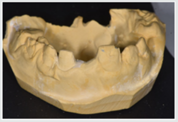
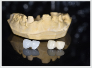
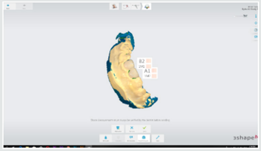
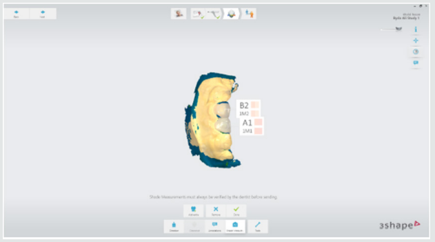
This study starting with collection of forty study casts of children aged 2-4 years old of both gender, with prematurely lost one of the maxillary central incisors and the adjacent lateral incisor was carious and considered to be abutment tooth were involved in this study for construction of cantilever bridges (Figure 1). For each cast, two bridges were constructed; 1st one is CAD/CAM acrylic bridge and the other one is manually performed acrylic bridge (Figure 2). Construction CAD/CAM bridge: The cast was 3D scanned by special scanner (710 3D) (smart optics Sensortechnik GmbH, Germany). The design of the bridge was carried out using Exocad Program (smart optics Sensortechnik GmbH, Germany). Acrylic block (Poly-methyl methacrylate) of classic shade A1 (Ivoclar vivadent, Switzerland) was used for fabrication of the bridge using CAD/ CAM machine (Charly dental, ZI Fonlabour, France). The bridge was finished and polished very well [4]. Construction of manually acrylic bridge: Wax pattern was fabricated on cast then followed the technique of typical wax loss; the heat-cure acrylic (Ivoclar vivadent, AG, FL-9494 Schaan/Liechtenstein) of classic shade A1 was used for bridge fabrication. Finally, surface finishing and polishing was done [7]. Saturated chocolate solution was prepared using 15g chocolate powder (MacChocolate TM, Malaysia) with 100ml distilled water. Baseline color readings for acrylic bridges were taken then immersed in chocolate solution for different time intervals (one week and two weeks) and maintained in incubator of 37 °C, Fresh chocolate solution was prepared every day. Before color measurements after one week and two weeks’ time intervals, the bridges were rinsed with distilled water for 30 seconds, cleaned with a soft bristle toothbrush and then dried with tissue paper [8]. Color measurement was carried out in the facial surfaces at the center third of the abutment and the center third of the pontic part of each bridge as shown in Figure 3. Color measurements of the bridges were measured using 3Shape scanner system (3 Shape A/S, Holmens Kanal 7.1060 Copenhagen K Denmark) and according to the software program of the system, Classic shade (Ivoclar vivadent, Switzerland) was depended.

Figure 1: One of the study casts involved in this study

lupinepublishers-openaccess-journal-pediatric-dentistry

Figure 2: One of the study casts involved in this study

lupinepublishers-openaccess-journal-pediatric-dentistry

Figure 3: Demonstrated the color shade measurement of the abutment and pontic portions of the acrylic bridge.

lupinepublishers-openaccess-journal-pediatric-dentistry

Results

Table 1 demonstrated the color shade of all samples at the baseline and after one-week and two weeks-time intervals. The results of the present study revealed that for all samples, the color measurement demonstrated that in CAD/CAM group even with using A1 shade acrylic block but at the baseline measurement the abutment revealed A0 shade while the pontic revealed A1 shade. Meanwhile, in manual group the abutment measured to be A1 shade and the pontic gave B1 shade. The results demonstrated that for all samples there were no changes in the color shade of CAD/CAM and manually fabricated acrylic bridges after one-week time interval, meanwhile, there were significantly color changes of all abutment and pontic portions of all samples of both bridge types after two weeks-time interval immersed in chocolate solution (Figure 4 & 5).

Figure 4: color shade measurement of CAD/CAM acrylic bridge after two weeks.

lupinepublishers-openaccess-journal-pediatric-dentistry

Figure 5: Color shade measurement of manual acrylic bridge after two weeks.

lupinepublishers-openaccess-journal-pediatric-dentistry

Table 1: Demonstrated the color shade of all samples at the baseline and after one-week and two weeks-time intervals.

lupinepublishers-openaccess-journal-pediatric-dentistry

Discussion

Restorations in the oral cavity are exposed to several factors that make them vulnerable to color changes, such as temperature, humidity, food and beverages. In the oral environment, restorative materials are also subjected to numerous other liquids, to temperature and load stress, and to tooth brushing. The success of restorations depends not only on mechanical and physical properties, but also on the esthetic appearance [9]. The color measurement in this study demonstrated that in CAD/CAM group even with using A1 shade acrylic block but at the baseline measurement the abutment revealed A0 shade while the pontic revealed A1 shade. These occurred because the thickness of the abutment was only about 0.5mm lead to that the color measured of the abutment was lighter than the pontic portion. Meanwhile, in manual group the abutment measured to be A1 shade and the pontic gave B1 shade. These results agreed with other studies those found the thickness of the material significantly affected the color shade of the prostheses [10,11].

The results demonstrated that the color changes demonstrated only after two weeks-time intervals immersed in chocolate solution. Even the color shades recorded in the CAD/CAM group considered to be lighter than in manual fabricated group, the discoloration from chocolate solution was probably due to adsorption of color colorant of chocolate solution at the surface of the prostheses.

The CAD/CAM bridges fabricated from blocks of pre-polymerized acrylic resin those had a hydrophobic surface that repels water [12]. As well as, perfect polishing surfaces of the bridges involved in this study revealed the limited discoloration that occurred agreed with other research [13]. As the duration of immersion increased, the color change values of both types of prostheses were recorded by 3Shape scanner system. Thus, the time is considered to be important factor in the staining of the dental prostheses and these results agreed with others [14,15]. Fabrication of dental prostheses with the help of CAD/ CAM technology is related to the advantages of high-density polymers based on highly cross linked polymethylmethacrylate [16]. Those advantages include; good esthetic, low water solubility and absorption, sufficient strength, low toxicity, easy repair with simple fabrication technique [17]. The using of hot cure acrylic for fabrication of dental prostheses even of some advantages but the main disadvantages include porosity with the presence of residual monomer which is a potential allergen, increased finishing time, brittle and uneven thickness [18]. A limitation of this study is that it was an in vitro study and need to be collected with in vivo study to measure the degree of color changes of the prostheses with presenting the effect of saliva and oral hygiene measures. Further clinical and in vitro studies are necessary to evaluate the susceptibility of CAD/CAM and manually acrylic bridges to discoloration by other beverages and nutrients.

Conclusion

Color considered as the most important factors for aesthetic appearance of dental restorations. In addition to the optimal chemo mechanical properties of acrylic resins, their availability in different color-shades has increased their application in fixed and removable prostheses. Acrylic resins can have acquired discoloration over time because of the process of adsorption and liquid molecules adhere to resin materials which was decreased their effect with using of CAD/CAM technology over conventional methods of acrylic resin prostheses fabrication.

Read More Lupine Publishers Pediatric Dentistry Journal Articles:
https://lupine-publishers-pediatric-dentistry.blogspot.com/

Friday, 2 October 2020

Lupine Publishers | Oral Squamous Papilloma on the Tongue of a 12-Year Old Female: Report of a Case with Human Papilloma Virus Literature Review

 Lupine Publishers | Journal of Pediatric Dentistry


Abstract

Human papillomavirus (HPV) infection that causes squamous papilloma is common in the oral cavity of adults but not in children. Although benign, the slow progressive growth is a concern to clinicians and parents as the lesion may clinically appear as an exophytic verrucous carcinoma or squamous carcinoma. This case report describes a squamous papilloma arising on the tongue of a 12-year old child.

Keywords:Child; Human Papillomavirus (HPV); Squamous Papilloma; Oncogenic Potential; Tongue; Koilocytes

Introduction

Human papillomavirus (HPV) are slow, benign proliferations of stratified squamous epithelium frequently observed in the oral cavity that are the viral etiologic agent for squamous papilloma [1,2]. Squamous papillomata are commonly observed in adults 30-50 years of age and is the fourth most common oral mucosal lesion in both children and adults [3]. Although the entire oral cavity may be affected, the viral lesion has a predilection for the laryngotracheobronchial complex in children and the lower lip, hard and soft palate of the maxilla and uvula in adults [1,4-6]. Clinically, the lesion appears as an exophytic mass and may cause anxiety to the clinician and parents, as the lesion can clinically appear like an exophytic verrucous carcinoma, squamous carcinoma or condyloma accuminatum [1,4,7,8]. This case report describes a squamous papilloma arising on the tongue of a 12-year old child.

Virology

The human papillomavirus is a 55nm non-enveloped icosahedral double-stranded deoxyribonucleic acid (DNA) virus that is a member of the papovavirus group [7-14]. There are over 150 genotypically different types and classified as either mucosal or cutaneous.10-12 The viral types with oncogenic potential include HPVs 16, 18, 31, 33, 35, 39, 45, 51, 55, 56, 58, 59, 66 and 68. Oral squamous papilloma infection is associated with HPV subtypes 6, 11 and 16 [7-13]. Papillomas induced by HPV types 6 and 11 are considered to have low oncogenic potential. However, approximately 85% of dysplastic lesions, carcinoma in situ and squamous cell carcinoma involve the DNA sequence of HPV 16 and 18. DNA replication of HPV occurs in the nuclei of epithelial cells. The HPV capsid proteins enter the host cell delivering the viral DNA to the nucleus which allows proliferation of the viral lesion [7,10,11,12,14].

Case Report

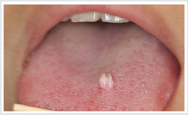
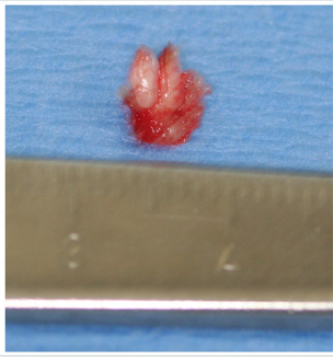
A 12-year old Asian female was referred to the office by her family dentist for evaluation of a soft tissue lesion localized to the dorsal surface of the tongue (Figure 1). The patient stated that the lesion has been present for at least six months. Her past medical history was unremarkable. She was not taking any medications and denied any allergy to medications. The patient also denies being sexually active. Sexual abuse was also ruled-out. Head and neck examination were negative for palpable neck masses and lymphadenopathy. Oral examination of the dorsal surface of the tongue revealed a pink-white colored exophytic lesion that was freely movable. The surface texture had a pebbly appearance that resembled, “cauliflower”. The remaining oral examination was unremarkable. Examination of the upper and lower extremities was negative for any soft tissue lesions resembling HPV. Based on the clinical appearance of the lesion, the differential diagnosis included squamous papilloma, verruciform xanthoma, papillary hyperplasia and condyloma accuminatum of the tongue. The father and patient were informed of the clinical findings and excisional biopsy (Figure 2) for a definitive microscopic diagnosis under local anesthesia was recommended.

Figure 1: Clinical photograph of squamous papilloma on left dorsal surface of the tongue in 12-year old Asian child.

lupinepublishers-openaccess-journal-pediatric-dentistry

Figure 2: Excised squamous papilloma specimen from dorsal surface of tongue of 12-year old Asian patient.

lupinepublishers-openaccess-journal-pediatric-dentistry

Histopathology

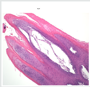
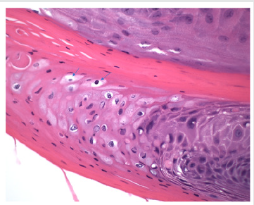
Excisional biopsy was completed under local anesthesia with 1.0mm margins of normal tissue and to the depth of the tongue musculature. The lesion was stained with hematoxylin and eosin for histopathological diagnosis. Histological examination demonstrated long, thin papillary projections of parakeratinized stratified squamous epithelium (Figure 3a). Localized areas of basilar hyperplasia with koilocytes were observed (Figure 3b). The histological findings were consistent with squamous papilloma.

Figure 3(a): Histopathology demonstrating proliferation of hyper keratinized stratified squamous epithelium finger-like projections with thin fibrovascular connective tissue core (Hematoxylin and eosin stain. Original magnification x 40).

lupinepublishers-openaccess-journal-pediatric-dentistry

Figure 3(b): Presence of koilocytes (arrows) in the spinous layer of the epithelium that are characteristic histopathologic findings of oral squamous papilloma (Hematoxylin and eosin stain, x 200).

lupinepublishers-openaccess-journal-pediatric-dentistry

Discussion

In children, viral transmission remains controversial. HPV transmission may occur by any number of different mechanisms, such as autoinoculation, heteroinoculation, perinatal transmission, sexual abuse and contact with fomites [1,4,5,9,10]. However, trauma is one mechanism that should be considered in our patient. Iatrogenic tongue biting may initiate an infection of the basal squamous epithelial cells that allows the entrance of the HPV into the tongue. Viral DNA then enters the nucleus of the infected epithelial stem cells and can replicate causing the HPV infection [15]. Histopathologic features of oral squamous papilloma demonstrate hyperkeratosis in the epithelium, proliferation of the spinous cells that result in long, thin finger-like projections above the mucosa [1- 3,8,14]. The characteristic feature of HPV infection is the presence of koilocytes (Figure 3b) which are virus-infected epithelial cells due to perinuclear cytoplasmic vacuolization of cells of the spinous layer of the epithelium. This results in the nuclei becoming pyknotic and cremated surrounded by an optically clear zone [1-10]. All the described histopathologic findings in our patient are characteristic of HPV infection. Although the prevalence of oral HPV infection is low, a bimodal distribution is observed. The highest prevalence is observed in children less than 1 year of age and the second peak occurs in adolescents, between 13 to 20 years old [9,16]. Despite the bimodal distribution, oral HPV infection is considered low in children [17]. In a study of 4140 children between the ages of 10 to 18 years old, the prevalence of oral HPV infection was 1% and the most common type was HPV 11 [18]. Treatment of HPV is by surgical excision [19]. The United States Food and Drug Administration (2016) approved Gardasil 9 human papillomavirus 9-valent vaccine to prevent infection against HPV types 6, 11, 16 and 18 (Merck & Co., North Wales, Pa). At present, only Gardasil 9 human papillomavirus 9-valent vaccine recombinant has been approved for vaccination in the United States (Center for Disease Control, 2019) [20]. To obtain the greatest clinical efficacy and cost effectiveness of vaccination, the Center for Disease Control (CDC) recommends that both male and female children between the ages of 11-12 years get two HPV vaccinations six months apart before individuals have been exposed to the human papillomavirus. It is also recommended that adult women up to age 26 years and men up to age 21 years also obtain the HPV vaccine [21].

Conclusion

A case of oral squamous papilloma on the tongue of a 12-year old female is presented to create awareness of this soft tissue lesion in the oral cavity of children due to the human papillomavirus. It is only after educating clinicians and parents about the HPV will we observe greater identification, management and treatment in the HPV infected child.

Read More Lupine Publishers Pediatric Dentistry Journal Articles:
https://lupine-publishers-pediatric-dentistry.blogspot.com/
Read More Lupine Publishers Articles: https://lupinepublishers.blogspot.com/

Friday, 11 September 2020

Lupine Publishers | Management of Internal Root Resorption with Bioceramic Material on Permanent Tooth-A Case Report

 Lupine Publishers | Journal of Pediatric Dentistry


Abstract

Internal root resorption (IRR) is a category of pulp disease characterized by the loss of dentine as a result of the action of clastic cells stimulated by pulpal inflammation. The objective of this case report was to account for the diagnosis and management of an internal root resorption without perforation. The patient, a 26-year-old male, came to Guru Nanak Institute of Dental Sciences and Research, West Bengal, without having symptoms in the tooth. Endodontic treatment was performed using the following methods: irrigation of the root canal with 2.5% of sodium hypochlorite, then calcium hydroxide (CH) was applied as intracanal medicament for one month. Complete instrumentation was done with Hyflex One File (Coltene) and obturation with corresponding guta-percha and Roeko Guttaflow Bio seal sealer (Coltene). The patient was checked after one week and then after six months. He did not have any symptoms and IOPA radiograph did not show any further progression of the lesion.

Keywords:Internal Root Resorption; Calcium Hydroxide (CH); Sodium Hypochlorite; MTA

Introduction

The Glossary of the American Association of Endodontists defines internal root resorption (IRR) as a condition associated with a physiological or pathological process that results in the loss of dentin, cement and bone [1]. Most teeth with internal root resorption are symptom free and are first clinically recognized through routine radiographs. However, when resorption actively progresses, the tooth is only partially vital and may present typical symptoms of pulpitis. Bell (1830) first reported about IRR. Mummery (1920) called it “pink tooth of Mummery” due to the presence of pink discoloration on the crown [2]. This condition, although rare, is more frequent in the male population. The IRR is more common in the presence of a periapical lesion. Its prevalence was estimated between 0.01% and 1% depending on the inflammatory condition of the pulp [3]. The IRR could be caused by several stimuli: trauma, chronic inflammation of pulp/periodontal ligament, heat created by the friction of drills during the preparation of cavities, cracked tooth syndrome, tooth reimplantation and orthodontic treatment [4]. There have also been reported cases of internal reabsorption caused by Herpes Zoster virus [5]. The IRR is caused by inflammatory stimuli which produce an alteration of the odontoclast inhibitory mechanism resulting in an alteration of the pre-dentine layer. The vascular change in the pulp produces hyperemia increasing oxygen tension and causing an acidic pH level that attracts multinucleated cells, odontoclasts and dentin clasts. Dominance of inhibitory substances such as OPG (osteoprotegerin) as activators of RANKL (receptor activator of factor kappa B ligand) followed by swelling, results in the rupture of protective coatings allowing the invasion of odontoclasts and initiating resorptive patterns. Connective, post-resorptive activity tissue transforms into metaplastic granulation tissue [6]. Generally, IRR detection is done by X-rays, however, the use of cone beams computed tomography (CBCT) has been reported to be highly useful for diagnosis in endodontics, since it shows the lesion in detail and includes information about adjacent anatomy, which X-rays does not provide [7]. The periapical radiography is limited because it provides a twodimensional image [8], whereas diagnosis by CBCT shows images in all their dimensions through tomographic slices, without image overlay [9]. Also, diagnosis by CBCT may improve the accuracy and efficiency in the prognosis of the tooth [10]. Therapeutically, the biomaterial employed can influence the prognosis of the nonsurgical endodontic treatment done for extensive internal root resorption [11]. MTA is most commonly used in these cases because of its sealing ability, biocompatibility and potential induction of osteogenesis and cement genesis and it can be used in a humid environment [12]. Another study using an experimental immature tooth model, demonstrated that the MTA also increased the fracture resistance of bovine incisors when submitted to different reinforcement treatments Recently bioceramics are widely used in endodontics. Roeko Guttaflow Bio seal (COLTENE) is a bioceramic endodontic sealer which claims to avoid shrinkage upon setting as it has Zirconium oxide is used as the radiopacifier, and the material is claimed to be aluminum-free, non-soluble and does not shrink during setting. It gives advantage of flow of material as well as sealing ability which better bond with the corresponding gutta percha used for obturation [13]. The purpose of this case report is to describe the diagnosis and clinical management of an internal root resorption with bioceramic material.

Case Report

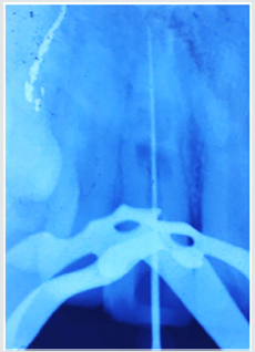
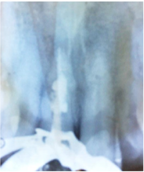
Male patient, 26 years old, treated at the post graduate department in Guru Nanak Institute of Dental Sciences and Research. The patient reported no pain at the time of appointment the chief complaint was discoloration of the front tooth which was traumatized 5 year back. Vitality tests using Endo-Ice (Coltene, Switzerland) were performed in [11]; the tooth gave negative response. The patient did not present tooth mobility and periodontal pockets. IOPA radiograph of the affected tooth#11 showed an oval enlargement (ballooning out) of the root canal space (Figure 1). The pulp chamber and canal cannot be followed throughout the lesion. Radiograph performed at different angulation to confirm the resorptive lacunae is a continuation of the distorted border of the root canal. Endodontic treatment was suggested; therefore, isolation protocol was performed to make the cavity opening later (Figure 2). Working length of the tooth was determined by IOPA radiograph using #15K file (Figure 3) and the result was confirmed with apex locator Canal Pro (COLTENE). After removing the pulp tissue properly chemical-mechanical instrumentation was performed with Hyflex One File (COLTENE) and irrigation was done with 1ml of 2.5% of sodium hypochlorite between each time instrumentation with 30-gauge side vented needle. This was followed by irrigation with normal saline to remove any remnants of hypochlorite, later canal was dried with absorbent points. Ca (OH)2 dressing was given for 1month and the medicament was changed weekly. After one month, temporary restoration was removed with [4] round diamond bur, canal was irrigated with 5 mL of 2.5% sodium hypochlorite (NaOCl) and 5 mL of 17% of ethylenediaminetetraacetic acid for removing the Ca (OH) dressing and then the canal was flushed with normal saline and dried.

Figure 1: .

lupinepublishers-openaccess-journal-pediatric-dentistry

Figure 2:

lupinepublishers-openaccess-journal-pediatric-dentistry

Figure 3:

lupinepublishers-openaccess-journal-pediatric-dentistry

Figure 4:

lupinepublishers-openaccess-journal-pediatric-dentistry

Figure 5: .

lupinepublishers-openaccess-journal-pediatric-dentistry

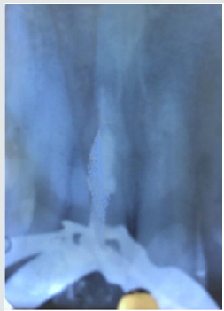
After removing the medication, obturation was done with Hyflex corresponding Gutta Percha and the remaining pulp chamber was obturated with Guttaflow Bio seal sealer (Figure 4). Access cavity restoration was done with light cure composite resin. The patient was recalled after 6 and 12 months (Figure 5, 6) for clinical and radiographic follow up. Clinical examination of tooth was functional without sensitivity to percussion or palpation [11].

Figure 6: .

lupinepublishers-openaccess-journal-pediatric-dentistry

Discussion

There is always a dilemma of whether to treat a tooth with a questionable prognosis endodontically or extract it and subsequently place an implant. Bell first reported a case on internal resorption in 1830. Since then there have been numerous reports in the literature [14]. Two types of internal root resorption are generally described: the internal root canal inflammatory resorption and the internal root canal replacement resorption. In the inflammatory resorption, the resorptive process of the intraradicular dentin progresses without adjunctive deposition of hard tissues adjacent to the resorptive sites. The phenomenon is associated with the presence of granulation tissues in the resorbed area and identifiable with routine radiographs as are radiolucent zone centered on the root canal. In the replacement resorption, the resorptive activity cause defects in the dentin adjacent to the root canal, with concomitant deposition of bone like tissue in some regions of the defect. It results in an irregular enlargement of the pulp space with partially or fully obliterated area of the pulp chamber. Internal resorption is the result of an inflamed pulp and the clastic precursor cells recruiting through the blood vessels. Treatment of internal resorption is quite predictable as it is easy to control the process of internal root resorption via severing the blood supply to the resorbing tissues with conventional root canal therapy. Intraoral X-ray of IRR is characterized by the radiographic appearance of an oval shape enlargement within the pulp chamber or the root canal. However, the early diagnosis of the IRR is difficult by examination of a conventional X-ray. If IRR is suspected, several shots under different angles of incidence are recommended. In the treatment of internal resorption, the use of calcium hydroxide also has two other important goals: to control bleeding, and to necrotize residual pulp tissue and to make the necrotic tissue more soluble to sodium hypochlorite. Because of the limited access by instruments to all areas of the resorption cavity, chemical means are needed to completely clean the canal. Studies on the effectiveness of sodium hypochlorite and calcium hydroxide to remove the resorptive and other tissues from the root canal indicate that they have an additive or even synergistic effect [15]. In cases where the resorption has not perforated, it is usually enough to use calcium hydroxide paste in the canal once from 1 to 2 weeks. This allows removal of the residual tissue at the next appointment by irrigation and instrumentation. In our treatment protocol, we choose Guttaflow Bio seal (COLTENE) sealer due to its versatile property of Bioceramic component & gutta-percha particles. Upon contact with fluids, this material provides natural repair constituents, such as silicates and calcium, which contribute to the activation of biochemical processes, providing additional support to the root canal regeneration. A novel material for root canal filling that combines gutta-percha in a powder form with a particle size of less than 30 μm and a sealer. The sealer has also showed least cytotoxicity as well as inflammatory reaction [15].

Conclusion

It is puzzling in diagnosing and treating a root resorption case, therefore a suitable management is perilous. Thorough investigations and discussion are required for the management especially when the prognosis of the tooth is poor upon consultation. Absence of periapical lesion and no signs and symptoms at the 12-months review provided a favorable outcome to once a tooth of hopeless prognosis.

Read More Lupine Publishers Pediatric Dentistry Journal Articles: https://lupine-publishers-pediatric-dentistry.blogspot.com/

Read More Lupine Publishers Articles: https://lupinepublishers.blogspot.com/

Friday, 4 September 2020

Lupine Publishers | Management of Mesiodens In Mixed Dentition- Molariform and Tuberculate: A Case Report

 Lupine Publishers | Journal of Pediatric Dentistry


Abstract

Timely intervention is the key to any setback in the mixed dentition. Teeth which are supplemental to the normal dentition are supernumeraries, the most common being mesiodens, present in the premaxillary region. Certain pathological consequences may arise due to mesiodens like unaesthetic midline diastema, rotation, displacement, root resorption and cyst formation. The current case report presents the management of developing malocclusion in the anterior region due to the presence of mesiodentes- a molariform and a tuberculate. The molariform mesiodens was impeding the eruption of the maxillary right central incisor, thus the surgical removal of both mesiodentes was planned and executed. In addition to this soft tissue uncovering was done for the unerupted maxillary central incisor. On follow up, uneventful healing was observed successfully.

Keywords:Mesiodens; Mesiodentes; Mixed Dentition; Molariform; Tuberculate

Introduction

Supernumerary teeth, or hyperdontia, is a term that describes teeth that are surplus in number when compared to the normal complement of teeth [1]. The etiology of supernumerary teeth remains unclear and not yet completely understood [2]. Among the various proposed theories that have attempted to explain the causes behind the development of supernumerary teeth, current literature favors the ‘lamina hyperactivity theory’ that states hyperdontia results from independent, locally conditioned hyperactivity of the dental lamina [1]. The most commonly occurring supernumerary tooth is the ‘mesiodens’, a term that was initially coined by Balk in 1917. As the name suggests, the mesiodens is usually located mesial to the central incisors in the premaxillary region [3]. With a frequency of prevalence between 0.15-3% in the permanent dentition and 0.02-1.9% in the primary dentition, it may occur as single or multiple, unilateral or bilateral, may be erupted or impacted and frequently found in conjection with cleft lip and palate and syndromes like Cleidocranial dysostosis, Gardner’s syndrome, Ellis-Van Creveld syndrome, Ehlers- Danlos syndrome, Incontinentia Pigmenti, and Tricia-Rhino- Phalangeal syndrome [4,5]. Also, mesiodentes may vary in shape from simple conical form to a larger, more complicated crown shape with several tubercles [6]. The dysfunctional nature of mesiodens is known to cause a variety of clinical complications such as being unaesthetic, pathological disturbances in the normal eruption and positions of adjacent teeth, altered growth and development in the area, retention of primary teeth, odontogenic cysts, caries, pulp necrosis of the adjacent teeth, dilaceration of developing tooth, nasal teeth, gingival and periodontal problems [2,7]. Accordingly, their early diagnosis and management is vital to waive off complications of such kind.

Case Report

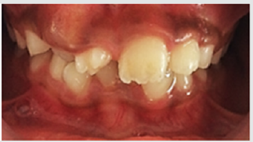
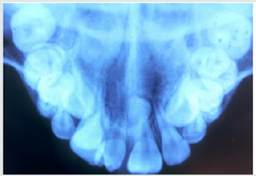
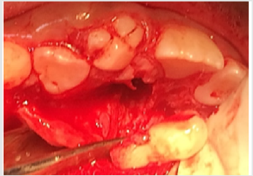
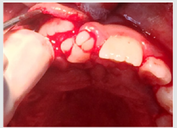
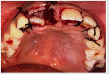
A 9-year old female patient reported to the department of Pedodontics and Preventive Dentistry with the complaint of irregularly placed upper front teeth and wanted it to be corrected. The patient was normal and healthy with non-contributory medical and dental histories. The extra oral examination did not reveal any abnormalities. Intraoral examination revealed a Class I mixed dentition with a missing upper right central incisor and an erupted molariform mesiodens in its place. In addition, there was a firm bulge palpable in the upper right central incisor area which suggests the impediment in the path of eruption of the central incisor by the erupted mesiodens (Figure 1). An occlusal radiograph was taken to rule out the possibility of multiple supernumerary teeth and surprisingly another unerupted and impacted inverted mesiodens with an incomplete root was found mesial to the upper left central incisor (Figure 2). The SLOB technique confirmed that the impacted mesiodens was present palatially. Both informed and written consent was obtained from the parents before initiating the treatment. We decided to extract both mesiodentes under local anesthesia. The erupted mesiodens was extracted by intra- alveolar extraction. The impacted mesiodens was surgically removed by raising a mucoperiosteal flap from maxillary first premolar to contralateral first premolar (Figure 3). Rotary cutting instruments with simultaneous irrigation were used for removing enough bone around the impacted mesiodens for its easy retrieval. Soft tissue uncovering was done for the unerupted right central incisor by placing an elliptical incision over the incisal portion of the palpable bulge (Figure 4). The extraction socket was checked for any pathological tissue and the flap was relocated and sutured with interrupted sutures (Figure 5). The patient was recalled after a week for suture removal and followed-up after 3 months. Uneventful healing with no associated symptoms was observed.

Figure 1: Preoperative view showing erupted molariform mesiodens along with unerupted maxillary central incisor.

lupinepublishers-openaccess-journal-pediatric-dentistry

Figure 2: Occlusal radiograph showing unerupted and impacted inverted mesiodens with an incomplete root.

lupinepublishers-openaccess-journal-pediatric-dentistry

Figure 3: Surgical removal of impacted mesiodens by raising a mucoperiosteal flap from maxillary first premolar to contralateral first premolar.

lupinepublishers-openaccess-journal-pediatric-dentistry

Figure 4: Soft tissue uncovering for the unerupted right central incisor by placing an elliptical incision over the incisal portion of the palpable bulge.

lupinepublishers-openaccess-journal-pediatric-dentistry

Figure 5: Interrupted sutures placed.

lupinepublishers-openaccess-journal-pediatric-dentistry

Discussion

The realm of pediatric dentistry incorporates the practice of interceptive orthodontics, thereby bestowing upon the pediatric dentist, opportunities of providing timely guidance in the development of occlusion. The current case report presents the management of developing malocclusion in the anterior region due to the presence of mesiodens. A mesiodens occurring in the primary dentition is a rarity even though, it being the most common dental abnormality in the permanent dentition [8]. Most cases of mesiodens are discovered during the first decade as maxillary central incisors are erupting and radiographic examinations are performed as an aid to screening for any other malformations and abnormalities [1]. Various theories regarding the etiology of mesiodens have been reported in the literature but the subject remains controversial [3]. Heredity has been suggested to be an etiologic factor based on the observation that supernumeraries are more common in family members; however, it does not follow a simple Mendelian pattern [9]. It was originally postulated that the mesiodens represented a phylogenetic relic of the extinct ancestors who had three central incisors. This is known as phylogenetic theory reversion (atavism) which has now been discarded by the embryologists [10]. The dichotomy theory states that, a mesiodens arises due to the splitting of the tooth bud. On the contrary, Taylor argued that splitting of the tooth bud may either form two equal sized teeth or one normal and one dysmorphic tooth [8]. The hyperactivity theory states that development of mesiodens is due to the hyperactivity of the dental lamina. The ‘field model’, proposes that a tooth bud which is forming at a given location develops according to its position within the field, further determining its shape. The ‘clone model’ postulates that that each tooth class is derived from a clone of ectomesenchymal cells which are programmed by epithelium to produce teeth of a given pattern. Depending upon the specific factors expressed from these ectomesenchymal cells, the shape of the accessory tooth germ forms in the vicinity of the incisors class of teeth becomes evident at the bell stage [11]. This case report presented with an erupted molariform mesiodens and an unerupted tuberculate mesiodens with an undeveloped root (Figure 6). Since the molariform mesiodens was impeding the eruption of the maxillary right central incisor, the surgical removal of both mesiodentes was planned and executed. Timing of interceptive treatment should be as soon as possible following clinical detection of an abnormal eruption pattern. It has been suggested that a tooth delayed in its eruption by more than six months with respect to its antimere should be radiographically investigated. Hogstrum and Andersson [12] suggested two alternatives exist.

The first option involves removal of the supernumerary as soon as it has been diagnosed. This could lead to an unpleasant experience that may have a psychological effect on a very young child and has been said to cause devitalization or deformation of adjacent teeth. Secondly, the supernumerary could be left until root development of the adjacent teeth is complete. The potential disadvantages associated with this deferred surgical plan include; loss of eruptive force of adjacent teeth, loss of space and crowding of the affected arch, and possible midline shifts. In the present case, since the roots of the adjacent teeth were completely formed and the child was at an age where she could sustain a surgical procedure, the surgery was undertaken with utmost attention to detail and caution. Access to mesiodens during surgery must dealt carefully considering the quantity of bone amputation and the possible damage to the adjacent teeth [7]. Follow up is indispensable in such cases since the eruption status should be monitored. The patient revealed satisfactory healing and suitable eruption of the maxillary central incisor. Pediatric dentists are the firsts to usually identify developing malocclusions and thus it is their responsibility to intervene and intercept in an apt manner to prevent future unfavorable sequalae.

Figure 6: Molariform and tuberculate mesiodens.

lupinepublishers-openaccess-journal-pediatric-dentistry

Acknowledgements

Authors are thankful to their colleagues and faculty.

Read More Lupine Publishers Pediatric Dentistry Journal Articles: https://lupine-publishers-pediatric-dentistry.blogspot.com/

Read More Lupine Publishers Articles: https://lupinepublishers.blogspot.com/