Showing posts with label Pediatric Dentistry. Show all posts

Wednesday, 17 March 2021

Lupine Publishers| Association of Mother’s Genetic Sensitivity to the Taste of 6-N-Propylthiouracil (Prop) and Their Children’s Dental Caries Status in Nepal

 Lupine Publishers| Interventions in Pediatric Dentistry Open Access Journal (IPDOAJ)


Abstract

Introduction: Dental caries is a multifactorial disease and is one of the most prevalent infectious disease that affects mankind. Young children present a unique risk for dental caries as their host-defense systems and bacterial flora are in the process of being developed and also because the newly erupted tooth surfaces are more susceptible to dental caries. Mothers are the persons who generally influence their children via their own food preferences. Hence the mother’s taste perception plays an important role in the development of dental caries in their children. The need of this study is to examine the association of mother’s taste perception to 6-n-propylthiouracil with caries prevalence in their young children as well as with other caries risk determinants such as mothers and their children’s oral hygiene practices.

Material and Method: 180 pair of mothers and their children in the age group of 3 to 6 years of both sexes were selected for the study & 6-n-propylthiouracil testing is done. A trained and calibrated examiner who did not have any knowledge of the mother’s PROP test performed a comprehensive clinical examination of the children to determine the presence or absence of DMFT/dmft.

Results: It is observed that nonstarter mother and children have higher caries prevalence than medium tasters and supertasters. Discussion- Genetic sensitivity to taste is an inherited trait in children from their parents, inheritance from mother being more pronounced.

Conclusion: Dental caries is multifactorial. No significant correlation between susceptibility of mother and child to genetic sensitivity exists, and genetic sensitivity is not the only criteria for severity.

Keywords: 6-n-Propylthiouracil; dental caries; dietary habits

Introduction

Dental caries is a multifactorial disease and is one of the most prevalent infectious disease that affects mankind. The development of dental caries depends on several critical interactions between a susceptible tooth surface, oral bacteria, fermentable carbohydrates and frequency of consumption of sugar [1,2]. Young children present a unique risk for dental caries as their host-defense systems and bacterial flora are in the process of being developed and also because the newly erupted tooth surfaces are more susceptible to dental caries [3]. Parents must also negotiate the dietary transition from bottle feeding to solid food and also take into consideration the child’s tastes. Several risk factors such as salivary counts of mutant’s streptococci, past carious experience and frequency of sugar intake have been evaluated to identify children at high risk of caries [1,4-7]. However, none of the currently available caries screening methods can identify children at high risk of caries quite effectively [1,8].

The role of diet as a direct cause of dental caries has been extensively reported [1,4,9,10]. A high intake of sugar has been correlated with a high dental caries in pre-school and school aged children. A high sugar intake reflects a preference for a sweet substance. Genetic sensitivity to taste may be associated with a preference or no preference of some food by children. Sensitivity to taste is an inherited trait in children [1,11]. In 1991 it was found that there is a genetic variation in the ability to taste the bitterness of the chemical 6-n-propylthiouracil (PROP) [1,12]. This variation was found to be associated with food preferences in children. Fox in 1931 found that some individuals perceived chemicals related to PROP as bitter (supertaster) while others could hardly perceive them at all (nonstarter). Those who are PROP super taster are typically supertasters in general and tend to dislike sweet while those who are PROP none tasters like sweet food and prefer strong tasting food products. Mothers are the persons who generally influence their children via their own food preferences. In many households’ mothers generally reward their children using sugary snacks which might increase their children’s preference for that kind of food. Hence the mother’s taste perception plays an important role in the development of dental caries in their children. This study was carries out with following aims and objectives:

Aim

To find the association between mother’s sensitivity to 6-n-propylthiouracil (PROP) and their children’s dental caries status

Objectives

To compare mother’s and child’s oral hygiene practices with their dental caries status. To determine the taste quality and taste preferences of food products among these study groups.

Materials and Methods

220 pairs of mother and children were randomly selected from consecutive children who were accompanied by their mothers visiting the Department of Pedodontics and Preventive Dentistry in the institution. Out of these 220 pairs of mother and children 180 pairs fulfilled the inclusion criteria. A single trained and calibrated examiner who did not have any knowledge of the mother’s PROP test performed a comprehensive clinical examination of the children to determine the presence or absence of DMFT/dmft. All examinations were performed with a mouth mirror and explorer in a lighted operatory after taking consent from the mother. Demographic information of mothers, their oral hygiene practices, their oral health status and the presence of grandparents in the household was collected by an open-ended questionnaire. A second questionnaire was used to collect data regarding the oral hygiene and feeding practices of the child (e.g. frequency of tooth brushing, intake of sugary food and frequency of intake of sugary food).

Inclusion Criteria

For children

a) Age groups of 3 to 6 years.
b) ASA physical status I/II and stable mental condition
c) Should be accompanied by mother.

For mother

a) Participant cooperation and acceptance of the study programmer.
b) Should be healthy and mentally stable.
c) Should be literate.

Exclusion Criteria

For children

a) Should not be less than three years and not over six years
b) Should not have any medical / hereditary conditions.
c) No acute dental diseases.

For mothers

a) Should not be pregnant
b) Should not suffer from systemic diseases
c) Should not be allergic to 6-n-propylthiouracil (PROP)
d) Should not be under any medications that could alter the taste sensation and affect salivary flow.

Prop testing

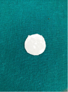
Figure 1: Filter paper with PROP solution.
Lupinepublishers-openaccess-pediatric-dentistry-journal

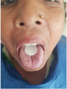
single examiner who did not have any knowledge of the mother’s and children’s oral health status had conducted the PROP test on the mothers. A small piece of filter paper (2 cm circle of Whatman’s grade 1 filter paper) containing approximately 1.6 mg (measured by calibrated dropper) of 6-n-propylthiouracil (PROP) was used to determine each mother’s taste type (Figure 1). The mother was asked to put the piece of filter paper in the mouth and moisten it with saliva for 30 seconds (Figure 2). After removing the filter paper, the mother was asked to quantify the intensity of bitter taste on the modified Green’s scale and was classified of supertasters (>60), medium tasters (12-60) and no tasters (<12). All examinations and PROP testing were carried out at the same time of the day (mid-morning) throughout the study. All the data gathered was tabulated and statistically analyzed. Student’s paired t test and chi square test was done.

Figure 2: PROP test on mother.
Lupinepublishers-openaccess-pediatric-dentistry-journal

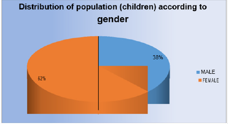
Figure 3: Distribution of population (children) according to gender.
Lupinepublishers-openaccess-pediatric-dentistry-journal

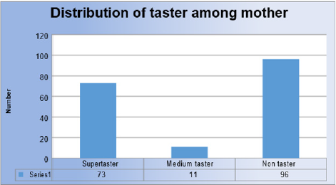
Figure 4: Distribution of taster among mother.
Lupinepublishers-openaccess-pediatric-dentistry-journal

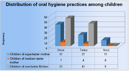
Figure 5: Distribution of oral hygiene practices among children.
Lupinepublishers-openaccess-pediatric-dentistry-journal

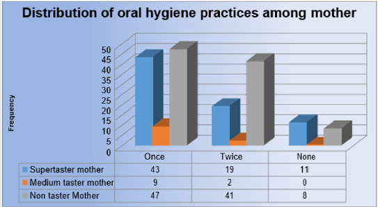
Figure 6: Distribution of oral hygiene practices among mother.
Lupinepublishers-openaccess-pediatric-dentistry-journal

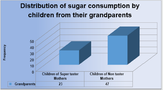
Figure 7: Distribution of sugar consumption by children from their grandparents.
Lupinepublishers-openaccess-pediatric-dentistry-journal

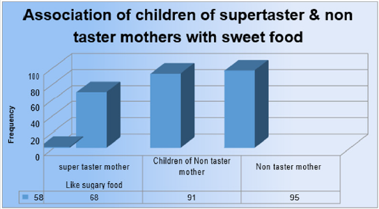
Figure 8: Association of children of supertaster & non taster mothers with sweet food.
Lupinepublishers-openaccess-pediatric-dentistry-journal

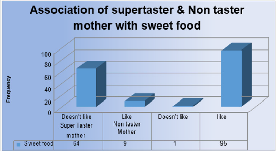
Figure 9: Association of supertaster & Non taster mother with sweet food.
Lupinepublishers-openaccess-pediatric-dentistry-journal

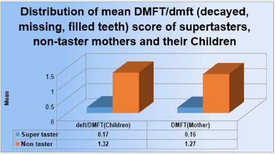
Figure 10: Distribution of mean DMFT/dmft score of supertasters, non-taster mothers and their Children.
Lupinepublishers-openaccess-pediatric-dentistry-journal

Table 1: Association of supertaster & non taster mothers and their children with sweet food.

Lupinepublishers-openaccess-pediatric-dentistry-journal

Table 2: Distribution of mean DMFT/dmft score of supertasters, non-taster mothers and their Children.

Lupinepublishers-openaccess-pediatric-dentistry-journal

Discussion

The present study comprised of 180 pairs of mother and child. The mothers were subjected to PROP testing and classified into super tasters, medium tasters and non-tasters according to the Green’s labeled magnitude scale. The mothers who were super tasters found the taste of PROP to be extremely bitter and the non-taster mothers described the taste of PROP to be tasteless while the medium taster mothers failed to categories the taste of PROP. The super taster mothers perceived the taste of PROP in lower concentration (1.6 mg per ml). This may be due to the presence of high-density fungiform papilla and taste receptors in the tongue compared to medium tasters and non-tasters. This may make the super taster mothers dislike sugary food. Mothers being the primary care givers to children influences the food habit of children. Mothers who prefers sugary foods are more likely to prepare and feed their children the same then those who do no prefer sugary food. In our study it was found that the mean dmft/ DMFT score among non-taster mothers (1.32) and their children (1.27) was higher than the mean dmft/ DMFT of super taster mothers (0.17) and their children (0.16) respectively. The influence of grandparents in the family cannot be neglected. Even in children of super taster mothers, 23 children reported they were given sugary snacks by their grandparents regularly while 47 children of non-taster mothers reported the same.
The pampering of grand parents may also be a causative factor for dental caries in children. The finding of the present study also showed that children who brushed their teeth once or twice per day experienced more dental caries if their mothers were non tasters whereas children of super tasters had less dental caries. This can be explained by the frequent intake of sugary food in case of children of non-taster mothers and also may be influenced by the food prepared by their mothers. Similarly, mothers who were non tasters had a higher dental caries experience than the super taster mothers. This may be due to their increased susceptibility to sugary food. In the present study when the super tasters and non-taster mothers were asked about their liking for sugary food, majority of the super taster mothers (64) stated that they did not prefer sweet food, while 95 non taster mothers stated that they preferred sugary food. In case of the children of super taster and non-taster mothers, the results were significant. The mother’s dietary habits and tastes probably affected the oral health status of the child. When the association between the preference of sugary food in super taster and non-taster mothers and their children was seen, it was found that the children of super taster mothers did not prefer sugary food while it was vice versa for the children of non-taster mothers. This may be due to genetic factors and also might be due to the dietary habits of the mothers which influenced the dental caries status of their children.

Conclusion

In the present study, the results suggested that the children of the mothers who are non-tasters or who prefers sweet food have higher dental caries experience. Factors such as the presence of grandparents in the household, frequency of brushing, preference of food and dietary habits may play an important role in the development of dental caries in children. Thus, mother’s genetic sensitivity to the bitter taste of prop can be used as a useful adjunct to identify children who are at risk of developing dental caries.

Read more about Interventions in Pediatric Dentistry Open Access Journal (IPDOAJ) please click on below link https://lupine-publishers-pediatric-dentistry.blogspot.com/


Friday, 4 September 2020

Lupine Publishers | Management of Mesiodens In Mixed Dentition- Molariform and Tuberculate: A Case Report

 Lupine Publishers | Journal of Pediatric Dentistry


Abstract

Timely intervention is the key to any setback in the mixed dentition. Teeth which are supplemental to the normal dentition are supernumeraries, the most common being mesiodens, present in the premaxillary region. Certain pathological consequences may arise due to mesiodens like unaesthetic midline diastema, rotation, displacement, root resorption and cyst formation. The current case report presents the management of developing malocclusion in the anterior region due to the presence of mesiodentes- a molariform and a tuberculate. The molariform mesiodens was impeding the eruption of the maxillary right central incisor, thus the surgical removal of both mesiodentes was planned and executed. In addition to this soft tissue uncovering was done for the unerupted maxillary central incisor. On follow up, uneventful healing was observed successfully.

Keywords:Mesiodens; Mesiodentes; Mixed Dentition; Molariform; Tuberculate

Introduction

Supernumerary teeth, or hyperdontia, is a term that describes teeth that are surplus in number when compared to the normal complement of teeth [1]. The etiology of supernumerary teeth remains unclear and not yet completely understood [2]. Among the various proposed theories that have attempted to explain the causes behind the development of supernumerary teeth, current literature favors the ‘lamina hyperactivity theory’ that states hyperdontia results from independent, locally conditioned hyperactivity of the dental lamina [1]. The most commonly occurring supernumerary tooth is the ‘mesiodens’, a term that was initially coined by Balk in 1917. As the name suggests, the mesiodens is usually located mesial to the central incisors in the premaxillary region [3]. With a frequency of prevalence between 0.15-3% in the permanent dentition and 0.02-1.9% in the primary dentition, it may occur as single or multiple, unilateral or bilateral, may be erupted or impacted and frequently found in conjection with cleft lip and palate and syndromes like Cleidocranial dysostosis, Gardner’s syndrome, Ellis-Van Creveld syndrome, Ehlers- Danlos syndrome, Incontinentia Pigmenti, and Tricia-Rhino- Phalangeal syndrome [4,5]. Also, mesiodentes may vary in shape from simple conical form to a larger, more complicated crown shape with several tubercles [6]. The dysfunctional nature of mesiodens is known to cause a variety of clinical complications such as being unaesthetic, pathological disturbances in the normal eruption and positions of adjacent teeth, altered growth and development in the area, retention of primary teeth, odontogenic cysts, caries, pulp necrosis of the adjacent teeth, dilaceration of developing tooth, nasal teeth, gingival and periodontal problems [2,7]. Accordingly, their early diagnosis and management is vital to waive off complications of such kind.

Case Report

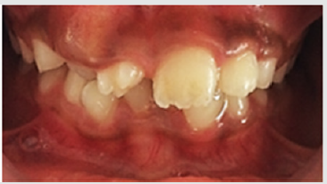
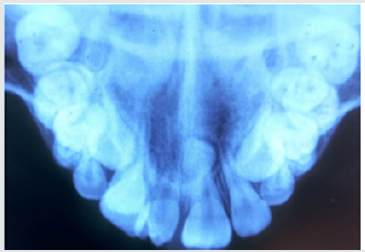
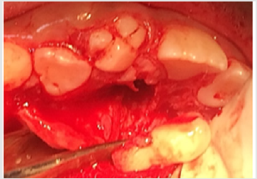
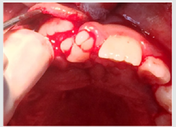
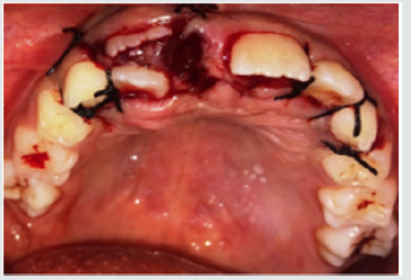
A 9-year old female patient reported to the department of Pedodontics and Preventive Dentistry with the complaint of irregularly placed upper front teeth and wanted it to be corrected. The patient was normal and healthy with non-contributory medical and dental histories. The extra oral examination did not reveal any abnormalities. Intraoral examination revealed a Class I mixed dentition with a missing upper right central incisor and an erupted molariform mesiodens in its place. In addition, there was a firm bulge palpable in the upper right central incisor area which suggests the impediment in the path of eruption of the central incisor by the erupted mesiodens (Figure 1). An occlusal radiograph was taken to rule out the possibility of multiple supernumerary teeth and surprisingly another unerupted and impacted inverted mesiodens with an incomplete root was found mesial to the upper left central incisor (Figure 2). The SLOB technique confirmed that the impacted mesiodens was present palatially. Both informed and written consent was obtained from the parents before initiating the treatment. We decided to extract both mesiodentes under local anesthesia. The erupted mesiodens was extracted by intra- alveolar extraction. The impacted mesiodens was surgically removed by raising a mucoperiosteal flap from maxillary first premolar to contralateral first premolar (Figure 3). Rotary cutting instruments with simultaneous irrigation were used for removing enough bone around the impacted mesiodens for its easy retrieval. Soft tissue uncovering was done for the unerupted right central incisor by placing an elliptical incision over the incisal portion of the palpable bulge (Figure 4). The extraction socket was checked for any pathological tissue and the flap was relocated and sutured with interrupted sutures (Figure 5). The patient was recalled after a week for suture removal and followed-up after 3 months. Uneventful healing with no associated symptoms was observed.

Figure 1: Preoperative view showing erupted molariform mesiodens along with unerupted maxillary central incisor.

lupinepublishers-openaccess-journal-pediatric-dentistry

Figure 2: Occlusal radiograph showing unerupted and impacted inverted mesiodens with an incomplete root.

lupinepublishers-openaccess-journal-pediatric-dentistry

Figure 3: Surgical removal of impacted mesiodens by raising a mucoperiosteal flap from maxillary first premolar to contralateral first premolar.

lupinepublishers-openaccess-journal-pediatric-dentistry

Figure 4: Soft tissue uncovering for the unerupted right central incisor by placing an elliptical incision over the incisal portion of the palpable bulge.

lupinepublishers-openaccess-journal-pediatric-dentistry

Figure 5: Interrupted sutures placed.

lupinepublishers-openaccess-journal-pediatric-dentistry

Discussion

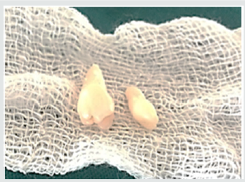
The realm of pediatric dentistry incorporates the practice of interceptive orthodontics, thereby bestowing upon the pediatric dentist, opportunities of providing timely guidance in the development of occlusion. The current case report presents the management of developing malocclusion in the anterior region due to the presence of mesiodens. A mesiodens occurring in the primary dentition is a rarity even though, it being the most common dental abnormality in the permanent dentition [8]. Most cases of mesiodens are discovered during the first decade as maxillary central incisors are erupting and radiographic examinations are performed as an aid to screening for any other malformations and abnormalities [1]. Various theories regarding the etiology of mesiodens have been reported in the literature but the subject remains controversial [3]. Heredity has been suggested to be an etiologic factor based on the observation that supernumeraries are more common in family members; however, it does not follow a simple Mendelian pattern [9]. It was originally postulated that the mesiodens represented a phylogenetic relic of the extinct ancestors who had three central incisors. This is known as phylogenetic theory reversion (atavism) which has now been discarded by the embryologists [10]. The dichotomy theory states that, a mesiodens arises due to the splitting of the tooth bud. On the contrary, Taylor argued that splitting of the tooth bud may either form two equal sized teeth or one normal and one dysmorphic tooth [8]. The hyperactivity theory states that development of mesiodens is due to the hyperactivity of the dental lamina. The ‘field model’, proposes that a tooth bud which is forming at a given location develops according to its position within the field, further determining its shape. The ‘clone model’ postulates that that each tooth class is derived from a clone of ectomesenchymal cells which are programmed by epithelium to produce teeth of a given pattern. Depending upon the specific factors expressed from these ectomesenchymal cells, the shape of the accessory tooth germ forms in the vicinity of the incisors class of teeth becomes evident at the bell stage [11]. This case report presented with an erupted molariform mesiodens and an unerupted tuberculate mesiodens with an undeveloped root (Figure 6). Since the molariform mesiodens was impeding the eruption of the maxillary right central incisor, the surgical removal of both mesiodentes was planned and executed. Timing of interceptive treatment should be as soon as possible following clinical detection of an abnormal eruption pattern. It has been suggested that a tooth delayed in its eruption by more than six months with respect to its antimere should be radiographically investigated. Hogstrum and Andersson [12] suggested two alternatives exist.

The first option involves removal of the supernumerary as soon as it has been diagnosed. This could lead to an unpleasant experience that may have a psychological effect on a very young child and has been said to cause devitalization or deformation of adjacent teeth. Secondly, the supernumerary could be left until root development of the adjacent teeth is complete. The potential disadvantages associated with this deferred surgical plan include; loss of eruptive force of adjacent teeth, loss of space and crowding of the affected arch, and possible midline shifts. In the present case, since the roots of the adjacent teeth were completely formed and the child was at an age where she could sustain a surgical procedure, the surgery was undertaken with utmost attention to detail and caution. Access to mesiodens during surgery must dealt carefully considering the quantity of bone amputation and the possible damage to the adjacent teeth [7]. Follow up is indispensable in such cases since the eruption status should be monitored. The patient revealed satisfactory healing and suitable eruption of the maxillary central incisor. Pediatric dentists are the firsts to usually identify developing malocclusions and thus it is their responsibility to intervene and intercept in an apt manner to prevent future unfavorable sequalae.

Figure 6: Molariform and tuberculate mesiodens.

lupinepublishers-openaccess-journal-pediatric-dentistry

Acknowledgements

Authors are thankful to their colleagues and faculty.

Read More Lupine Publishers Pediatric Dentistry Journal Articles: https://lupine-publishers-pediatric-dentistry.blogspot.com/

Read More Lupine Publishers Articles: https://lupinepublishers.blogspot.com/

Wednesday, 12 September 2018

Caries Survey in 3-5 Year Old Children in Dubai Schools (IPDOAJ) - Lupine publishers


Dental caries is considered the most prevalent chronic disease in the world, which affect individual from all ages. Host acceptability, cryogenic bacteria, fermentable carbohydrates and time are the four elements that are essential for the caries to start. dmft index is a measurement used to determine the degree of caries in any given population, in which it could help in setting aims and strategies to reduce and prevent this disease.