Thursday, 19 March 2020

Lupine Publishers | Midshaft Clavicle Malunion with an Atypical Posterior Apex Deformity

Lupine Publishers Orthopedics and Sports Medicine: open access journal


Abstract

Purpose: We are presenting this pattern of a rare variant of a clavicle malunion with an apex posterior-inferior deformity. This occurred in an elite major junior hockey player during his draft season. This illustrates that such a deformity will most likely result in shoulder weakness, altered shoulder mechanics and may cause brachial plexus neurological findings. In addition, this can cause associated sterno-clavicular deformity which can lead to sternoclavicular joint subluxation secondary to the increased strain placed on the sternoclavicular joint from an apex posterior inferior malunited clavicle. Deformity of > 20 degrees in any direction interferes with normal motion and normal cortical strength even in a young patient.
Introduction: Symptomatic malunion is fortunately less frequently observed (4) since the significant shift to operative treatment for displaced shortened mid shaft clavicle fractures. Symptomatic patients are typically those with marked displacement and significant shortening at the fracture site. Patient’s report weakness of the involved shoulder with rapid fatigability plus an increased deformity comes with an increased risk of recurrent fractures. Although not commonly described in the literature, clavicle malunion usually has a very consistent deformity pattern. As illustrated by McKee et al, the patient usually presents with a complex three dimensional deformity with shortening, an anterior apex at the fracture site and associated joint pain around the shoulder or sternum (6). The influence of the coraco-clavicular and a cromio-clavicular ligaments on the fracture fragments is hypothesized to cause an effect on the displacement of these fractures which involves the lateral segment of the clavicle being carried forward by virtue of its retained a cromio-clavicular and residual coraco-clavicular attachments. Angulations are more acute the closer the break is to these pivot points. This has had associated significant alteration in normal clavico-scapular motion.
Method: Case report and literature review.
Conclusion: Symptomatic clavicular malunion is rare but definitely higher with non-operative management and can cause discomfort and shoulder weakness. Neurological symptoms and signs are more likely to occur in inferior malunited clavicle, particularly with an inferior-posterior deformity. We illustrated the steps necessary to correct all deformities and lengthen the clavicle using a long working length precountored plate construct. This has improved the clinical symptoms of the patient and illuminated the risk of repeat fracture due to deformity. Plate removal is planned but is still an unanswered question.
Keywords: Mid Shaft; Clavicle Symptomatic; Malunion; Nonunion; Deformity

Case

An 18 year old elite Canadian Hockey player presented with a new fracture to his left clavicle and associated pain at the sternoclavicular joint with an obvious deformity. He had sustained a previous injury to his left mid shaft clavicle two years ago playing hockey. This was treated on operatively and went on to heal with a 25 degree posterior-inferior deformity. A review of his initial injury films, from two years ago, illustrated a moderately displaced mid shaft clavicle with a significant amount of shortening (2 cm)due to inferior apex deformity( 25 degrees).However, it was decided to treat him on operatively as it was a closed injury in a relatively young male and he was neurovascularlyintact. His fracture healed with 2.5 cm of shortening, slight scapular inward rotation and a 25-30 degree posterior-inferior deformity. The sternoclavicular joint deformity on the left side stopped him from playing hockey at an elite level for about two months but a steroid injection seemed to remove most of his symptoms and allowed him to compete. He also complained of an ongoing occasional shoulder weakness and an occasional fleeting numbness in his arm and hand. This was significant enough to warrant a CT of the chest to rule out thoracic outlet syndrome.n 2010, the Czech Republic participated in the World Health OrgAn 18 year old elite Canadian Hockey player presented with a new fracture to his left clavicle and associated pain at the sternoclavicular joint with an obvious deformity. He had sustained a previous injury to his left mid shaft clavicle two years ago playing hockey. This was treated on operatively and went on to heal with a 25 degree posterior-inferior deformity. A review of his initial injury films, from two years ago, illustrated a moderately displaced mid shaft clavicle with a significant amount of shortening (2 cm)due to inferior apex deformity( 25 degrees).However, it was decided to treat him on operatively as it was a closed injury in a relatively young male and he was neurovascularlyintact. His fracture healed with 2.5 cm of shortening, slight scapular inward rotation and a 25-30 degree posterior-inferior deformity. The sternoclavicular joint deformity on the left side stopped him from playing hockey at an elite level for about two months but a steroid injection seemed to remove most of his symptoms and allowed him to compete. He also complained of an ongoing occasional shoulder weakness and an occasional fleeting numbness in his arm and hand. This was significant enough to warrant a CT of the chest to rule out thoracic outlet syndrome.nization WHO Research to determine the quality of medical decAn 18 year old elite Canadian Hockey player presented with a new fracture to his left clavicle and associated pain at the sternoclavicular joint with an obvious deformity. He had sustained a previous injury to his left mid shaft clavicle two years ago playing hockey. This was treated on operatively and went on to heal with a 25 degree posterior-inferior deformity. A review of his initial injury films, from two years ago, illustrated a moderately displaced mid shaft clavicle with a significant amount of shortening (2 cm)due to inferior apex deformity( 25 degrees).However, it was decided to treat him on operatively as it was a closed injury in a relatively young male and he was neurovascularlyintact. His fracture healed with 2.5 cm of shortening, slight scapular inward rotation and a 25-30 degree posterior-inferior deformity. The sternoclavicular joint deformity on the left side stopped him from playing hockey at an elite level for about two months but a steroid injection seemed to remove most of his symptoms and allowed him to compete. He also complained of an ongoing occasional shoulder weakness and an occasional fleeting numbness in his arm and hand. This was significant enough to warrant a CT of the chest to rule out thoracic outlet syndrome.
This 18 year old male continued to play elite major junior hockey (prime pathway to the NHL in Canada) then unfortunately sustained another injury where he was checked into the boards during an elite hockey game. He felt immediate pain and tenderness along his clavicle and therefore presented to the hospital emergency. Interestingly, since his initial incident, he had never been free of symptoms and he subsequently fractured his clavicle with relatively low trauma within 18 months of his last fracture. Plus he had significant sterno-clavicular associated symptoms with pain and anterior subluxation of the ipsilateral sterno-clavicular joint
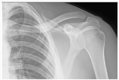
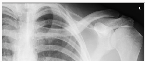
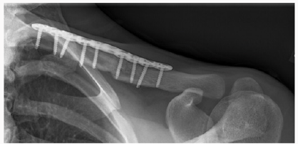
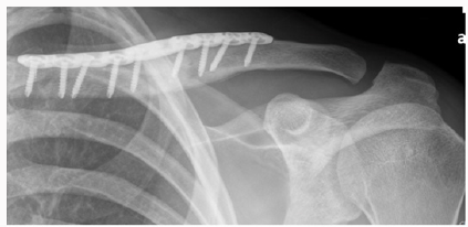
In the Emergency Department he was evaluated by the ER physician and the orthopaedic on call team. He had normal vital signs and good air entry bilateral chest, his neurological exam of both motor and sensory nerves of his left upper extremity showed no deficit, no signs of thoracic outlet syndrome and he illustrated a normal vascular exam. His investigation included x ray of his left clavicle with a contra lateral clavicle x ray for comparison. Both clavicles had an AP and orthogonal clavicular views (see images below). His clavicle demonstrated a more pronounced posterior-inferiorapex deformity (30-35 degrees), shortening and malrotation plus a significantly deformed (anterior subluxation) sternoclavicular joint as noted over the last year.
A detailed discussion with the patient about the findings was complete along with the possible operative and non operative treatment modalities available. Given the latest research and paper by McKee et al on the increased fracture rate in significantly deformed clavicles, an operative approach was chosen. This choice was also enhanced by the history of increased discomfort generally around the shoulder girdle discomfort plus the significant shoulder weakness, sterno-clavicular pain, neurological symptoms and reduced maximal function. We, therefore, elected to book him for a corrective osteotomy to restore length, alignment, rotation and angulations to augment the mechanics of his shoulder and the biomechanical ability of this clavicle to absorb an impact without re-fracturing.

Operative Procedure

The patient underwent general anaesthesia and was placed in a beach chair position in a 45 degree semi sitting position with a small pad behind the left shoulder blade and the involved upper extremity was draped freely with the distal arm placed in a sterile extremity drape. An oblique incision was made along the superior surface of the clavicle to expose the nonunion site. The skin and subcutaneous tissue was raised as a flap, and the underlying myofascial planes identified. This layer was raised as contiguous flaps and was preserved so that a two-layered closure could subsequently be achieved. Next, the malunion site was identified, and a long oblique, superior to inferior, osteotomy was performed. This provided a long osteotomy surface to correct the inferior apex deformity while allowing for the three dimensional correction with excellent bone to bone contact.
The osteotomy was performed with a, well irrigated, cooled, micro sagital saw. After careful dissection a small blunt Haworth elevator was placed underneath the clavicle to protect the neurovascular structures during the osteotomy and elevation of the deformity. Very importantly, the medullar canal was re-established, on both sides of the osteotomy, with a 3.5-mm drill-bit plus very aggressive curettage of the sclerotic bone in order to obtain an excellent opening in the medullar canal in the proximal and distal segments.
However, we have a very novel solution in the Czech Republic - whetSmall reduction clamps were then utilized to perform a reduction that would allow lengthening of the clavicle along with rotational and ambulatory correction utilizing the precountored plate as a reduction tool. First, shortening was corrected and held by translating the medial lateral fragment over the large surface osteotomy area to gain the planned length of 2.5 cm based on our preoperative planning. This was accomplished almost entirely by deformity correction. Secondly, rotation was corrected by rotating the lateral fragment about forty degrees clockwise until the flat surface of the lateral fragment was facing superior as desired. We then placed a long 10 whole precountored clavicle plate on the superior surface of the clavicle using the construct, with its long working length, to help gradually realign the bone back to the plate. This was and should be done very slowly and carefully as the underlying neurovascular structures can be tethered to the deformed bone. This was then held using absolute stability fixation with non locking screws on each side of the osteotomy. The screws were then gradually tightening of screws on either side of the deformity.r you are an individual patient crippled and dying for legal or iSmall reduction clamps were then utilized to perform a reduction that would allow lengthening of the clavicle along with rotational and ambulatory correction utilizing the precountored plate as a reduction tool. First, shortening was corrected and held by translating the medial lateral fragment over the large surface osteotomy area to gain the planned length of 2.5 cm based on our preoperative planning. This was accomplished almost entirely by deformity correction. Secondly, rotation was corrected by rotating the lateral fragment about forty degrees clockwise until the flat surface of the lateral fragment was facing superior as desired. We then placed a long 10 whole precountored clavicle plate on the superior surface of the clavicle using the construct, with its long working length, to help gradually realign the bone back to the plate. This was and should be done very slowly and carefully as the underlying neurovascular structures can be tethered to the deformed bone. This was then held using absolute stability fixation with non locking screws on each side of the osteotomy. The screws were then gradually tightening of screws on either side of the deformity.
Intra operatively, significant improvement in the shoulder contour was obvious as well as a noticeable reduction in the anterior subluxation of the sternoclavicular joint. Screw length was checked with an image at the end of the procedure. Deformity correction usually necessitates some screw changes as the initial screws can be long once the deformity is reduced. Wound closure was done in layers closing the myofascial flap over the plate and subsequently the subcutaneous tissue and the skin was re approximated with narrow skin staples.
Post operatively the patient was placed in a shoulder sling for comfort and scheduled for early physio to initiate shoulder and elbow function. His post op exam confirmed intact neurovascular status of his left upper extremity. Chest x ray taken in recovery room confirmed we had not created a pneumothorax. The operative procedure was performed as an outpatient. The patient went home on the same day and returned at 10 days for wound examination and staple removal. Aggressive physio was initiated that day following the initial gentle ROM and pendulum exercises which were initiated immediately post op (Figures 1-9).
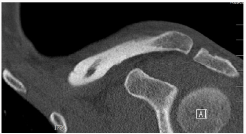
Figure 1: Axial CAT scan of the chest delineating the sternoclavicular deformity related to the clavicle malunion.
Figure 2: Coronal CT showing the direction of malunited clavicle.
Figure 3:
Figure 4: (a) Comparison right (normal)(b) Left (Malunited) clavicle
Figure 5:
Figure 6:
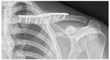
Figure 7: Early post operative.
Figure 8:
Figure 9: Three months post-operative (signs of radiographic healing).

Discussion

Clavicles fractures are common injuries and are reported to represent 2% to 5% of all adult fractures [1]. More recent evidence suggests that specific subsets of patients may be at higher risk for nonunion, symptomatic malunion, or suboptimal functional outcomes [2]. A recent meta-analysis suggests that the incidence of clavicle nonunion after nonsurgical treatment is approximately 5.9%, but can be as high as 15%for some fracture subtypes [3]. Nonsurgical treatment universally results in some degree of malunion; however, symptomatic malunion is fortunately quite low and is usually used particularly in very young patients [4]. Symptomatic patients are typically those with marked displacement at the fracture site, with shortening of >2 cm. Patients that are symptomatic may report weakness of the involved shoulder, rapid fatigability, numbness and paresthesia of the hand and forearm with elevation of the limb, and an asymmetric, “droopy,” “ptotic,” or “driven in”shoulder [5].
McKee et al performed a review of a cohort of patients to analyze the functional results of corrective osteotomy of a mal united clavicular fracture in patients with chronic pain, weakness, neurologic symptoms, and dissatisfaction with the appearance of the shoulder. Fifteen patients (nine men and six women with a mean age of thirty-seven years) who had amalunion following non operative treatment of a displaced mid shaft fracture of the clavicle were reviewed both preoperatively and postoperatively. The mean time from the injury to presentation was three years (range, one to fifteen years).Follow-up, at a mean of twenty months (range, twelve to forty-two months) postoperatively, illustrated that the osteotomy site had united in fourteen of the fifteen patients. All fourteen patients expressed satisfaction with the result. There was one nonunion, and two patients had elective removal of their plates. With regards to the patho anatomy of the deformed clavicle, McKee et al. noted that the deformity of the clavicle was a complex three-dimensional problem with all their patients illustrating a superior-anterior apex deformity. In his series there were certain consistent features seen in patients who presented with symptoms following non operative treatment and a healed clavicle. The hall mark characteristic is shortening in the medial-lateral dimension, with inferior displacement of the distal fragment and superior displacement of the proximal fragment. They, therefore, concluded that the shortening in the medial-lateral plane had a negative effect on muscle-tendon tension, and muscle balance. The anatomic boundaries of neurovascular structures were of paramount importance in the development of symptoms [6].
In a study by Edelson et al, he studied the bony anatomic details in 73 cadaver specimens which had clavicle malunions in different regions of the clavicle. According to the Allman classification. Edelson found that in the middle-third fractures, similar anterior angulations to the lateral third fracture malunion was indeed present. The most consistent finding at the middle-third level was that the lateral shaft fragment was almost invariably displaced posterior to the medial shaft fragment. The author also commented that initial anterior-posterior radiographs of clavicle fractures are often dominated by inferior displacement or ptosis of the lateral fragment. However, in the cadaveric specimens, anterior angulations rather than drooping of the lateral fragment were the predominant deformity. Although often initially displaced in a down ward direction, the lateral fragment does not usually heal in this position, unless it is a greenstick fracture as occurred in our patient.
Therefore the literature concludes that the principle deformity in a healed malunionis anterior, superior angulations. In this series there were only 4 cases in which the lateral clavicle healed with downward angulations of 20° or more at the fracture site as occurred in our young patient with his greenstick type of fracture. The author hypothesized that inferior displacement of the lateral fragment, which predominates on the initial radiographs, is most likely due to post-traumatic muscle a tony, principally of the deltoid and trapezius, similar to that which can cause the glenohumeral joint to appear subluxed after fractures of the humeral head and claimed that as soon as the muscle tonus returns, the clavicle resumes a horizontal orientation, and fracture position is then dominated by the pronators and internal rotators of the scapula and upper arm, which reposition the fragments into the anteriorsuperior apex position [7].
We believe that corrective osteotomy can lead to predictably good results (> 95%), however one should be careful with the inferior dissection as it can and has produced neurological and vascular issues in the past. So which fracture requires surgical correction? In general principles, according to the Canadian Orthopedic Trauma Society (COTS)and the McKee et al papers, “symptomatic deformity” with significant shortening of 2-3 cm , angulations deformity >30 degree or translation of >1cm . This has been supported in numerous repeated studies since 2008. In addition softer indications would be symptoms of thoracic outlet syndrome, weakness or rapid fatigability with overhead activity, a relatively weak arm at over a year from the fracture or more commonly a combination of all of these symptoms, should be considered for an operative correction [6].
Another area of controversy between surgeons who treat this type of injury is the need for hardware removal to decrease the risk of re-fracture. Some surgeons prefer to remove the implant in all patients after clavicle fracture union, whereas others plan for additional surgery only if the patient complains of symptomatic hardware. In either case, adolescent patients undergoing surgical fixation for clavicle fracture must be warned of the possibility of return to the operating room to remove the implant.

Conclusion


Malunion of the clavicle with > 20 to 30 degrees of deformity and symptoms of weakness and malfunction should be considered for corrective osteotomy. The success rate is very high (.95%) and results in excellent patient satisfaction. This again supports McKee’s initial study that highlighted the clinical impact of mid shaft clavicle deformity and the importance of surgical reconstruction with an absolute stability. We also believe that if a surgeon carefully follows the steps of the surgical technique described in this case report; the incidence of vascular and neurological injuries can be mitigated although not entirely illuminated as a risk.

Monday, 16 March 2020

Lupine Publishers | Statistical Software: A Risk for Medical Science?

Lupine Publishers | Journal of Otolaryngology Research Impact Factor


Opinion

pinion This editorial is based on my personal professional experiences of some four decades work in health and business applications of statistics. I am fully aware that these experiences cannot be interpreted as a random sample and there is no warranty of any kind for the absence of possible biases. You might well be familiar with the US-FDA view that there is no unbiased data in the scientific universe available. Historically, the use of statistics in medicine is a well-documented fact since about two centuries. My personal experiences cover the transition period from electronic desktop calculators with paper and pencil, to the omnipresence of cheap computing power and sophisticated statistical software of today. I remember my early professional study design activities as a statistician as heavily impacted by cost considerations: Human work time was and still is quite expensive. During the last period of about some two decades I got the strong impression that the medical profession, especially those doctors who are working scientifically – showed a quite strong trend to higher statistical understanding and knowledge as compared to my early working times. This positive trend is highly appreciated by me and I see it as an enormous economic advantage that computers took over the tedious workload of numbers crunching nowadays. There is a negative trend involved over the last two decades as well: I observed a growing trend of numbers of “so called” statisticians who actually are experts in using available statistical software packages only but have little or no statistical expertise. I see a professional statistician as a human who understands the primary objective of the study’s objective and to assess the medical question under consideration and to decide the statistical model selection for the very question based on the scope of the scientific question and all of the constraints and limitations in practice. I know that this is an “ideal world” assumption and we all are but humans with our limitations. My experience that it pays to strive for perfection is illustrated by some examples which I consider to be potentially useful and beneficial for your work as doctor:

Project Planning Phase

a) Overall considerations of project management and quality control, legal requirements.
b) Definition of research question(s).
c) Definition of data selection criteria, sampling variables and observation time(s) schedule.
d) Aspects of data collection and documentation, ongoing project quality controls.
e) Feasibility considerations of various project designs.
f) Administrative aspects: financials, selection of partners, estimation of required realization times.
g) Statistical methods for data analysis, feasibility of pilot- (sub)-projects.
h) Final definition of data selection criteria.
i) Aspects of results publication.
j) Aspects of possibly necessary actions in case of emergencies.

Project Realization Phase

a) Ongoing control of “plan vs actual” progress.
b) Ongoing communication between all project partners.
c) Maintenance of open minds for early signs of project’s critical developments.
d) Ongoing monitoring for possible emergency actions.
e) The KISS principle: Keep everything as simple and stupid as possible.
I’d like to recommend using the items in the above project management as guidance for your project but under no condition as a comprehensive cookbook! As ENT scientist you should permanently remember that you are doing research in humans and not in bolts and nuts!

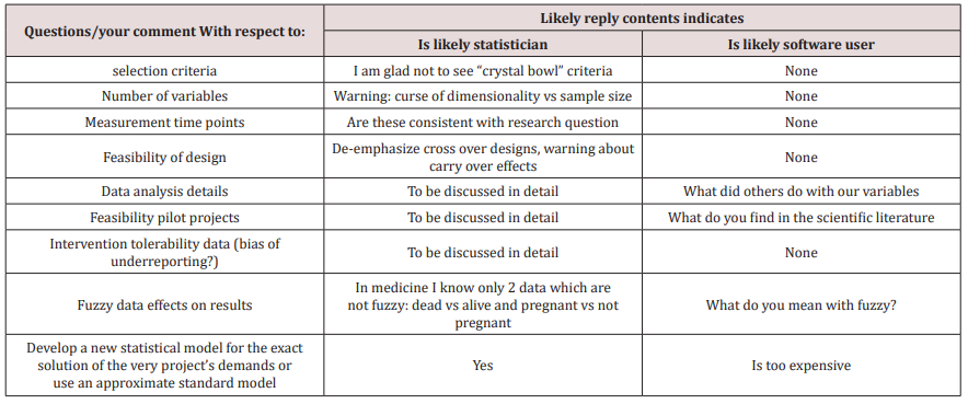
How you could easily detect the difference between a “user of statistical software” or a “real” statistician

                         The next table provides you with selected questions/topics to distinguish between professional statistician and software user (Table 1).


Friday, 13 March 2020

The Optimal Pain Management Methods Post Thoracic Surgery: A Literature Review| Lupine Publishers

Abstract


Post-operative pain control is one of the key factors that can aid in fast and safe recovery after any surgical interventions. Thoracic surgery can cause significant postoperative pain which can lead to delayed recovery, delayed hospital discharge and possibly increased risk of chest complications in the form of atelectasis and even lower respiratory infections. Therefore, appropriate pain management following thoracic surgery is mandatory to prevent development of such morbidities including chronic pain.
Keywords:Thoracic Surgery, Analgesia, VATS, Robotics, Thoracotomy

Introduction

Thoracic surgical procedures can result in severe pain which can present as a challenge to be appropriately managed postoperatively. In particular, thoracotomies are well known for their severity of pain due to the incision, manipulation of muscles and ligaments, retraction of the ribs with compression, stretching of the intercostal nerves, possible rib fractures, pleural irritation, and postoperative tube thoracotomy [1]. Recognition of this has contributed to the development of minimally invasive techniques such as video assisted thoracoscopic surgeries (VATS) and lately robotic surgery [1]. These techniques not only aim to produce better aesthetic results, but also reduce post-operative pain and enhance recovery without compromising the quality of treatment offered. Poor pain management can lead to several and serious complications such as lung atelectasis, hypostatic pneumonia due to avoidance of deep breathing in these patients as a result of pain and superimposed infection [1]. Pain management as a result, does not only lead to greater patient satisfaction, but it also reduces morbidity and mortality in patients undergoing thoracic surgery [2]. Historically, post-operative pain management for thoracic surgery involved the use of narcotics alongside parenteral or oral anti-inflammatory agents [2]. Post chest tube removal patients typically are transitioned to oral analgesia. Multiple additional pain control adjuncts were also implemented with differing levels of success [1]. Over time, intra-operative techniques have been developed which aims to target pain reduction postoperatively [2]. As our understanding of both pain management and the factors that play a role in the development of pain has increased, we have been able to target these and improve postoperative pulmonary morbidity and pain scores [1,2]. We aim to review different means of pain control in this paper in order to assess their effectiveness in achieving optimum results.

Thoracotomy

The mechanism of pain in thoracotomy involves the innervation of the intercostal, sympathetic, vagus and phrenic nerves [3]. Additionally, shoulder pain may result from stretching of the joints during the operation.
After a thoracotomy, pain can persist for two months or more, and in certain incidences it recurs after a period of cessation. The incidence of chronic pain post thoracotomy is reported to be 22-67% in the population [4]. Good surgical technique and effective acute post-operative pain treatment are evident means of preventing post-thoracotomy pain and consequent pulmonary complications [4]. Due to the multifactorial character of the pain, a multimodal approach to target pain is advised. Typically, both regional and systemic anaesthesia are administered. A combination of opioids such as fentanyl or morphine are typically used [5]. A variety of techniques for the administration of local anaesthetics are available at present, and the effectiveness of each is assessed in this paper.
a) Thoracic Epidural Analgesia (TEA)
TEA was the most widely used method of means of analgesia. It was the gold standard means of pain relief [6,7]. It is typically inserted prior to general anaesthesia, at the level of T5-T6, midway along the dermatomal distribution of the thoracotomy incision. A study by Tiippana et al. [8] measured the visual analogue scale (VAS) in order to assess the presence of pain during rest and at the time at which they coughed in 114 patients of whom 89 had TEA and 22 who had other methods of pain control. TEA was effective in alleviating pain at rest and during coughing. In TEA patients, the incidence of chronic pain of at least moderate severity was 11% and 12% at 3 and 6 months, respectively. The study found that at one week after discharge, 92% of all patients needed daily pain medication. The study advised for extended postoperative analgesia for up to the week post-discharge to be administered in order to manage this. The study however concluded overall, that TEA was effective in controlling evoked post-operative pain. However, the study did encounter problems of technical form in 24% of the epidural catheters. The incidence of chronic pain, however, was lower compared with previous studies where TEA was not used. Several other studies support that TEA is superior to less invasive methods. According to Shelley B. et al. [9] TEA was preferred by 62% of the respondents over paravertebral block (PVB) with 30% and other analgesic techniques with 8%. Limitations of this technique included hypotension and urinary retention. Certain patients with active infection and on anticoagulation are excluded from epidural placement.
b) Paravertebral Block (PVB)
PVB is considered an effective method for pain management and its use has been increased in the recent years. This technique involves injecting local anaesthetic into the paravertebral space and it is able to block unilateral multi-segmental spinal and sympathetic nerves. Previous studies have shown that it is effective in achieving analgesia and is associated with a lower incidence of side effects such as nausea, vomiting, hypotension and urinary retention [10,11]. As the lungs are collapsed, it is associated with a lower risk of pneumothorax.
In a study by Davies R.G. et al. [10] there was no significant difference in pain scores, morphine consumption and supplementary use of analgesia between TEA and PVB. The rate of failed technique was lower in PVB (OR =0.28, p=0.007). Respiratory function was improved at both 24 and 48 hours with PVB but only significantly improved at 24 hours.
c) Intercostal Nerve Block (ICNB)
ICNBs are generally administered as single injections at least two dermatomes above and below the thoracotomy incision [12]. It is performed percutaneously or under direct vision, using single injections or through placement of an intercostal catheter. It can also be formed using cryotherapy. It is associated with reduced post-operative pain scores; however, it is less effective than TEA in controlling chronic pain [12]. This was illustrated by a study by Sanjay et al. [12] which found that patients that underwent ICNB had higher pain scores 4 hours post-operatively, than those who received epidural anaesthesia using 0.25% bupivacaine (p<0.05). The study concluded that in the early post-operative period there was significant impact in pain relief for both techniques, but thereafter, epidural anaesthesia was proven to significantly reduce post thoracotomy pain over ICNB. Due to the multifactorial nature of post-thoracotomy pain, various approaches are required in order to target pain. ICNBs are useful in the blockade of intercostal nerves, whilst PVB and TEA appear to block the intercostal and sympathetic nerves. Due to the inability of regional anaesthesia to block the vagus and phrenic nerves which are implicated in the pathophysiology of pain, NSAIDs and opioids are required as adjuncts. TEA is proven to be the most effective means of treating pain alongside PVB; however, it is associated with more side effects than PVB. At present, there are a limited number of studies directly comparing pain control and post-operative outcomes between PVB and TEA. There is no conclusive evidence that either method is superior to the other regarding pain control.

Video-Assisted Thoracoscopic Surgery (VATS)

Existing evidence supports the noninferiority of thoracic PVB when compared to TEA for postoperative analgesia [13]. PVB is versatile and may be applied both unilaterally or bilaterally. It can be used to avoid contralateral sympathectomy, consequently minimising hypotension. This is an apparent advantage it has over thoracic epidural. Furthermore, it offers a more favourable side effect profile when compared to epidural anaesthesia. At present, the factors taken into consideration when selecting a regional technique include tolerance of side effects associated with TEA, consensus on best practice/technique, and operator experience [13]. A randomised controlled trial by KosiÅ„ski et al. [14] compared the analgesic efficacy of continuous thoracic epidural block and percutaneous continuous PVB in 51 patients undergoing VATS lobectomy. The primary outcome measures were postoperative static (at rest) and dynamic (coughing) visual analogue pain scores (VAS), patient-controlled morphine use and side-effect profile. The study found that pain control (VAS) was superior in the PVB group at 24 hours, both at rest (1.7 vs3.3, p=0.01) and on coughing (5.8 vs 6.6, p=0.023), and control of pain at rest was also superior in the PVB group at 36 hours (3.0 vs 3.7 (p=0.025) and at 48 hours (1.2 vs 2.0, p=0.026). There were no significant differences in the postoperative morphine requirements. In regard to side-effect profile, the study showed that the incidence of postoperative urinary retention (defined as no spontaneous micturition for 8 hours or ultrasound-assessed volume of the urinary bladder >500ml) was greater in the epidural group (64.0% vs 34.6%, p=0.0036), as was the incidence of hypotension (32.0% vs 7.7%, p=0.0031). There was no significant difference in the incidence of atelectasis (4.0% vs 7.7%, p=0.0542). However, the incidence of pneumonia was significantly more frequent in the PVB group (3.8% vs 0%, p=0/0331). KosiÅ„ski et al. concluded that PVB is as effective as thoracic epidural block in regard to pain management as it offers a superior safety profile with minimal postoperative complications. A further randomised controlled trial by Okajima et al. [15] compared the requirements for postoperative supplemental analgesia in 90 patients who received wither a PVB or thoracic epidural infusion for VATS lobectomy, segmentectomy or wedge resection. The main outcome measures were pain scores at rest (verbal rating scale 0= none and 10=maximum pain), blood pressure, side effects and overall satisfaction scores relating to pain control (1=dissatisfied and 5=satisfied). The study found a similar frequency of supplemental analgesia (50mg diclofenac sodium suppository or 15mg pentazocine intramuscularly) for moderate pain in both groups, with 56% of those in the PVB group requiring ≥2 doses, compared to 48% in the epidural group (p=0.26). Hypotension, defined as a systolic blood pressure <90mmHg, occurred more frequently in the epidural group (21.2% vs 2.8%, p=0.02). There was no difference in the incidence of pruritus (3.0% vs 0%, p=0.29) and post-operative nausea and vomiting (30.3% vs 25.0%, p=0.62) between both groups. The study found no statistical difference between patient-reported satisfaction in pain control between epidural and PVB using the verbal rating scale (5.0 vs 4.5, p=0.36). The study concluded that PVB offered additional to equivalent analgesia to epidural, a lower incidence of haemodynamic instability postoperatively. A further study by Khoshbin et al. [16] performed an analysis on 81 patients undergoing VATS for pleural aspiration +/- pleurodesis, lung biopsies or bullectomy. The main outcome was postoperative pain levels, documented every 6 hours and scored against the Visual analogue Scale (0= no pain, 10= worst possible pain). In both PVB and epidural groups, bupivacaine 0.125% was the local anaesthetic of choice, with clonidine added to the epidural infusion at 300μg in 500ml. The study showed that there was no significant difference in mean pain scores between PVB or EP (2.1 vs 2.9, p=0.899), therefore concluding that PVB is as effective as epidural in controlling pain post-VATS.

Robotic Lung Surgery

Minimally invasive techniques are considered advantageous over open surgical approaches due to their shorter recovery times, reduced perceived levels of pain post-operatively and shorter postoperative length of stay in hospital [17-19]. Robotic surgery has become a popular method in recent years. Debate remains regarding whether robotic surgery is superior to VATS in regard with pain reduction. A case control study by Louie et al. [19] compared 45 robotic assisted lobectomies (RAL) to 34 VATS lobectomies. The study showed that both groups had a similar mean ICU stay (0.9 vs 0.6 days) and a mean total length of stay (4.0 vs 4.5 days). The study showed that patients that underwent robotic lobectomies had a shorter duration of analgesic use post-operatively (p=0.039) and a shorter time resuming to normal everyday activities (p=0.001). A limitation in this study was an inaccurate record of the amount of pain relief used by the patients, ultimately working as a confounding factor when interpreting the results. In a separate study by Jang et al. [18] 40 patients undergoing RAL were compared retrospectively to 80 VATS patients (40 initial patients and 40 most recent patients), all with resectable non-small cell lung cancer. The study showed that the post-operative median length of stay was significantly shorter in RAL patients compared to the initial VATS patients. The rate of post-operative complications was significantly lower in the RAL group (10%) compared to the initial VATS group (32.5%) and similar to the recent VATS group (17.5%). Post-operative recovery was easier for patients in both the RAL and VATS group due to earlier mobilisation, allowing them to return to their everyday activities quicker. In a retrospective review by Kwon et al. [17] 74 patients undergoing robotic surgery, 227 patients undergoing VATS and 201 patients undergoing anatomical pulmonary resection were assessed and compared with regard to acute (visual pain score) and chronic pain (Pain DETECT questionnaire). The study showed that there was no significant difference in acute or chronic pain between patients undergoing robotic assisted surgery and VATS. Despite no significant difference in pain scores, 69.2% of patients who underwent robotic-assisted surgery felt the approach affected their pain versus 44.2% of the patients who underwent VATS (p=0.0330). These results all support the superiority of robotic surgery over VATS and open approaches with regard to pain, length of hospital stay and recovery times. Both robotic surgery and VATS have their benefits i.e. two-versus three-dimensional view, instrument manoeuvrability, and reduced post-operative pain.

Conclusion

Since post-thoracotomy pain is multifactorial, a multimodal approach is required. In particular, ICNB blocks the intercostal nerves, and PVB and TEA appear to block the intercostal and sympathetic nerves. NSAIDs and opioids are required as valgus and phrenic nerve cannot be blocked by regional anaesthesia. TEA is evident to be the most effective in treating pain alongside with PVB. It is however associated with more side effects than PVB.


Read More Lupine Publishers Journal of Surgery Articles : https://surgery-casestudies-lupine-publishers.blogspot.com/
Read More Lupine Publishers blogger Articles: https://lupinepublishers.blogspot.com

Lupine Publishers | The Prevention and Treatment of Malaria in Traditional Medicine of Tetun Ethnic People in West Timor Indonesia

Lupine Publishers | Open access Journal of Complimentary and Alternative Medicine


Abstract


Native people in West Timor Indonesia have been exposed to malaria since long time ago. Because of this experience, it is believed that this community has developed their local concept about malaria, and how to manage it. This research was intended to document and analyze local knowledge and practices of malaria prevention and treatment developed by Tetun ethnic people in West Timor. The research was a field study, conducted through some interviews, discussions and observations. The results of this study showed that this community has long been developing various methods to prevent and threat malaria. The prevention and treatment of malaria in traditional medicine of Tetun ethnic people consists of both herbal and non-herbal methods and supported by some prohibitions and restrictions. The results also showed that the practice of traditional medicine for prevention and treatment of malaria by Tetun ethnic people can be explained scientifically. Medicinal plants that widely used like Strychnos ligustrina, Carica papaya, Momordica sp., Cleome rutidosperma, Physalis angulata, Alstonia spectabilis, Alstonia scholaris and Melia azedarach have been proven to have antimalarial activities as anti-plasmodial, antipyretic, analgesic, anti-inflammatory and immunostimulant.

Keywords: Local Knowledge; Traditional Medicine; Malaria Prevention and Treatment; Tetun Ethnic; West Timor


Introduction

Traditional communities in ancient times developed their local knowledge about the prevention and treatment of a disease based on their experience interacting with the disease for a long time. This local knowledge was then become a guidance for them to establish strategies to prevent and treat the disease, which were practiced widely in the community, and become their traditional medicine [1,2]. Traditional medicine is a term imposed on pre-scientific medical systems, and defines as a sum total of knowledge, skills and practices based on theories, beliefs and experiences of different cultural customs used in health care, disease prevention and increased physical and mental performance, which have been used for generations from one generation to the next [3,4]. Malaria is an ancient disease that has not been fully eradicated until this time [5]. Since long time ago, malaria was the main infectious disease that often attacks Timorese people, especially in Belu and Malaka Districts in West Timor (Indonesia). Several old manuscripts noted that Timorese people in early of 19th century were suffered from malaria which caused many deaths [6,7]. Until this time, Belu and Malaka Districts are still hyper-endemic areas of malaria. According to the Global Fund report, in 2014, Belu and Malaka Districts were classified as high malaria endemic areas, with the Annual Parasite Insidence (API) of 12.87o/oo and 11.58o/oo respectively, higher than Indonesian average API 1.38o/oo. Various programs for malaria prevention and eradication sponsored by the Indonesian Ministry of Health and World Health Organization such as insecticide-impregnated net, fogging, mass blood survey for early diagnosis and prompt treatment, and treat malaria patient using Artemisinin Combination Therapy (ACT) have been implemented, but decreasing of the API value is still not too convincing [8]. Cultural factors that influence public attitudes and acceptance on the programs of prevention and treatment of malaria are estimated to be one of the obstacles to the success of these programs. The implementation of various disease control programs and strategies often faces major challenges stemming from the social and cultural situation of the community. The social and cultural situation of a community in a particular place can negatively influence the choice, acceptance and use of interventions in disease control. Many programs of disease control and eradication are unsuccessful because of these social and cultural barriers. Therefore, it is very necessary to understand the local knowledge of the community, including an understanding of the health-illness concept that they believe in. An understanding of this can help policy makers in designing a sustainable and more effective disease control programs [9]. The Tetun ethnic is one of native communities that inhabit territories from the central part of Timor island (in Belu and Malaka districts, Indonesia) to the east (in Republic Democratic de Timor Lester, RDTL). Tetun people are still using traditional medicines to date, and often running various traditional medication rituals [10]. Because of their long-time interaction with malaria, it should be assumed that they have developed their own local knowledge about malaria and methods to prevent and treat it. Therefore, this research was intended to study the local knowledge of the Tetun ethnic people regarding malaria and the methods they have developed for the prevention and treatment of this disease.

Study Design

This study is a kind of research in the field of medical anthropology. This study was conducted as a qualitative exploratory research, with a field study as main technique, supported by a literature study.

Profile of Study Site and People

This research was conducted in Belu and Malaka Districts located in the central part of Timor island. These areas are located at 9°15’ S-9°34’ S and 124°40’ E-124°54’ E. Belu and Malaka are two of Indonesian territories that border directly with the Republic Democratic Timor Leste (RDTL). The topography of Belu Districts is mainly hilly, while Malaka is generally a stretch of flat land. Some areas of Malaka at the south part meet the rainy season twice in a year, while the areas of north part and also Belu areas are only have one rainy season. The main rainy season takes place between November-March due to wind that brings rain from the Indonesian Ocean. This rain occurs evenly in Malaka and Belu regions. The additional rainy season in April-June, which is limited in some areas of Malaka, is affected by wind from Australia that carries moisture from the Timor Sea. Based on the ethnolinguistics, there are four indigenous ethnic groups that live in Belu and Malaka Districts, namely Tetun, Dawan, Kemak, and Bunaq (Marae). Tetun ethnic is the majority ethnic group in Belu and Malaka, consists of approximately 80% of the population. They scaterred in almost all sub-districts of Belu and Malaka [11].

The Informants

The informants of this study were people of Tetun ethnic who have lived for long time in Belu or Malaka Districts. They were people with good knowledge and experiences of traditional medicine practices. The informants were selected through the purpossive and snowball tehniques. A total of 94 informants (42 men and 52 women) with the age of 40-90 years old were involved in this study. They came from 15 vilages of five sub-districts in Malaka (Wewiku, Malaka Barat, Weliman, Malaka Tengah and Kobalima Timur Subdistricts), and 14 vilages of ten sub-districts in Belu (Raimanuk, Tasifeto Barat, Nanaet Duabesi, Tasifeto Timur, Lasiolat, Raihat, Lamaknen, Kakuluk Mesak, Atambua Barat and Atambua Selatan Sub-districts). These informants consist of traditional public healers, home healers, and traditional medicine users.

Data Collection

Data were collected through several interviews, discussions, and observation. Interviews were conducted with a semi-structured questionnaire. Interviews were intended to collect informations about local knowledge on health-illness concept, symptoms, signs and causes of malaria, traditional methods for the prevention and treatment of malaria, and medicinal plants used for the prevention and treatment of malaria. More deep questions were developed spontaneously based on the answers given by the informants to the previous questions. Interviews and discussions were conducted in Tetun (local language) and Indonesian. We recorded the contents of every interview by wrote a detailed essence of the conversation, but not fully word by word. Several interviews were recorded with audio and video recorder. In this field study, we were assisted by several local guides to search for informants, accompanied in the interviews, to interpreted specific local terms that strange for us, and help us to search, document and collect plant specimens. All plants mentioned by informants were collected in-situ and documented by making photographs and herbaria for taxonomic identification. This field study was conducted from April 2017 to December 2017.

Data Analysis

Data obtained from interviews, discussions and observations were analyzed qualitatively, and presented in narrative or qualitative descriptions [12]. The steps of qualitative analysis are as follows:
a) Transcription of data: first of all, the interview data, discussions and field observation records were well-transcribed in a neat text.
b) Data reduction: transcripts were analyzed to marked meaningful parts, and then grouped based on the same characteristics into certain categories, i.e. the local knowledge about health-illness, local concepts about malaria, methods for the prevention and treatment of malaria, and plants used for the prevention and treatment of malaria.
c) Presentation of data: data that has been grouped were arranged regularly according to each category to make them easy to understand. Data of plants used in malaria prevention and treatment were presented in a table.
d) Verification and conclusion: determined the meaning of the data presented.

Local Concepts about Health-Illness

The concept of health and illness in Tetun community is very simple. Tetun people define health as a condition of normal, good and not sick. Illness is interpreted as a condition in which someone feels unwell or sick or has a disease in the body. Tetun traditional people state a condition as health or ill by seeing physical signs. A person is said to be health if he/she looks physically strong, fresh, agile, has a bright face and good appetite; and vice versa, if the physical performance seems weak, lethargic, pale face, lack of appetite, then the person is said to be sick or has an illness in the body. Someone is said to have recovered from illness when showing physical signs such as being able to get up, not feel dizzy anymore, being able to walk quickly and to work again, and his/her appetite is back and improved. The concept of Tetun people about health and illness is also associated with the ability to carry out daily life activities. Someone who is still able to work or move without feeling bad or pain in his body, then that person is not said to be sick. People who are clinically suffering from a certain disease but not feel sick and still able to carry out daily activities without being disturbed by the disease, then that person is not considered sick. WHO and Indonesian Ministry of Health define health as a state of complete physical, mental and social well-being, and not merely the absence of diseases or infirmity [13]. Comparing the concept of health according to Tetun people’s understanding with this official definition, it can be concluded that the concept of health of Tetun ethnic people is inadequate to describes whole condition called health, because for this community, health and illness are more related to physical performance than psychological and social performance.

Local Concept about Symptoms and Signs, and Causes of Malaria

The indigenous people of Tetun know malaria as is in mana’s (hot body, fever) with primary signs and symptoms are high fever, shivering, intermittent fever, headache, muscle and joint pain, pale, yellow eyes, and abdominal pain and/or diarrhea. Many informants did not know that swollen spleen (splenomegaly) is also one of the signs of malaria that is already severe, but they assumed that the swollen spleen can cause fever (they say “malaria”). In general, almost all the informants assumed that malaria is a common, mild and not serious disease, only a sick of hot body or fever. This local concept seems to greatly influences people’s perceptions of the danger of malaria and result in reduction of their alertness on malaria and the seriousness of managing this disease. In the local knowledge of Tetun ethnic people, the causes of malaria are: sweet food and drink, chilled, sunburn, fatigue, presence of other disease in the body, magic, cold food and drink, lack of sleep, inadequate post-natal care, spicy food, alcohol, and oily or fatty food. Tetun ethnic people assumed that sweet food and drink, sunburn, magic, spicy food, alcohol, and oily or fatty food cause an excessive heat in the body, and as a result, someone will get high fever malaria. Chilled, cold food and drink, lack of sleep is assumed to cause cold entering the body, and as the result, someone will get shivering malaria. The fatigue, presence of other disease in the body and inadequate post-natal care for mother and infant are assumed to destroy the equilibrium of hot and cold in the body and result in malaria with high fever and/or shivering. According to some informants, mosquito as malaria transmitter was a new knowledge that coming from outside, introduced by the Catholic missionaries from Europe. According to Foster dichotomous on causes of disease [14], the causes of malaria in the local concept of Tetun people are naturalistic, not personalistic. Factors such as sweet foods or drinks, long time in rain, water or cold places, long working under the hot sun, fatigue and the presence of other diseases in the body are naturalistic properties that cause heat-cold balance in the human body to be disrupted, and then causes someone to get malaria. Many Tetun people do not consider mosquito as carrier of malaria, causing them to have low awareness of the threat of mosquitoes. This may be one of the causes of the still high endemic of malaria in Belu and Malaka until this time [15].

Methods for The Prevention and Treatment of Malaria

The Tetun ethnic people have their own patterns or habits of life that they do for generations to prevent malaria attacks. The methods that are considered effective in preventing attacks of malaria are: luli or hale’u, drink medicinal concoction of bitter herbs, eat bitter food, and drink tua moruk. Luli or hale’u means avoiding things that can cause malaria (according to their local concepts about the cause of malaria), which are: not eating sweets frequently, not working for long time under the rain or hot sun, and not too tired at work or physical activities. Eating bitter foods, especially papaya and bitter melon, and drinking bitter palm sap tua moruk are also considered effective to prevent someone from being attacked by malaria. Some informants who previously linked malaria with mosquitoes stated that repelling mosquitoes using smoke of burned aromatic plants and sleeping under mosquito nets are effective for malaria prevention. The treatment of malaria in traditional medicine of Tetun ethnic consists of herbal and non-herbal methods. Herbal method consists of drinking herbal concoction, inhaling the vapor of boiled medicinal plant, massage with paste of medicinal plant, bath with water of boiled medicinal plant, and attach the paste of medicinal plant as a cataplasm on the swollen spleen. A non-herbal method is sunu kok, that is burning the waist above the swollen spleen using a piece of coconut shell coal or a heated metal. The results of the interviews showed that most traditional medication for malarial patient usually combine two or more methods. It was found also that the role of traditional healer in the treatment of malaria patient is not so important. Tetun ethnic people assumed that malaria is a common and not a serious disease, thus the treatment of malaria does not require a high competency healer. Several informants stated that they usually conducted self- and home-medication for malaria complaint. In the traditional medicine of Tetun ethnic people, the treatment of malaria is a simple treatment for reducing heat or fever [15]. The assumption of malaria as a common, mild and not a serious disease results in lack of awareness about dangers of malaria. It was found that in many cases, health workers often complain of disobedience of patients who stop taking antimalarial drugs immediately after they feel cured (being able to get up, not feel dizzy anymore, being able to work again, and the appetite is improved), even though Plasmodium in their blood has not been completely eliminated. As the result, the success of the malaria eradication program in this area has increased very slowly [8].

Plants Used for The Prevention of Malaria

Tetun ethnic people believe that consumption of bitter food or drink can prevent someone from malaria attacks. Therefore, small children are often forced by their parents to eat stew and drink decoctions of flowers, leaves and young fruit of Carica papaya, or young fruit of Momordica sp. (M. charantia or M. balsamina). Some informants gave information that if they feel tired, achy and lack of appetite, they will drink decoction of Carica papaya leaves, fruit of Momordica charantia, Melia azadarach leaves, Alstonia scholaris, Alstonia spectabilis or Strychnos ligustrina stem bark. Consumption of these plants’ decoction is believed to restoring body freshness, increasing appetite, eliminating fatigue, and thus, preventing from malaria attack. Some informants also believed that drinking tua moruk is effective in malaria preventon. Tua moruk is a traditional drink made by fresh tapped palm sap soaked with the stem bark of Alstonia scholaris, Alstonia spectabilis or Strychnos ligustrina soaked in it. Several publications of other previous studies showed that the bitter plants used by Tetun people to prevent malaria has been shown to have pharmacological activities as antiplasmodium and immunostimulant [16-18].

Plants Used for The Treatment of Malaria

In this study, we recorded a total of 96 species from 39 families used by Tetun people in various formula for drink, massage, bath, inhalation and cataplasm (Table 1). Strychnos ligustrina, Carica papaya, Cleome rutidosperma, Physalis angulata, Alstonia spectabilis, Alstonia scholaris and Melia azedarach are some of the most widely plants used in various formula for drink. For massage, Garuga floribunda, Jatropha curcas, Acorus calamus, Allium cepa, Drynaria quercifolia, Ocimum sp. and Ruta graveolens are common. For bathing, people use Tamarindus indica, Psidium guajava, Melicope latifolia and Blumea balsamifera. Leaves of Brucea javanica, Annona muricata and Annona reticulata are used in inhalation method. Root of Moringa oleifera and leaves of Ficus hispida are used as cataplasm to reduce the swollen spleen [19]. Several plants were found in various formula for more than one mode of application. Several previous publications showed that most of these plants are also used in other traditional medicine for the same purpose in many areas of Indonesia and the world [16,18], and have been scientifically proven to have pharmacological activities as true antimalarial (antiplasmodial) and/or indirect antimalarial such as antipyretic, analgesic, anti-inflammatory and immunostimulant [20].
Table 1: Plants used by Tetum ethnic people for the treatment of malaria.

Lupinepublishers-openaccess-complementary-alternative-medicine-journal


Conclusion

The practice of preventing and treating malaria in the traditional medicine of Tetun ethnic people is a direct implementation of their local knowledge about malaria. The local concept of signs and symptoms and the causes of malaria encourage traditional people to create methods to prevent and treat malaria. The local concept of the Tetun ethnic people about malaria is the main reference in the creation of rules regarding prohibitions and restrictions, and recommendations for preventing attacks of malaria. The local concept of the causes of malaria determines the choice of plants for the treatment of malaria. Scientifically, these plants have been proven to have activities as true antimalarial and indirect antimalarial. The local concept of malaria as a common, mild and harmless disease causes that the role of traditional healer is not always needed in the treatment of malaria. Methods for the prevention and treatment of malaria developed by Tetun ethnic people consist of both herbal and non-herbal methods and supported by the implementation of several prohibitions and restrictions to provide healing for the sufferers of malaria.


Acknowledgement

We thank to Indonesian Ministry of Research, Technology and Higher Education, for financial support (Research Contract No. 0668/K8/KM/2018).

For More Lupine Publishers Open Access Journals Please visit our Website:

To Know More About Open Access Publishers Please Click on Lupine Publishers
Follow on Twitter:  https://twitter.com/lupine_online