Showing posts with label Recent and Future Trends in Pediatric Dentistry. Show all posts

Thursday, 11 February 2021

Lupine Publishers | Anterior Open Bite Using Simões Network in Growing Patient: A Case Report

 Lupine Publishers | Journal of Pediatric Dentistry


Abstract

The anterior open bite is characterized by the negative vertical overhang occurring in the anterior region. It consists of a discrepancy in the vertical direction and is one of the malocclusions with greater aesthetic-functional impairment, besides dental and skeletal alterations. It has a high prevalence in the deciduous and mixed dentition and its etiology is multifactorial, highlighting the deleterious oral habits as the most prevalent. The objective of this study was to present the clinical case of a growing female patient presenting an anterior open bite associated with thumb sucking, by means of treatment with the functional orthopedic device Simões Network (SN3). During the first 12 months follow-up, we observed facial and intraoral oral changes and forward, the improvement of functional and craniofacial relationships, observed through complementary tests.

Keywords: Open Bite; dentition, mixed; orthopedics

Introduction

The balance of soft tissue growth and facial changes are important for the craniofacial development. Mineralized bone is formed through a process known as ossification by the membrane activity providing the function of remodeling and displacement. Breath, chewing, phonation and swallowing functions are prior to the regular growth development [1]. Thumb or pacifier sucking, and tongue thrusting may cause a disorder knowing as an anterior open bite. The severity of the malocclusion will be according to the magnitude, frequency and time of the habit [2]. Prolonged breastfeeding will be recommended to avoid nonnutritive sucking habits, as the sucking of fingers, pacifiers and bottle feeding [3,4]. Anterior open bite (AOB) is defined as the lack of incisal contact between anterior teeth in centric relation. AOB creates aesthetics problems, speech disorders and tongue thrusting habit [5]. This malocclusion requires early treatment due to all the etiological factors mentioned before. The stability will be achieved in a long term; thus, the pediatric dentist must be alert and minimize the problem as soon as possible in attempt to decrease the time of the treatment and to maintain the stability 5. The auto correction index is low when the correct habits are achieved [6,7]. The prevalence in the population ranges from 1,5% to 11%. Some authors also describe that 17% to 36% of those seeking orthodontic treatments feature AOB [8-11]. This malocclusion may also occurs due to a skeletal component classified open bite into dental and skeletal, associated with excessive molar height, divergent upper and lower occlusal planes, steep mandibular plane angle, increased gonial angle, short mandibular ramus, downward rotation of posterior part of the maxilla or palatal plane tipped up anteriorly, increased lower anterior facial height and decreased upper anterior facial height. According to severity, modalities of treatment are required: growth modulation; orthodontic mechanotherapy and the combination with orthognathic surgery [12-14]. Orthopedics devices is a therapy to readapt the muscular system which is very efficient in growing patients resuming the facial balance [15]. This article presents a clinical case of growing female patient, with anterior open bite treated with the functional orthopedic device Simões Network (SN3) [16].

Case Report

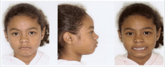
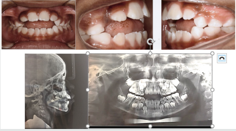
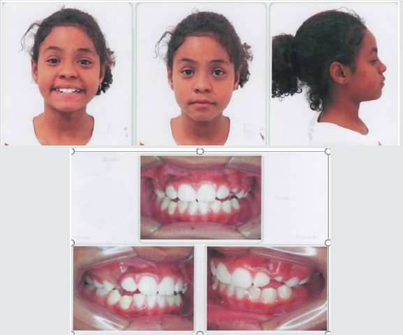
A female patient, 8 years and 4 months of age, melanoderma, came for treatment at the Postgraduate Course in Orthodontics of Brazil University (São Paulo, SP, Brazil). A facial analysis detected convex facial profile, lack of lip closure, and a decrease in nasolabial angle (Figure 1). The patient exhibited thumb sucking habit, mixed breathing, atypical swallowing and speech. shows angle class II malocclusion, 6 millimeters of an anterior open bite, mild crowding and a supernumerary Figures 2&3 tooth in the anterior lower jaw with mandibular midline deviation to the right. Cephalometric Rx shows proclined upper incisors due to the thumb sucking the objetives

Figure 1: Convex facial profile, a decrease in nasolabial angle and the upper lip covering the incisor.
Lupinepublishers-openaccess-pediatric-dentistry-journal

Figure 2: Anterior open bite, mild crowding and a supernumerary tooth in the anterior lower jaw and proclined upper incisors.
Lupinepublishers-openaccess-pediatric-dentistry-journal

Figure 3: Angle class II malocclusion, supernumerary tooth in the anterior lower jaw and an 6mm open bite.
Lupinepublishers-openaccess-pediatric-dentistry-journal

for the first phase of the treatment were to eliminate the thumb sucking, the open bite, dental deviations, provide arch expansion and the extraction of the supernumerary tooth.

Treatment Progress

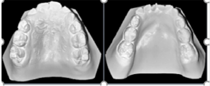
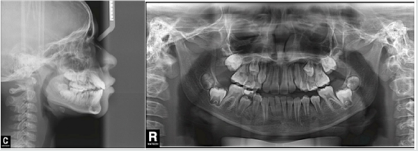
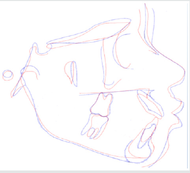
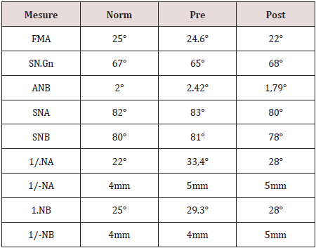
A removable appliance known by Simões Network (SN3) composed with a stainless steel bimaxillary grid (“lower winglets model”) that simulates the incisors occlusion and provides the correct tongue position [15] (Figure 4). The screw was expanded with one-quarter turn biweekly. After 3 months of the treatment beginning o, we added a lip bumper to improve lip seal (Figure 5). We recommended the use for 10 or 12 hours a day. The supernumerary extraction was performed 7 months of the treatment beginning. Figure 6 shows the final of the first stage. Figure 7 shows Angle class I malocclusion and Figure 8 shows the Cephalometric and panoramic Rx after 23 months with the orthopedic appliance. Cephalometric superimposition (Figure 9) and analysis (Table 1) indicated dentoalveolar open bite pretreatment and the correction posttreatment.

Figure 4: SN3 appliance, bimaxillary anchorage with “lower winglets model”.
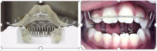
Lupinepublishers-openaccess-pediatric-dentistry-journal

Figure 5: A lip bumper was added to improve lip seal.
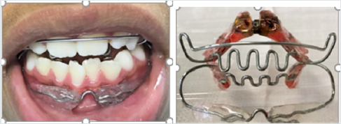
Lupinepublishers-openaccess-pediatric-dentistry-journal

Figure 6: The correction of open bite and the improvement of the alignment.
Lupinepublishers-openaccess-pediatric-dentistry-journal

Figure 7: The arch expansion and molar Class I.
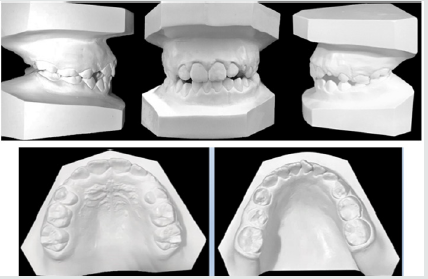
Lupinepublishers-openaccess-pediatric-dentistry-journal

Figure 8: Cephalometric and panoramic Rx after 23 months of treatment.
Lupinepublishers-openaccess-pediatric-dentistry-journal

Figure 9: Cephalometric superimposition revealed maxillary incisor retrusion and mandibular incisor in normal bite.
Lupinepublishers-openaccess-pediatric-dentistry-journal

Table 1: Summary of cephalometric measures.
Lupinepublishers-openaccess-pediatric-dentistry-journal

Discussion and Conclusion

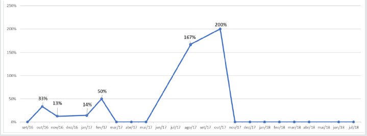
According to the authors, AOB is a challenge daily faced because and it can result speech and swallows’ problems, tongue posture and imbalance between jaw postures [17-19]. Bonna 2016 alerts that the orthodontics or orthopedic devices are fundamental but without family support the habit suppression will not be achieved [20]. The objective for the first phase of treatment were to eliminate thumb sucking, open bite and arch expansion with orthopedic appliance Simões Network SN3 and after a lip bumper was included to improve seal lip. These goals were achieved during the first stage. Graphic 1 shows best fit reduction open bite from May to November 2017. Even pubertal increments offer best time for orthopedic treatment helping determine the predictability, growth direction, patient management and total treatment time, we did not wait to treat because the disadvantages of the open bite [21]. This reported case was successfully treated with SN3 remained stable after the AOB correction. For the second phase with fixed orthodontic treatment will be necessary [22-29].

Graphic 1: Closure open bite variation during time.
Lupinepublishers-openaccess-pediatric-dentistry-journal


Friday, 22 January 2021

Lupine Publishers | Treatment of Infected Primary Teeth using Modified Antibiotic Paste

Lupine Publishers | Journal of Pediatric Dentistry


Abstract

Objectives: Treatment of pulpectomized primary molars with chronic infection using a mixture of three antibiotics: Metronidazole, Ciprofloxacin, and Doxycycline mixed with Macrogol or Propylene Glycol (modified 3MIX-MP) as an intracanal medicament before the complete cleaning and shaping and obturation.

Study design: A 7 years old child with infected primary molar came to our clinic for treatment. A detailed medical history and drug allergy were taken. Ciprofloxacin (500mg), Metronidazole (500mg) and Doxycycline (100mg) tablets divided in the proportion of 1:3:3 (one part of Ciprofloxacin, three parts of Metronidazole, and three parts of Doxycycline) and mixed with propylene glycol to form an ointment. Biomechanical preparation was done. The modified 3MIX-MP paste placed in the pulp chamber then temporary filling. The patient was recalled after 2 weeks. The tooth was obturated and restored then a stainless-steel crown placed. Then reevaluated at 3rd, 6th, and 12th months.

Results: Excellent clinical and radiographic success when compared to conventional pulpectomy and non-instrumentational lesion sterilization tissue repair therapy.

Conclusion: Treatment of Primary molar with modified 3MIX-MP, followed by instrumentation and obturation provided excellent clinical and radiographic success when compared to non-instrumentational lesion sterilization tissue repair therapy.

Keywords: Pulp infection; Pulpectomy; Modified antibiotic paste; Primary molars; Chronic, infected pulp; Modified 3 MIX-MP; Pulpectomy; Triple antibiotic paste; Primary teeth

Introduction

The first topical antibiotic introduced to endodontics was Grossman’s polyantibiotic paste in 1951, later many topical antibiotics have been introduced with varying combinations, few of those include Septomixine forte; PBSC (Combination of Penicillin, Bacitracin, Streptomycin and Caprylate sodium), and Clindamycin. However, none of these combinations has proven to be 100% successful in eliminating all the bacterial strains from the root canal system [1-5].

Materials and Methods

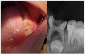
A child aged 7 years old with chronic infection related to the lower left primary molar came to our clinic for treatment of the infected molar (Figure 1). Treatment was explained to the parents and written informed consent was taken from parents before start of the study. A detailed medical history and previous illness with a history of drug allergy were taken from the parents, then the mentioned primary molar was diagnosed clinically, the molar was badly decayed with signs of chronic infection such as: gingival swelling and tenderness to percussion. A radiographic examination was done and a per radicular radiolucency was found, with no excessive root resorption. Commercially available chemotherapeutic agents such as Ciprofloxacin (500mg) (Omacip, NPI Pharma, Oman), Metronidazole (500mg) (Anazol, JPI, Saudi Arabia), and Doxycycline (100mg) (Tabocine, TPMC, Tabuk) tablets were obtained [6,7], then these tablets were crushed into fine powder using sterile porcelain mortar and pestle. These powdered drugs were transferred into three separate sterile glass containers, capped tightly and stored in the refrigerator until its use. Just before use, each powdered drug was divided in the proportion of 1:3:3 (one part of Ciprofloxacin, three parts of Metronidazole, and three parts of Doxycycline) and were mixed with propylene glycol and polyethylene glycol to form an ointment. Reddy GA et al. Trairatvorakul and Detsomboonrat, Jaya et al., Cruz et al. also followed the similar protocol of preparation of 3MIX antibiotic paste [8-11].

Figure 1: Preoperative illustration.

Lupinepublishers-openaccess-pediatric-dentistry-journal

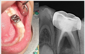
Figure 2: Postoperative illustration.

Lupinepublishers-openaccess-pediatric-dentistry-journal

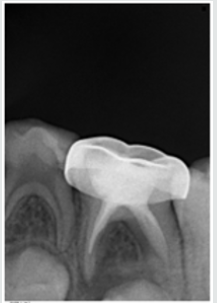
Preoperative clinical and radiographical signs and symptoms were recorded. The tooth was anesthetized using 2% Xylocaine with 1:80,000 adrenalin and isolated with rubber dam. Access opening was performed using round bur, Biomechanical preparation was done using k files from size 10–25. The root canals were chemically cleaned with 1% sodium hypochlorite solution and dried with paper points. The 3MIX-MP paste placed in the pulp chamber and pressed with dampened cotton pellet and temporized with Cavit. The patient was recalled after 2 weeks for evaluation. The tooth was obturated with reinforced zinc oxide eugenol (IRM, Dentsply) using lentulo spirals. Then restored with glass ionomer restorative material (Riva self-cure, SDI) and reinforced by placing stainless steel crowns (Figure 2). Further, the treated tooth was reevaluated both clinically and radiographically at 3rd, 6th, and 12th months intervals postoperatively (Figure 3). At the time of revisits, the tooth was examined clinically for any signs of failure that includes a report of spontaneous pain, presence of swelling, sinus tract and mobility. Radiographic evaluation was done to check the radiolucency and signs of resorption. The tooth was asymptomatic without pain, swelling, sinus tract and mobility also there was no increase in furcation radiolucency or development of root resorption which is abnormal for the age of the child.

Figure 3: 12 months Follow up.

Lupinepublishers-openaccess-pediatric-dentistry-journal

Results

Excellent clinical and radiographic success when compared to conventional pulpectomy and non-instrumentational lesion sterilization tissue repair therapy.

Discussion

This study was approved by “Research Ethics Committee, Taibah University, College of Dentistry, TU CD-REC”. The concept of Non-Instrumentation Endodontic Therapy introduced by Niigata university school of dentistry; Japan has gained reputation as it proved to attain 100% sterility in the root canal system [12- 15]. They recommended a technique similar to pulpotomy where debriding only the pulp chamber of chronically infected primary teeth and placing medicament (ciprofloxacin, metronidazole, and minocycline) near the root orifice without preparing the radicular portion. Cruz et al. suggested vehicles such as macrogol and propylene glycol (3MIX–MP) and demonstrated that these vehicles will carry the medicament deep into the dentinal tubules, thus aid in effective eradication of bacteria [11]. Metronidazole (Nitroimidazole compound) due to its wide spectrum of antibacterial action against anaerobes (Ingham et al. 1975) gained importance as the 1st choice drug for triple antibiotic paste preparation [16,17]. Metronidazole binds to the DNA and disrupts its helical structure and thus leads to rapid cell death. However, metronidazole even at higher concentrations could not eradicate all the bacteria thus indicating the necessity of some additional drugs to sterilize these lesions [15]. The two other antibacterial drugs, i.e. ciprofloxacin, and minocycline, in addition to metronidazole (3MIX) were added in an effort to eliminate all bacteria [8,10,15,18]. The 2nd choice of drug ciprofloxacin is a synthetic fluoroquinolone with rapid bactericidal action. It inhibits the enzyme DNA gyrase of bacteria. It exhibits very potent activity against Gram-negative bacteria but very limited activity against Gram-positive bacteria. Most of the anaerobic bacteria are resistant to ciprofloxacin. Hence, it is often combined with metronidazole in treating mixed infections. The 3rd choice of drug was minocycline. It is a semisynthetic derivative of tetracycline, primarily bacteriostatic, inhibiting protein synthesis by binding to 30S ribosomes in susceptible organisms and exhibits broad spectrum of activity against Gram-positive and Gramnegative microorganisms [3].

In our present study we replaced Minocycline with Doxycycline due to the difficulty in obtaining Minocycline, and before using the Doxycycline as a replacement we have done further searches for previous studies to ensure that both medications have the same effect and this replacement will not affect the efficacy of the mentioned mix. The already done studies concerning the difference between both Doxycycline and Minocycline revealed that still no statistically significant differences had been demonstrated in clinical trials when comparing Minocycline with Doxycycline, and investigators had concluded that both are equally effective. And they differ in their adverse event profile [19]. Considerably fewer adverse effects have been reported for Doxycycline than Minocycline; the adverse effects for Minocycline are 5 times more common than for Doxycycline [19]. We have followed the same protocol of Reddy GA et al. of extirpation of both necrotic coronal as well as all accessible radicular pulp tissue and then complete obturation, which is reported successful clinically over 16th month follow-up [9]. Although the previous studies have demonstrated that the LSTR (Lesion Sterilization Tissue Repair) technique as one of the successful techniques for management of chronically infected primary teeth, the controversies aroused about the duration of therapeutic activity of the medicament and leaving the infected material in the radicular region. So that the present study planned where in treated tooth were revisited after 2 weeks for medicament removal and obturation.

Conclusion

All the primary teeth with chronic infection which were treated using modified 3MIX-MP, followed by the instrumentation and obturation provided excellent clinical and radiographic success when compared to conventional pulpectomy and noninstrumentational lesion sterilization tissue repair therapy.

Read More Lupine Publishers Pediatric Dentistry Journal Articles: 
https://lupine-publishers-pediatric-dentistry.blogspot.com/

Friday, 16 October 2020

Lupine Publishers | Bilateral Dens in Dente in Maxillary Laterals: A Case Report

 Lupine Publishers | Journal of Pediatric Dentistry


Abstract

Dens invaginates is a defect categorized by a prominent lingual cusp and centrally located fossa. It occurs due to early invagination of the enamel epithelium into dental papilla of the underlying tooth germ. The affected teeth show a deep invagination of enamel as well as dentin initiating from foramen caecum or tip of the cusps and may extend even into the root. The teeth that are most frequently involved teeth are the maxillary lateral incisors, there might also occur a bilateral involvement. In this anomaly there can be seen several morphologic variations and it may lead to early pulpal involvement from the caries progressing into the pulp from lingual pit. The treatment varies from a preventive restoration to endodontic therapy, depending on the severity of the case. The present case report refers to one such case having a deep lingual pit bilaterally in both the maxillary permanent lateral incisors.

Introduction

Dens invaginatus is a rare malformation of teeth, showing various morphological variations. Radiographically the teeth show an invagination of enamel and dentin extending up to the pulp cavity or the roots. This malformation was described earliest by Ploquet [1], who discovered this anomaly in a whale’s tooth [2]. Dens invaginatus in a human tooth was first described by a dentist Socrates’ in 1856 [3]. ‘Anomalous cavities in human teeth were reported by Mühlreiter [4] in 1873, Baume [5] in 1874 and Busch [6] in 1897 reported about this anomaly as well. In 1887 Tomes [7] described the dens invaginatus as: The enamel of the coronal portion is generally well developed but we tend to find a small depression in its centre that appears to be a dark spot. On division of the tooth longitudinally, there occurs a dark centre of depression that is a blocked orifice within the tooth. If the section be a fortunate one, we shall be able to trace the enamel as it is continued from the exterior of the tooth through the opening into the cavity, the surface of which is lined completely with this tissue’ [7]. There are various reports on cases of dens invaginatus malformation in the dental literature [8-11]. Synonyms for this anomaly include:

a) Dens in dente,

b) Invaginated odontome,

c) Dilated gestant odontome,

d) Dilated composite odontome,

e) Tooth inclusion,

f) Dentoid in0 dente.

Case Report

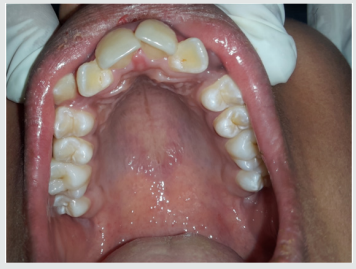
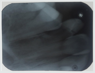
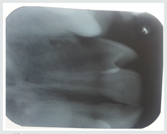
A 12-year-old female patient referred to the Pedodontics department with the complaint of irregularly arranged teeth. General health of the patient was normal and medical history was not relevant. Clinical examination revealed teeth 12 and 22 with a lingual pit. The pre-operative photographs were taken for the same (Figure 1). An intraoral Periapical radiograph was also taken for both the teeth, which revealed deep lingual pits involving the coronal part of teeth (Figure 2). As the teeth showed only incisal invaginations, and no pulpal involvement preventive treatment was selected. Also, if the pits were left untreated, it would harbor irritants and microorganisms further leading to caries. And as these pits were deep enough, the caries would progress rapidly into the pulp and cause pulpal infection leading to the need for endodontic treatments.

Figure 1: Pre-operative photograph.

lupinepublishers-openaccess-journal-pediatric-dentistry

Figure 2a: IOPA.

lupinepublishers-openaccess-journal-pediatric-dentistry

Figure 2b: IOPA.

lupinepublishers-openaccess-journal-pediatric-dentistry

Treatment Done

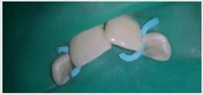
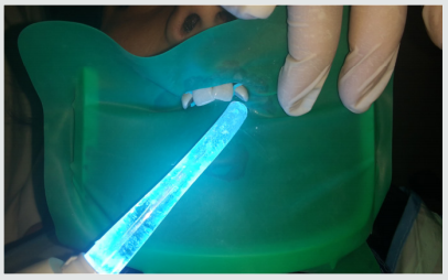
Following the oral prophylaxis, rubber dam placement was done for 12 and 22 (Figure 3). The teeth were initially washed, cleaned and dried. After that acid etching was performed using 37% phosphoric acid for 20 seconds. It was then washed and dried. Following that dentin bonding agent was applied with the applicator tip and cured for 20 seconds. The pits were then restored with flowable composite bilaterally (Figure 4). The teeth were then examined for any excess material and later finishing was done. The occlusion was also checked for any interference followed by post-operative photographs (Figure 5). Furthermore, the child was referred for orthodontic consultation.

Figure 3: IOPA.

lupinepublishers-openaccess-journal-pediatric-dentistry

Figure 3: Rubberdam placement.

lupinepublishers-openaccess-journal-pediatric-dentistry

Figure 4: Composite restoration.

lupinepublishers-openaccess-journal-pediatric-dentistry

Figure 5: Post-operative view.

lupinepublishers-openaccess-journal-pediatric-dentistry

Discussion

Dens Invaginatus has several variations of it. Oehlers gave a classification for various types of dens invaginatus [12]:

a) Type I: an enamel-lined minor form occurring within the confines of the crown not extending beyond the amelocemental junction.

b) Type II: an enamel-lined form which invades the root but remains confined as a blind sac. It may or may not communicate with the dental pulp.

c) Type III: a form which penetrates through the root perforating at the apical area showing a ‘second foramen’ in the apical or in the periodontal area. There is no immediate communication with the pulp. The invagination may be completely lined by enamel, but frequently cementum will be found lining the invagination.

Prevalence of Dens Invaginatus

The teeth mainly affected are maxillary lateral incisors and bilateral occurrence is not rare, they occur in almost 43% of all cases [13]. Swanson & McCarthy (1947) were the foremost to present bilateral dens invaginatus malformation, Conklin (1968) presented a patient with both maxillary central and lateral incisors affected bilaterally, and Burton et al. (1980) reported a case with six teeth involved, the maxillary incisors as well as the maxillary canines. Krolls (1969) also detected dental invaginations in maxillary central incisors and in many maxillary and mandibular premolars in a single patient. Conklin (1978) found the malformation in four mandibular incisors in one patient. There are many theories described in the literature to explain about the dental coronal invaginations some of them being:

a) The bulking of the enamel organ was thought to be caused due to the growth pressure of the arches [14,15].

b) Focal failure of the growth of the inner enamel epithelium leads to the invaginations; however, the surrounding epithelium continues to grow and engulfs the static area [11].

c) The invagination is due to the aggressive proliferation of a part of the inner enamel epithelium invading the dental papilla. It was thought as a ‘benign neoplasm of limited growth [16].

d) Protrusion and distortion of the enamel organ is thought to cause an enamel lined channel terminating at the cingulum or at the incisal tip. This might also lead to an irregular crown formation [17,18].

e) The ‘twin-theory’ [18] suggested a fusion of two toothgerms to be the causative factor.

f) Infection was also considered to be responsible for the anomaly [19].

g) Trauma was thought to be a reason, but they could not adequately explain why just maxillary lateral incisors were affected and not central incisors [20].

h) Most authors consider dens invaginatus as a deep folding of the foramen coecum during tooth development which may even result in a second apical foramen [3].

i) On the other hand, the invagination also may start from the incisal edge of the tooth. Genetic factors cannot be expelled (Grahnen 1962, Casamassimo et al. 1978, Ireland et al. 1987, Hosey & Bedi 1996).

Clinical Features

Due to the invagination there is just a thin layer of enamel and dentin left, which thus allows several microbes into the pulpal space easily. In some of the cases there is a complete enamel lining present. Also, channels may be present between the invagination and the pulp chamber. Pulp necrosis hence occurs in the earlier stages, within a few years of eruption and may be even before the root closure. The sequelae of untreated coronal invaginations can be as follows:

a) Abscess formation.

b) Retention of neighboring teeth.

c) Displacement of teeth.

d) Cysts, or Internal resorption.

Treatment Modalities

Preventive and restorative treatment should be opted for teeth with deep palatal or incisal invaginations and should be treated by fissure sealing [21]. Also, composite resin restoration followed by pit and fissure sealant can be done in the required cases [21]. Root canal treatment, surgical treatment and extraction of teeth should be done in severe cases wherein there is radicular invagination and pulpal involvement.

Conclusion

The clinician should be aware of occurrence of such tooth anomalies and should do a thorough clinical diagnosis as these anomalies are not rare. Earlier diagnosis and treatment of such cases can prevent further complications such as endodontic therapies and extractions. Hence the needed treatment should be done as early as possible and a periodic follow up should be maintained.

Read More Lupine Publishers Pediatric Dentistry Journal Articles:
https://lupine-publishers-pediatric-dentistry.blogspot.com/
Read More Lupine Publishers Articles:
https://lupinepublishers.blogspot.com/

Tuesday, 30 October 2018

Tuesday, 9 October 2018

Proposal of dental attention model for children with disabilities in Ecuador (IPDOAJ) - Lupinepublishers


Children with disabilities are a vulnerable group that needs special attention and care in dentistry (because of physical, developmental, mental, sensory, behavioral, cognitive or emotional impairment). It is known that disability generates different levels of risk for oral diseases. Ecuador is a country that is at the forefront of global care for people with disabilities, the Catholic University of Cuenca-Ecuador, has proposed an early intervention model (based on evidence) to the control of caries lesions in these patients, through a school based intervention developed at ADINEA school in the same city. This model is currently being applied since August 2017. The purpose of this paper is to present, academically the conceptual model of our intervention. This proposal has 4 principal components parents support, minimal intervention approach, tooth decay control and behavior management.

https://www.lupinepublishers.com/pediatric-dentistry-journal/abstracts/IPDOAJ.MS.ID.000125.php

https://www.lupinepublishers.com/pediatric-dentistry-journal/pdf/IPDOAJ.MS.ID.000125.pdf

Wednesday, 3 October 2018

Focal Fibrous Hyperplasia: A Rare Case Report and Literature Review (IPDOAJ) - Lupinepublishers


Intra-oral fibrous type soft tissue overgrowth is common in oral cavity. It can be either benign or neoplastic lesions. Nonneoplastic type of reactive lesion seen in the oral cavity is caused by the chronic irritation. Focal fibrous hyperplasia is one such lesion which is seen in the line of occlusion. Focal fibrous hyperplasia is also known as irritational fibroma or traumatic fibroma. Proper oral hygiene, removal of etiologic factors and conservative excision will prevent the focal fibrous hyperplasia and its recurrence. Proper diagnosis, management and follow up should be done in these cases.

https://www.lupinepublishers.com/pediatric-dentistry-journal/abstracts/IPDOAJ.MS.ID.000120.php

https://www.lupinepublishers.com/pediatric-dentistry-journal/pdf/IPDOAJ.MS.ID.000120.pdf

Friday, 28 September 2018

Epidemiology and Control of Lassa fever (IPDOAJ) - Lupinepublishers


The Lassa Virus (an Arenavirus) is found in West Africa, where it sometimes a severe hemorrhage illness called Lassa fever. The antivirus drug ribavirin is effective, but only if administered early in the course of illness. Because of its expense, need for intravenous administration, potential toxicity, and teratogenicity, empiric therapy with ribavirin is undesirable. Laboratory diagnosis of acute Lassa virus infection has traditionally employed the indirect fluorescent-antibody (IFA) test. However, enzymelinked immunosorbent assays (ELISAs) for Lassa virus antigen and immunoglobin M (IgM and IgG) antibodies have been developed that are thought to be more sensitive and specific likely represent underestimates with. The high sensitive and specificity, capability for early diagnosis, and prognostic value of the ELISAs make them the diagnostic tests of choice for detection of Lassa fever.

https://www.lupinepublishers.com/ipdoaj/abstracts/IPDOAJ.MS.ID.000116.php

https://www.lupinepublishers.com/ipdoaj/pdf/IPDOAJ.MS.ID.000116.pdf

Wednesday, 26 September 2018

Mesiodens: A Case Report and Literature Review (IPDOAJ) - Lupinepublishers


Supernumerary teeth are considered to be one of the most significant dental anomalies affecting the primary and early mixed dentition and may cause a variety of pathological disturbances to the developing permanent dentition and also resulting in poor dental and facial esthetics. Mesiodens are the erupted or unerupted extra tooth in the midline of maxilla, along with normal teeth (between the central incisors). Early diagnosis and prompt treatment are necessary for prevention of deleterious effects of mesiodenson dentoalveolar structures. The aim of this case report is to share knowledge about management of mesiodens which might assist the clinician in decision-making.

https://www.lupinepublishers.com/ipdoaj/abstracts/IPDOAJ.MS.ID.000113.php

https://www.lupinepublishers.com/ipdoaj/pdf/IPDOAJ.MS.ID.000113.pdf


Tuesday, 25 September 2018

Biosynthesis of Copper Nanoparticles by using Aloe Barbadensis Leaf Extracts (IPDOAJ) - Lupinepublishers


Development of green nanotechnology is generating interest of researchers toward eco-friendly biosynthesis of nanoparticles. In this study, biosynthesis of stable copper nanoparticles was done using aloe barbadensis leaf extract. First, we prepared leaf extract of aloe barbadensis in deionized water. This extract added to 1mMol of copper sulfate solution and we observed the change in color of the solution from colorless to colored solution. The present study tracing of an object is a green synthesis of copper nanoparticles by the interaction of leaf extract and copper salt. Biosynthesis of nanoparticle put forward a cost-free and environmentally suitable method of nanoparticle synthesis. Powder X-ray diffraction and SEM analysis showed that synthesized CuO nanoparticles with an average particle size of 60nm. Shape of copper nanoparticles was spherical and cubic and their range was 80-120nm. EDX of synthesized nanoparticles showed copper 38%. UV spectrophotometer confirms peak of the copper nanoparticle.

https://www.lupinepublishers.com/ipdoaj/abstracts/IPDOAJ.MS.ID.000110.php

https://www.lupinepublishers.com/ipdoaj/pdf/IPDOAJ.MS.ID.000110.pdf


Wednesday, 19 September 2018

Oral Mucosal Lesions in Children (IPDOAJ) - Lupinepublishers


Soft tissue lesions of the oral cavity are common in children, have various clinical presentations as colored lesions, mucosal ulceration or Nodular lesions and so. Some are a symptomatic and some are symptomatic or even disturb the daily activities of the child. When evaluating soft tissue lesions, it is important to distinguish between findings that are normal and those that are indicative a pathological condition, and normal anatomical variations. Most of adult oral soft tissue lesions may present in child oral cavity with predilection for some lesions. The literatures regarding oral mucosal lesions in children are relatively few. The most common oral disease, found was caries in priority and cancer therapy–related mucosal disorders.