Tuesday, 4 August 2020

Lupine Publishers | Variant of Takotsubo Cardiomyopathy with Co-Existent Severe Coronary Artery Disease – A Case Report

Lupine Publishers | Advancements in Cardiovascular Research

Abstract

TakotSubo Cardio Myopathy (TSCM) is a reversible form of cardiomyopathy characterized by transient left ventricular (LV) systolic dysfunction. Clinically often mimics acute myocardial infarction (AMI) with chest pain or breathlessness with electrocardiographic, cardiac marker(s) and LV wall motion abnormalities. In a third of patients, significant emotional or physical stress precedes the presentation. Often, these patients have normal epicardial coronaries or non-flow obstructive coronary disease. In most patients, LV function shows complete recovery within weeks with favorable outcomes. We report a unique case of TSCM following sudden emotional stress with typical transient apical ballooning; nevertheless, with severe co-existent triple vessel coronary artery disease and complete recovery of cardiac function on medical management.
Keywords: Tako-Tsubo Cardiomyopathy; Apical Ballooning; Coronary Artery Disease

Case Presentation

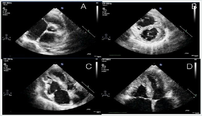
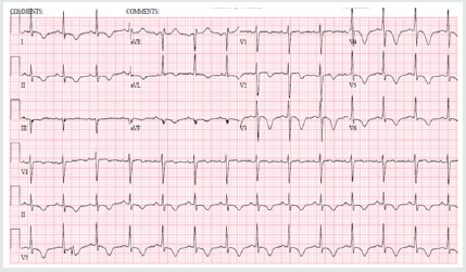
73 year old hypertensive female admitted with sudden onset intermittent chest pains and shortness of breath (< 24 hours after the death of her granddaughters in a road traffic accident). There were no other significant past medical history of note and she never smoked. On arrival, she was tachycardic (heart rate 102) in sinus rhythm, normotensive and clinical examination was unremarkable. Her initial ECG (Figure 1) demonstrated sinus tachycardia with deep T wave inversions in both precordial and limb leads. Her bloods revealed high sensitivity Troponin-T of 395ng/L [normal reference range <15ng/L]. Her bedside echocardiogram revealed typical pattern of “apical ballooning” with severe hypokinesis in the anterior, lateral, septal and inferior walls of the left ventricle, sparing the basal walls. Her left ventricular systolic function was moderately impaired with an ejection fraction (EF) of 40% (Figure 2). Both her clinical presentation and echocardiographic appearances were highly suggestive of “Takotsubo Cardiomyopathy”. Nevertheless, she was conventionally treated for presumed “Non-ST elevation myocardial infarction (NSTEMI)” at the initial stages [1].
Figure 1: ECG on admission showing sinus rhythm with deep T-wave inversions in both precordial and limb leads.
Lupinepublishers-openaccess-cardiology
Figure 2: 2-D Transthoracic echocardiogram (apical view) revealing typical apical ballooning with akinetic apical wall and preserved basal contraction of left ventricle.
Lupinepublishers-openaccess-cardiology
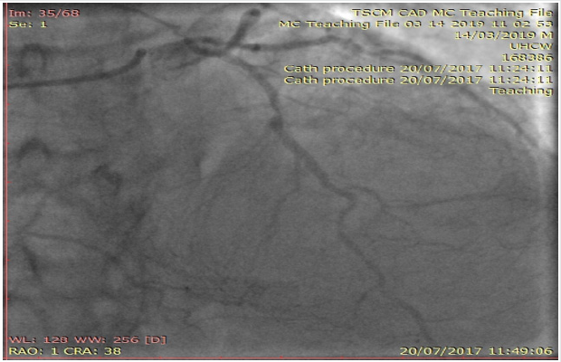
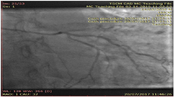
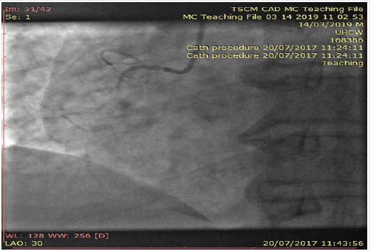
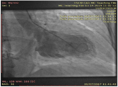
Interestingly, her inpatient coronary angiogram revealed severe triple vessel coronary artery disease (CAD). This was evident by the presence of mild disease in the left main stem, severe mid vessel calcific LAD lesion, severe obtuse marginal (OM1) disease (Figure 3) and chronic total occlusion of her mid right coronary artery (Figures 4a & 4b). However, her distal RCA appears well collateralized from contralateral LC system. The left ventriculogram revealed moderately impaired systolic function with moderate mid to apical anterior and inferior wall hypokinesis, sparing the basal segments consistent with “apical ballooning”. Figure 5, LV in systole] Given her clinical presentation (albeit coronary artery disease and LV dysfunction), multidisciplinary consensus favored medical management as NSTEMI. Expectedly patient agreed and preferred this option too. She was discharged on appropriate treatment and secondary prevention. Interestingly, her Cardiac MRI (CMR) scan at 1-month post discharge showed near normalization of LV function with calculated EF 50% with no evidence of infarction or myocardial oedema or fibrosis. Thence, improvement in her LV function with no evidence of infarction on CMR confirms the diagnosis of Takotsubo Cardiomyopathy, despite her significant CAD. At 12-month follow up, patient still remains stable and symptom free; therefore, with plans to consider surgical coronary revascularization, if worsening symptoms on top of medical therapy [2,3].
Figure 3: Coronary angiogram of left system revealing severe mid calcific left anterior descending artery disease with contralateral collaterals to distal right coronary artery.
Lupinepublishers-openaccess-cardiology
Figure 4a: Coronary angiogram of left system showing severe branch vessel disease in obtuse marginal branch of left circumflex artery.
Lupinepublishers-openaccess-cardiology
Figure 4b: Angiogram of right coronary artery showing chronic total occlusion at the proximal segment.
Lupinepublishers-openaccess-cardiology
Figure 5: Left ventriculogram at systole confirming the typical pattern of “apical ballooning” sparring the basal anterior and inferior segments.
Lupinepublishers-openaccess-cardiology

Discussion

With her established macrovascular risk factors for CAD (hypertension and age) and clinical presentation together with abnormalities in ECG, cardiac enzymes and echocardiogram, the working differential diagnoses were:
a. Acute Coronary Syndrome (ACS) i.e., Non-ST elevation myocardial infarction
b. TakotSubo Cardiomyopathy or transient apical ballooning syndrome or stress induced cardiomyopathy or broken heart syndrome.
Conventional electrocardiogram (ECG) still remains a costeffective important tool in the assessment of CAD. Nevertheless, its low sensitivity (<50%) and specificity (<80%) in predicting coronary disease still remains a major limitation. Clinicians mostly rely on surface ECGs to localize ischaemic myocardial territory (highly specific) prior to coronary revascularization of the culprit vessel(s) routinely in ACS patients. Clinically TSCM (typical LV appearance of Japanese “Octopus trap” with apical ballooning) often mimics AMI (chest pains, elevated cardiac enzymes and ECG changes) and is characterized by a transient left ventricular dysfunction. Though its incidence and prevalence doesn’t appear to have vastly changed over the years; however clinicians are more comfortable in diagnosing TSCM with better access to imaging (echocardiogram, Cardiac MRI) and invasive investigations (coronary angiogram) nowadays. The Revised Mayo Clinic Criteria suggested the presence of the following to confirm the diagnosis of TSCM [4,5].

a. Transient hypokinesis, akinesis or dyskinesis of the left ventricular mid segments with or without apical involvement– distinctively the extent of regional wall motion abnormalities observed will be beyond the distribution of single epicardial coronary vessel and with often a stressful trigger (but not always present).
b. Absence of obstructive coronary artery disease or angiographic evidence of plaque rupture
c. New electrocardiographic abnormalities or modest elevation in cardiac Troponin
d. Absence of pheochromocytoma and myocarditis.

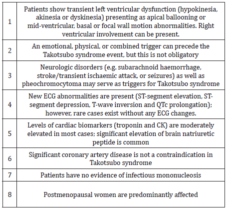
However, concomitant coronary artery disease (CAD) has been reported in 10 – 29% of patients who have been diagnosed with TSCM. In the International Takotsubo Registry (n=1750) 15.3% had some degree of coronary artery disease, although the extent of coronary artery involvement was not discussed in TSCM patients. The new Inter TAK diagnostic criteria highlight the clinical entity that TSCM can exist with concomitant CAD too (Table 1). Our patient meets with these diagnostic criteria for “Takotsubo Cardiomyopathy” together confirming that there was no evidence of infarction in her Cardiac MRI (no sub-endocardial late gadolinium enhancement in her LV or microvascular oedema) as well as complete recovery of her LV function rather than the initially presumed diagnosis of ACS [6].
Table 1: International Takotsubo Diagnostic Criteria (8).
Lupinepublishers-openaccess-cardiology
The pathophysiology for the condition is not well completely understood yet; nonetheless, stress related acute catecholaminergic surge, multi-vessel epicardial coronary spasm, micro-vascular dysfunction, abnormal fatty acid metabolism, myocardial stunning, micro-infarction, direct catecholamine toxicity to myocytes and possible reperfusion injury were proposed as possible mechanisms. On top of the typical transient apical ballooning, other atypical variants including inverted or reversed, mid ventricular or localized have been reported in the literature. In the majority of cases of suspected TSCM, it is appropriate to perform coronary angiography to exclude an acute coronary syndrome given the overlap of symptoms. Also, there is little evidence to support the clinicians in the management of TSCM with co-existent coronary artery disease as highlighted in this case and the clinical dilemma encountered. Therefore, we believe long term follow-up would be really helpful in understanding the disease as well as their management and prognosis for these TSCM patients [7,8].

Conclusion


We report an unusual case of Takotsubo Cardiomyopathy with co-existent severe triple vessel coronary artery disease. This case illustrates the importance of good history taking and the judicious use of diagnostic imaging to arrive at a prompt diagnosis and appropriate management. With limited evidence in literature to support clinicians as how best to manage these patients with severe coronary artery disease with Takotsubo Cardiomyopathy, we highlight the need for International registries with long term follow-ups.

Read More Lupine Publishers Journal of Cardiology & Clinical Research Articles :

Monday, 3 August 2020

Lupine Publishers | Peer Review of Statistics in Surgical Research: Identify The X-Factor or Toss a Coin!

Lupine Publishers | Advancements in Cardiovascular Research

Opinion

Professor Peter Bacchetti’s excellent article [1], highlighting “the other problem of peer review of finding flaws that are not really there based on unfounded statistical criticism, and its demoralizing effect on authors”. I wish to add some thoughts to the debated issues. Professor David Horrobin’s original classics on the subject [2,3]. have not yet been surpassed. It was updated recently [4] and prompted some contributory thoughts [5]. Having enough experience as author of reject articles and some as peer reviewer, I find the most devastating effect to author’s morale is making no comment, giving no reason for rejection or not replying all. The BMJ is guilty on this account as an article of mine was rejected that was accepted elsewhere after minor editing [6]. The BMJ, however, is in the good company of most biomedical journals who apply the COPE rules. The article lacked statistics of any kind that perhaps might be one of the reasons it was disliked at BMJ. To Editors’ credit, however, it took about a month to say ‘No’ that caused no momentum loss, unlike other Journals who reach the same verdict on other articles after 6 months or a year that drag another year or two before the author could recover and gather enough time, interest and energy to face the damn thing again. One subtle aim of that article [6], mentioned to BMJ Editors, was an attempt to say that “there is science and in particular evidence based medicine without statistics”.
It is a devil’s advocate to say statistics has not only been made into a “big lie” but also ‘false God’. It was invented elsewhere but currently worshiped only at most medical and surgical journals. A look at Science and Nature testifies such prestigious magazines have reduced statistics to real size and value as a “tool for testing a hypothesis”. It is not too basic a question for every biomedical peer reviewer to find out the exact role, aim and limitations of statistics. Some was mentioned in an article [7], nobody noticed save the late great Professor GD Chisholm editor of Br J Urology. It was based on a study that was rejected by a grant committee. It aimed at resolving 2 of the most serious puzzles of current clinical practice, postoperative hyponatraemia and the multiple vital organ dysfunction or failure syndrome [8]. However, giving data and statistics [7,8]. before clarifying the theories [9]. has proved as wrong as putting the cart in front of the horse. Einstein’s methods on proposing the special and general relativity theory is the correct way. When statistics was haled in the sixties everyone thought it was the only mean to discover “The Unifying Theory”.
This has proved both immensely costly and wrong. The basic fact is ‘statistics cannot, was not intended to and will never could, make a discovery’. Observation, mental experiments and the X factor are the only way to make a discovery long before it is verified and proved by practical studies and statistical tests. Before explaining the X-factor please allow me tell a relevant true story that symbolizes the current problem with statistics. Two friends of mine in UK had a disagreement, made a bit on a round of drinks and decided the first person to enter the hospital club will be the judge. Guess who did? I did but having no clue on how to resolve the conflict suggested that a flip of a coin might be the best way. They agreed also to my condition that while head or tail will determine the winner among them, if the coin stood on edge the judge should be the winner of all. It did and I won. Another conflict started on: Who should buy the 3rd round of drinks? Both agreed that it was my turn. I explained that buying the 3rd round will gain good company but lose all winnings, and my turn should be the 5th round! The point is statistics can tell the probability of head or tail and exclude the odd but when evaluating to either 0 or 100% and the truth is known, instead of expiring it generates residual arguments. Professor Richard Smith contributed to this debate by quoting Dr Hedge on Professor Robert Fox’s famous thought that “swabbing the rejects with the accepts does not make a difference.”

He added that perhaps it has already been done at BMJ” and asked “How can you know?” With due respect Sir, I frankly think nobody can. Despite a proven incremental value of an average article it does not make a noticeable difference or great loss to scientific advances. Statistically speaking that means a quality article submitted to BMJ has 50% chance of being accepted or rejected. So, why not save everybody the trouble and toss a coin? Here is where statistics has shot itself in the foot. It gives an average chance to the average and an odd chance to the odd but can’t tell which is important. The odd chance of a tossed coin to stand on edge matches that of a breakthrough scientific or medical article coming an editor or peer reviewer’s way but detecting such article makes all the difference. Some call it a hunch or gut feeling. Others qualify it by the three-pronged tests of quality, relevance and civility. Identifying the “X-factor” that makes such an article stand out is worth all the trouble. I honestly do not know but it is the arresting beauty found in Einstein’s famous papers, Newton’s laws, Mozart’s music and Shakespeare’s writing among many examples that include medicine [2-4]. I wrote 2 articles on such para-scientific para-medical stuff to identify the X-factor, “Rules and lures of the science game” and “The Mozarts of Science” sent to journals nearly two years ago and have not received a reply yet. I think a message of “Ignore the big headed bustard” arrived. Qualified people to find out the X-factor are COPE members. Another question that requires a ‘Yes’ or ‘No’ answer would be: if any of Einstein’s papers is evaluated using the current peer review standard and statistics adopted by most biomedical journals, would it be accepted?

Read More Lupine Publishers Journal of Cardiology & Clinical Research Articles :

Wednesday, 29 July 2020

Lupine Publishers | Hypertrophic Cardiomiopathy in Children: The Need of Heart Transplantation

Lupine Publishers | Advancements in Cardiovascular Research


Abstract

Hypertrophic cardiomyopathy (HCM) is the most common cardiac disease affecting the cardiac muscle. It can manifest in different forms with or without left ventricular outflow obstruction, with or without right ventricle involvement. Forms with biventricular hypertrophy seem to have poor prognosis. In our case, we describe a young patient with sarcomeric biventricular hypertrophic cardiomyopathy (MYH7 mutation), the poor prognosis of this form and strategies options adopted after failure of medical treatment. It is not always easy the management of hypertrophic cardiomiopathy, after medical treatment failure, especially in children. In some cases, heart transplantation is the only one therapeutic option.
Keywords: Hypertrophic Cardiomiopathy; Right Ventricular Hypertrophy; Heart Transplantation

Introduction

Hypertrophic cardiomiopathy (HCM) is the most common cardiac disease affecting the cardiac muscle and is characterized by heterogeneous genetic, morphological, functional, and clinical features. It is also one of the main causes of sudden cardiac death (SDC) in the young. Left ventricular hypertrophy with left ventricular outflow obstruction (LVOTO) is the most characteristic feature of HCM. There are also variant of HCM without LVOTO, with apical hypertrophy, with medio-ventricular obstruction and with right ventricular hypertrophy. The treatment and the prognosis of HCM seem to be variable on the basis of different forms, the age at presentation, sarcomeric gene mutations or rare phenocopies. Heart transplantation (HT) is the only therapeutic option for selected patients with HCM and refractory heart failure. In effect ESC guidelines recommend heart transplantation in eligible patients who have an LVEF < 50% and NYHA functional Class III–IV symptoms despite optimal medical therapy or intractable ventricular arrhythmia (II a); in eligible patients with normal LVEF (50%) and severe drug refractory symptoms (NYHA functional Class III–IV) caused by diastolic dysfunction (II b)[1].
Right ventricular hypertrophy (SRVH) is a relatively rare subtype of HCM. The anatomic, genetic, clinical, and prognostic characteristics of patients with SRVH and the clinical relevance of these characteristics have not been described widely in the literature [2,3]. MYBPC3 gene mutations have previously been described in two patients with RV hypertrophy. In a recent study, 90% of HCM patients with SRVH were found to possess relevant sarcomere protein mutations and variations in the MYH7 (Myosin heavy chain 7) and TTN genes, followed by variations in MYBPC3. Always in this study 73% of HCM patients with SRVH and multiple sarcomere gene mutations had poor prognosis. 7 In addiction MYH7 mutations can cause hypertrophic cardiomyopathy or skeletal myopathies with or without cardiac involvement, on the basis of the side of mutation. In our case, we describe the poor prognosis and treatment strategies of a young patient with biventricular hypertrophic cardiomyopathy and MYH7 mutation.

Case Report

A 12-year-old young woman with familiarity for hypertrophic cardiomyopathy (mother and mother’s twin with biventricular hypertrophic cardiomiopathy and MYH7 mutation) was hospitalized in our hospital for dyspnea after mild-moderate efforts and reduced functional capacity (NYHA Class II). Mother and aunt of the patient were asymptomatic with good functional capacity. Patient had the same genetic mutation of mother and aunt (p.Asn696Ser heterozygosis MYH7) but with increased and poor phenotypic expression [4]. Echocardiography and cardiac magnetic resonance were performed showing a hypertrophic cardiomyopathy with right ventricular involvement. Precisely, cardiovascular imaging showed left ventricle asymmetric hypertrophy especially at the level of anterior and inferior wall (basal and mild anterior wall =14 mm, z score= 3,5; antero-lateral basal wall = 12 mm, z score 2,78; mild inferior wall = 14 mm and apical inferior wall = 12 mm) with normal ejection fraction (FE = 62% at CMR) and moderate diastolic dysfunction (panel B and D). In addiction wall thickness of right ventricle outflow and basal-mild free wall were increased (= 13 mm) with apical obstruction and development of maximum gradient of 10 mmHg (PANEL A and C) [5,6] (Figure 1).
Figure 1.
Lupinepublishers-openaccess-cardiology
The function of right ventricle was at inferior limits (FE = 51% at CMR, TAPSE = 16 mm at echocardiography). Thus the patient had an interesting right ventricle involvement and moderate diastolic dysfunction of left ventricle. She had not arrhythmia at ECG-Holter but she had reduced functional capacity. also demonstrated at stress test. Stress test was suspended at 6 min (Bruce Protocol) after pre-syncopal symptoms: lack of adaptation of the blood pressure to the effort was observed. In addition, from several months she had pre-syncopal episodes at the peak of the effort. ECG showed left ventricular hypertrophy and biatrial enlargement. Pro BNP was increased = 5841 pg/ml. Considering clinical situation, we decided to start medical treatment with betablockers (bisoprolol) but the patient didn’t tolerate medical treatment. Thus, we decided to start low dose of captopril without improvement of symptomatology. Also, treatment with diuretic was not tolerate by patient [7,8]. Therefore, considering symptom refractory to medical therapy, the poor prognosis and the impossibility to optimize medical treatment, we decided to plan cardiac transplantation, the only option possible at this moment.
Thus right catheterization was performed and patient was inserted in heart transplantation list. ICD implantation was not considered in the absence of ventricular arrhythmia and other factors. Discussion: hypertrophic cardiomyopathy associated with MYH7 mutation and right ventricle involvement seems to have poor prognosis, especially if right ventricle hypertrophy is severe [9]. In effect the young patient had a greater right ventricular hypertrophy compared than mother and aunt. In these cases, after medical treatment failure, heart transplantation seems to be the only strategy to improve symptomatology and quality of the life of the patient. Especially in pediatric population, it is not always easy the management of hypertrophic cardiomiopathy after medical treatment failure and heart transplantation seems to be the only one therapeutic option. Other study are needed to study some variants of HCM with right ventricle hypertrophy, their treatment and prognosis.


Tuesday, 28 July 2020

Lupine Publishers | Brackish Water Desalination Using Solar Desalination Pannel

 Lupine Publishers | Current Investigations in Agriculture and Current Research


Abstract

The origin and continuation of mankind is based on water. Water is one of the most abundant resources on earth, covering three-fourths of the planet’s surface. Water is one of the earth’s most abundant resources, covering about three-quarters of the planet’s surface. The reason for this apparent contradiction is, of course, that - 97.5% of the earth’s water is salt water in the oceans and only 2.5% is fresh water in ground water, lakes and rivers and this supplies most human and animal needs. It would be feasible to address the water-shortage problem with seawater desalination; however, the separation of salts from seawater requires large amounts of energy which, when produced from fossil fuels, can cause harm to the environment. Therefore, there is a need to employ environmentally-friendly energy sources in order to desalinate seawater. Solar distillation systems are increasingly attractive in the areas suffering from shortages of fresh water but where solar energy is plentiful and where operational and maintenance costs are low.

Introduction

Water is essential for life. Many countries around the world, especially developing countries and countries in the Middle East region, suffer from a shortage of fresh water. The United Nations (UN) Environment Programme stated that one-third of the world’s population lives in countries with insufficient freshwater to support the population [1]. Consequently, drinking water of acceptable quality has become a scarce commodity. The total global water reserves are _1.4 billion km3, of which around 97.5% is in the oceans and the remaining 2.5% is fresh water present in the atmosphere, ice, mountains and ground water. Of the total, only _0.014% is directly available for human beings and other organisms [2]. Thus, tremendous efforts are now required to make available new water resources in order to reduce the water deficit in countries which have shortages [3]. According to World Health Organization (WHO) guidelines, the permissible limit of salinity in drinking water is 500ppm and for special cases up to 1000ppm [4]. Most of the water available on the earth has a salinity up to 10000ppm and seawater normally has salinity in the range of 35 000-45 000ppm in the form of total dissolved salts [5]. Desalination is a process in which saline water is separated into two parts, one that has a low concentration of dissolved salts, which is called fresh water, and the other which has a much higher concentration of dissolved salts than the original feed water, which is usually referred to as brine concentrate [6]. The desalination of seawater has become one of the most important commercial processes to provide fresh water for many communities and industrial sectors which play a crucial role in socioeconomic development in a number of developing countries, especially in Africa and some countries in the Middle East region, which suffer from a scarcity of fresh water.
There is extensive R&D activity, especially in the field of renewable energy technologies, to find new and feasible methods to produce drinking water [7,8]. Currently, there are more than 7500desalination plants in operation worldwide producing several billion gallons of water per day. Fifty-seven per cent are in the Middle East [9] where large-scale conventional heat and power plants are among the region’s most important commercial processes, they play a crucial role in providing fresh water for many communal and industrial sectors, especially in areas with a high density of population. However, since they are operated with fossil fuel, they are becoming very expensive to run and the environmental pollution they produce is increasingly recognized as very harmful to the globe. Moreover, such plants are not economically viable in remote areas, even near a coast where seawater is abundant. Many such areas often also experience a shortage of fossil fuels and an inadequate electricity supply. The development of compact, smallscale systems for water desalination is imperative for the population in such areas [7,8]. Thermal solar energy water desalination is known to be a viable method of producing fresh water from saline water [2] in remote locations; conventional basin solar stills with a relatively large footprint are an example of such simple technology. And using a clean natural energy resource in water desalination processes will significantly reduce the pollution that causes global warming. This article aims to present a review of the published literature on the various desalination technologies and their advantages and disadvantages in addition to their economics.
Water and energy are two inseparable commodities that govern the lives of humanity and promote civilization. The history of mankind proves that water and civilization are two inseparable entities. This is proved by the fact that all great civilizations were developed and flourished near large sources of water. Rivers, seas, oases, and oceans have attracted mankind to their coasts because water is the source of life. History proves the importance of water in the sustainability of life and the development of civilization. Maybe the most significant example of this influence is the Nile River in Egypt. The river provided water for irrigation and mud full of nutrients. Ancient Egyptian engineers were able to master the river water and Egypt, as an agricultural nation, became the main wheat exporting country in the whole Mediterranean Basin [10]. Energy is as important as water for the development of good standards of life because it is the force that puts in operation all human activities. Water is also itself a power generating force. The first confirmed attempts to harness waterpower occurred more than 2000 years ago in which time the energy gained was mainly used to grind grain [11]. The Greeks were the first to express philosophical ideas about the nature of water and energy. Thales of Militus (640-546BC), one of the seven wise men of antiquity wrote about water [12,13] that it is fertile and moulded (can take the shape of its container). The same philosopher said that seawater is the immense sea that surrounds the earth, which is the primary mother of all life. Later on, Embedokles (495-435BC) developed the theory of the elements [12] describing that the world consists of four primary elements: fire, air, water and earth. These with today’s knowledge may be translated to: energy, atmosphere, water and soil, which are the four basic constituents that affect the quality of our lives [14].
Aristotle (384-322), who is one of the greatest philosophers and scientists of antiquity, described in a surprisingly correct way the origin and properties of natural, brackish and seawater. He wrote for the water cycle in nature [15]: “Now the sun moving, as it does, sets up processes of change and becoming and decay, and by its agency the finest and sweetest water is every day carried out and is dissolved into vapor and rises to the upper regions, where it is condensed again by the cold and so returns to the earth. This, as we have said before, is the regular cycle of nature.” Even today no better explanation is given for the water cycle in nature. Really, the water cycle is a huge solar energy open distiller in a perpetual operational cycle. For the seawater Aristotle wrote [16]: “Salt water when it turns into vapor becomes sweet, and the vapor does not form salt water when it condenses again. This is known by experiment.”Water demand and consumption.

Materials and Methods

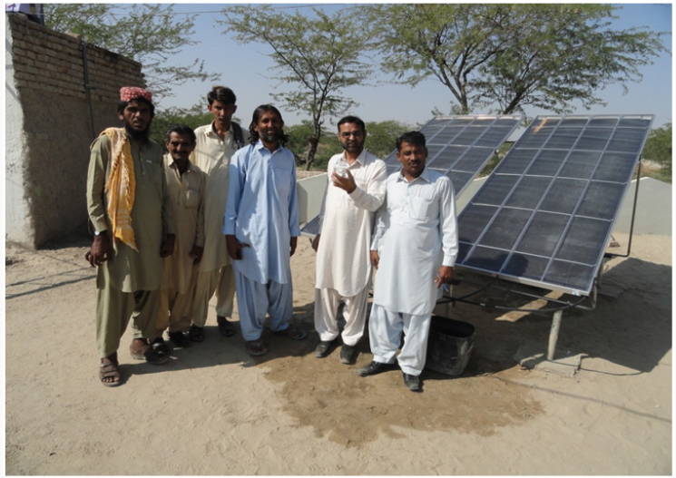
The following are the description of solar powered water desalination system AROCELL solar water purifies Australian technology. It only use sun energy, there are no moving parts, no electronics. It is robust and easy to setup, low maintenance and very low in operating cost because the water purifier only need solar energy. The feed water is supplied by gravity or pressure pump CAROCELL direct solar powered desalination technology, working at ambient temperature, heats the input water causing vapors condensation change precluding all bacteria and pathogens, therefore eliminating all water borne diseases Exposure to ultra violet light and extreme heat from solar energy through the advanced composite panels enhances the germ killing process. CAROCELL’s increased efficiency (65% with peak efficiencies above 80%) over other solar distillation products (30-40%) is a combination of the proprietary materials used to dramatically increase the temperature of the feed water on the solar collector which enhances the evaporation/condensation processes inside the panel. Additionally, this sophisticated geometrical design has easy maintenance, optimum performance and a self-controlling natural convection loop enabling widely superior energy recovery (Figure 1). Under this study 04 solar desalination units were installed at National Agricultural Research Centre, Islamabad, Pakistan to check the efficiency of cleaning the brackish water through desalination solar techniques. Further the discharging volume of cleaning water was also determined during 09am to 4pm in the month of February, 2014.
Figure 1: Train the farmers for operation and maintenance of solar desalination system.
Lupinepublishers-openaccess-Agriculture

Results and Discussion

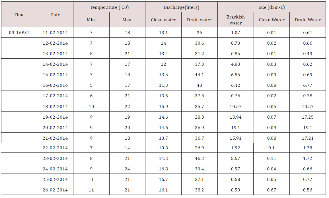
Approximately 70% underground water is brackish. This water is unfit for drinking. The removal of excess salts from brackish water is the utmost requisite for the supply of drinkable water at the remote areas having dense saline water. Water quality is the main issues to save the whole humanity from epidemic diseases. Clean drinkable water is the basic right of the whole world. So this experiment was launched at National Agricultural Research Centre, Islamabad, Pakistan. Electrical conductivity is the main criteria to evaluate the water fitness for drinking purpose. Data presented in Table 1 showed the performance of desalinization solar units installed. Brackish water having different ECw levels under various sunlight temperatures at National Agricultural Research Centre, Islamabad, Pakistan removed the toxic salts through solar desalinization technique and reduced it from 0.01to 0.11dSm-1. Water having so minute salts is fit for drinking purpose. Utilization of sunlight for the reclamation of brackish water through solar desalinization is the friendly environment, most economical and easily installed with local training. Maintenance cost of these solar units is also very minute.
Table 1: Effect of solar desalination on cleaning of brackish water at NARC, Islamabad, Pakistan.
Lupinepublishers-openaccess-Agriculture

The use of solar irradiation for treatment of chemically and biologically contaminated water is not a new trend [17-23]. Pure water is the basic necessary for all living organism. Now days, the availability of clean water resource is a major issue for mankind. A lack of infrastructure for water storage and distribution is also a factor in the developing world. More than71% of the earth surface is covered with the water, but only 1% clean drinkable water is available with the international standards [24]. The purification of brackish water into drinkable water depends upon the intake saline water capacity of each desalinization solar unit. Data indicated in Table 1 showed that the discharge volume of cleaning water was increasing as well as the temperature of the sunlight hours increasing. Solar radiation removes a wide range of organic chemicals and pathogenic organisms by direct exposure, is relatively economical, and avoids cohort of harmful byproducts of chemically driven technologies [17]. More prominently, the economics of the process are approximately capacity self-reliant [25].


Monday, 27 July 2020

Lupine Publishers | Farmer’s Perspective on Importance and Constraints of Seaweed Farming in Sri Lanka

 Lupine Publishers | Current Investigations in Agriculture and Current Research


Abstract

Seaweed cultivation is identified as a catalyst for social progression in coastal communities. Despite the potentials, the seaweed cultivation introduced to resettled coastal districts in Sri Lanka seems not performing to the expectations owing to various reasons. Farmers’ perspective as the leading stakeholders would facilitate the understanding of such a complexity. Therefore, the study attempted to assess the values and constraints related to seaweed farming as perceived by the seaweed farmers. Two-stage stratified random sampling technique was employed to draw a sample of 160 seaweed growers from the purposely-selected coastal areas of northern Sri Lanka. A perceived ordinal ranking method was exercised to assess the perceived importance, whilst Garrett’s ranking technique to detect the judgment of the farmers about the constraints. Next to fishing, seaweed farming received the highest perceived importance of the respondents. Favourable income and employment generation, the ability to easily manage with fishing, supportive role in empowering women and the existence of a favourable contract growing system were among the major causative responses contributed to the perceived importance. Major constraints identified in sea weed farming were adverse weather pattern (19.6%), poor quality of existing planting materials (16.68%), distortions prevailing in the purchasing mechanism (14.7%) and improper aquatic environments (13.74%). Thus, the study concluded that seaweed farming is perceived as an important livelihood option for the coastal communities and developing strategies to mitigate the impact of adverse environmental changes would promote seaweed cultivation.
Keywords: Constraints; Perceived importance; Seaweed farming; Sri Lanka

Introduction

Seaweed cultivation has become increasingly popular as one of the most important economic activities that can be practiced in the coastal regions. The potential role that seaweed could play in rural development being as a catalyst of social progression [1] by rendering extensive employment opportunities [2] to the coastal communities has already been identified. Accordingly, the government of Sri Lanka has introduced seaweed farming to resettled coastal districts in the northern part of the country to improve socio-economic conditions in vulnerable regions through the promotion of sustainable livelihood development. However, the available sporadic information in this regard imply that the level of performance seems to be suppressed by issues such as harsh weather conditions [3-5] and improper aquatic environments, which were commonly associated with seaweed farming. In this backdrop, assessing the value and constraints related to seaweed farming as perceived by the beneficiaries would uncover trustworthy, qualitative and in-depth information that would otherwise not become known. Such information would be essential for future planning by the policy makers for a better design to meet the farmer needs and can be useful both to fine-tune and enhance the effectiveness of the existing seaweed cultivation system.

Methodology

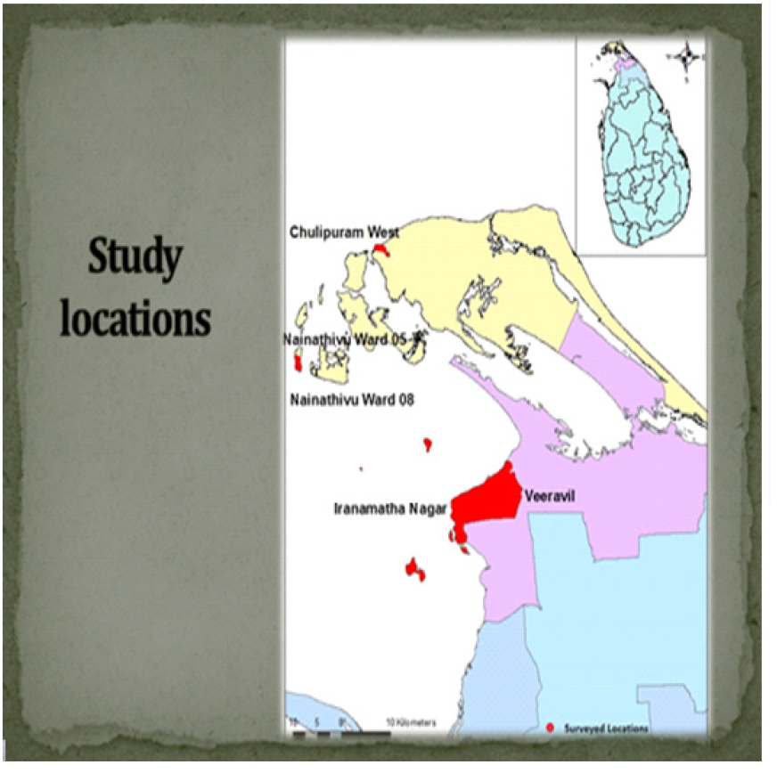
This study was conducted in purposely-selected coastal areas of Jaffna and Kilinochchi districts, located in the Northern Province of Sri Lanka (Figure 1). Two-stage stratified random sampling technique was employed to select 03 Divisional Secretariat (DS) divisions and 04 Grama Niladhari (GN) divisions respectively and finally to draw a sample of 160 seaweed growers. The required primary data were mainly collected through a structured and pre-tested questionnaire, in-depth interviews and focus group discussions. The perceived ordinal ranking method as previously adopted by Crawford [6] was exercised to assess the perceived importance of seaweed growers towards different livelihood options, whilst Garrett’s ranking technique [7-9] was exercised to detect the judgment of the farmers about the constraints faced by them in seaweed cultivation.
Figure 1: Study locations (marked in red).
Lupinepublishers-openaccess-Agriculture

Results and Discussion

Farmers’ perceived importance towards seaweed farming

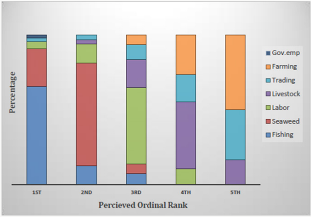
Next to fishing, seaweed farming received the highest perceived importance of the respondents. Accordingly, sea weed farming was perceived by 94% of farmers as either first or second in importance (Figure 2). The reasons behind the perceived importance were satisfactory income and generation of employment, easily manageable nature with fishing, cooperative role in women empowerment and the prevailing promising contract growing system (Figure 2). Being an economically viable alternative livelihood option [10], seaweed farming has diversified the livelihood options of the farming communities and thereby has provided a stable annual average income, which improves household economic resilience [11] enabling a sustainable way of life. This finding is in conformity with the findings of Zacharia [12] and Tobisson [13] on the role of seaweed farming in coastal livelihood improvement. On the other hand, previous studies have identified the possibility of developing employment-incomeconsumption relationships related to the seaweed farming [2]. This employment potential [6,14] is highlighted by the participants and viewed that harvesting and initial preparation stages render extensive employment opportunities. Especially, the shallow water seaweed farming allows more involvement of women and renders more employment opportunities for women during initial preparation and harvesting stages.
Figure 2: Perceived importance towards different livelihood activities.
Lupinepublishers-openaccess-Agriculture
The interviewees identified that the seaweed farming is instrumental in empowering women [13,15] within the study area. Further, it was reported that without being much disturbing to the primary economic activity, the seaweed farming can be easily integrated with conventional fishing. Moreover, it was identified that seaweed farming requires lesser time for its maintenance after planting and allows farmers to engage in other activities as well. Altogether, these major causative responses led to the perceived importance and will assist in promoting the seaweed cultivation within the study area. Apart from these major causes the provision of rapid return on investment, the requirement of simple farming techniques and an alternative for deprivation of terrestrial lands for cultivation also affected for the perceived importance towards seaweed farming.

Constraints for seaweed farming

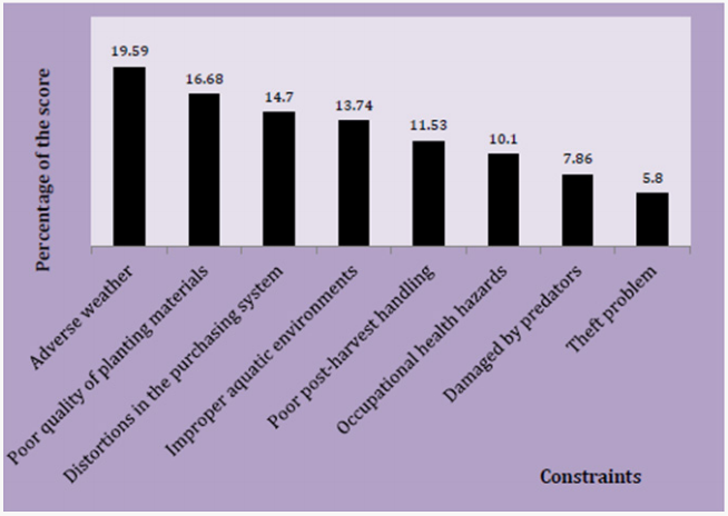
The results indicated that the adverse weather pattern (19.6%) is the major constraint faced by the seaweed farmers within the study area and obtained the highest rank. In conformity to the findings of Zamroni [3], the seasonal changes associated with monsoonal weather pattern is critically affecting the seaweed cultivation by limiting it to four cultivation seasons per year. Further, heavy rains and severe weather conditions like prolonged higher temperature periods regularly disturb the seaweed cultivation. These unfavorable changes would ultimately affect both quality and quantity [16] of seaweed harvest (Figure 3). Consisting with the findings of Zamroni [3], the poor quality of planting materials (16.68) obtained the second highest rank as a major constraint faced by the seaweed farmers. Farmers ordinarily use traditional self-propagation techniques using cuttings of the previous harvest. However, inferior strains created by overutilization of the old seaweed stocks may have negatively affected the growth rate and quality of seaweed and avoided growers from optimizing their yield.
Figure 3: Constraints ranked by seaweed growers.
Lupinepublishers-openaccess-Agriculture
The study identified that the buyers adopted a contract growing system as the purchasing mechanism of seaweed. Such mechanisms were further reported in the studies of Krishnan [3,17] and Hurdato (2013) related to seaweed farming. As identified by the study, low farm gate price, sporadic payments, and defective weighing processes have distorted (14.7) the qualities of the existing purchasing system. The respondents also viewed that the buyers regularly justify these low prices, highlighting the importance of technical provisions like raw materials and advisory support, which are mainly provided by them. Conversely, few growers believed that non-existence of layers of intermediaries and protection from time to time price fluctuations would still make the contract growing system more favourable.
The ideal aquatic environment provided in near-shore areas is favourable for sea weed farming. However, due to high competition and legal cut-offs imposed by the local authorities’ farmers haveshifted the cultivation to less fertile improper aquatic environments (13.74). Seaweed-farming locations were co-managed by coastal villagers and idled seaweed farms also contributed to this problem. Participants also indicated that seaweed cultivation locations away from the near-shore areas increased the overall production cost due to the expenses incurred for extra transportation.

Conclusion and Recommendations


Based on the logical interpretation of the findings, it can be concluded that seaweed farming is perceived as an alternative livelihood option for the coastal communities. This social acceptability implies the possibility of further expanding seaweed farming to other matching locations as a commercial enterprise. The adverse weather pattern was the major constraint perceived by the seaweed growers. In order to promote the existing system, the establishment of a weather damage relief program from the government, early warning system of sudden environmental changes and commercial level seaweed nurseries are recommended. Further, the collaborative action between key stakeholders is identified as a necessity to promote the seaweed industry.


Monday, 15 June 2020

Lupine Publishers | Evaluation of Ear, Nose and Throat Foreign Bodies in the Department of ENT-Head and Neck Surgery in a Teaching Hospital

Lupine Publishers | Journal of Otolaryngology


Abstract

Objective: FB (foreign bodies) in ear, nose and throat are often encountered by otolaryngologists in their daily practice and is commonly seen in both children and adults. Depending upon the type and location of FB, it may have serious impact on individual’s health if instant appropriate action is not taken. That’s why, there’s frequent visits to ED (emergency department) on having FB in ear and aerodigestive tracts. The objective of this study was to evaluate the nature, common sites, modes of presentation, modes of management of FB, age and gender distribution.
Materials and Methods: A retrospective hospital-based study was done in Universal College of Medical Sciences, Bhairahawa, Rupandehi, Nepal from March 2014 to September 2017. The information was obtained from hospital record books.
Results: Out of 483 total patients, 287 (59.42%) were male and 196 (40.57%) were female. Most of them were less than 10 years old. Of the 483 patients, 202 (41.82%) had FB in the ear, 132 (27.32%) in the nose and 149 (30.84%) in the throat. Living FB were found in 54 (26.73%) patients out of 202 in the ear, 10 (7.57%) patients out of 132 in the nose and none in the throat. Of the total patients, 97 (20.08%) required general anesthesia (GA) to remove FBs and the rest 427 (88.4%) patients were dealt with or without local anesthesia. Most of the FBs were removed promptly on presentation otherwise within 24 hours of presentation in the hospital.
Conclusion: FB in ENT were found more commonly in the children and the commonest site was ear. Timely presentation, prompt diagnosis and needful management in a center with otolaryngology practice reduces the morbidity and mortality. Most of the FB in ENT can be removed in outpatient department (OPD) or emergency room (ER) with or without local anesthesia (LA).
Keywords: Ear; Nose; Throat; Foreign Bodies; Local Anesthesia

Introduction

A foreign body (FB) is any object or substance that is not derived from the individual’s own body part and can cause harm by its mere presence if prompt medical care is not provided [1,2]. They may be found in Ear, Nose and Throat. They are very common in otorhinolaryngological clinical practice. It can be introduced spontaneously or accidentally by both children and adults. However, children are common victims as they have habit of inserting nearby objects in their nose, ear or mouth, imitation and also other contributing factors are like boredom, playing, mental retardation, insanity, and attention deficit hyperactivity disorder, along with availability of the objects and absence of watchful caregivers. Consequently, it may cause minor irritation to life threatening problem. A proper technique, good light, appropriate instrument, a co-operative or fully restrained patient and a gentle approach by the related doctor or health worker are required for the removal of FB. One should have a clear diagnosis before making attempts to remove the FB so as to lessen the morbidity [3,4]. FB may be classified as animate (living) and inanimate (nonliving). The inanimate FB can again be classified as vegetative (organic) and non-vegetative (inorganic) FB, and hygroscopic (hydrophilic) and non-hygroscopic (hydrophobic) [1,2]. The objective of this study was to evaluate the nature, common sites, modes of presentation, modes of management of FB, age and gender distribution.

Materials and Methods

A retrospective study was conducted in the Department of ENT – Head and Neck Surgery, Universal College of Medical Sciences, from March 2014 to September 2017. The data were obtained from the hospital record books. Otoscopy and anterior rhinoscopy were performed to diagnose FB of the ear and nose respectively. Instruments such as Jobson Horne probe, FB hook, Tilley’s forceps, and crocodile forceps were used in FB removal from the nose and ear. Syringing and suctioning were also done for FB ear removal. Plain X-ray of the neck was done in patients with a history of FB ingestion. Flexible nasopharyngo laryngoscopy (NPL) and flexible upper gastrointestinal (UGI) endoscopy were done in cases where the FB was not visible in X-ray to rule out presence of a FB or to determine its site of impaction and in selected cases UGI endoscopy was used for FB removal too. It was followed by removal of the FB from the oropharynx/hypopharynx and esophagus with direct laryngoscopy or rigid esophagoscopy, respectively under general anesthesia (GA). FB struck in the oropharynx or parts of hypopharynx were confirmed with the help of Lack’s tongue depressor and head light or indirect laryngoscopy and removed with the Tilley’s forceps under local anesthesia (LA) in the OPD with patient co-operation.

Results

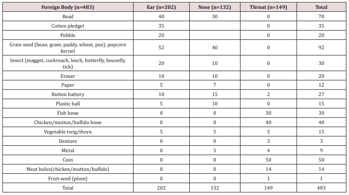
There were 483 patients recorded, out of which 287 (59.42%) were male and 196 (40.57%) were female with male to female ratio 1.46:1. The number of FB (Table 1 & Figure 1) in ear was 202 (41.82%), 132 (27.32%) in nose and 149 (30.82%) in the throat. Out of 202 FB in ear, 54 (26.73%) was animate and 148 (73.26%) inanimate. Out of 132 nasal FB, 10 (7.57%) was animate and 122 (92.42%) was inanimate. The FB encountered in throat was entirely of inanimate nature.
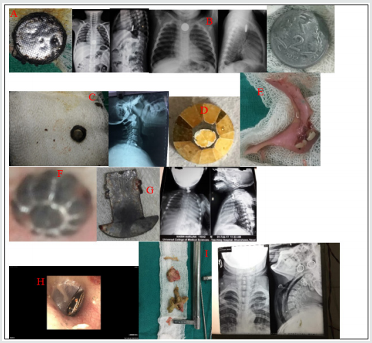
Figure 1. Few examples of ENT foreign bodies. A) FB toy battery in esophagus and removed with rigid esophagoscope, B) FB coin in esophagus, C) FB button battery in esophagus, D) FB bead removed from ear, E) FB denture, F) FB bead removed from nose, G) FB metal hook in esophagus, H) FB insect in ear and I) FB chicken bone with meat bolus in esophagus.
lupinepublishers-openaccess-journal-otolaryngology

FB in Ear

Total of 202 patients were recorded with FBs in the ear. The most common type of FB in the ear was grain seed 25.74% (52) followed by FB bead 19.8% (40). Fifty-four (26.73%) patients had animate (living) FBs. These were mostly insects in the form of maggots, cockroach, grasshopper, butterfly, housefly and ticks. Hundred and forty-eight (73.26%) patients had inanimate (nonliving) FBs. Out of 148 patients, fifty-two (35.13%) had hygroscopic FBs in the form grain seed in the form of bean, pea, wheat, paddy and gram as shown in Table 1. The rest ninety-six (64.86%) patients had non-hygroscopic FBs in the form of bead, cotton pledget, pebble, eraser, paper, button batteries, plastic ball and vegetable twig or thorn. Majority of cases were seen in 0-10 years age group i.e. 72% (147). The most common site of FB lodgement was found to be the external auditory canal. Most of these FBs were removed in the OPD or in the ER with or without local anesthesia (LA). In 4 children, the FB were found impacted in the deeper part of EAC (3 button batteries and 1 plastic ball) and had to remove under GA via post-aural approach.

FB in the Nose

Hundred and thirty-two (27.32%) had FB lodgement in the nose. The most common was grain seed 40 (Out of it, only 10 (7.57%) patients had living FBs i.e., 9 had maggots and 1 had leech. The rest 122 (92.42%) patients had nonliving FBs as shown in Table 1. Forty FB (30.3%) were of hygroscopic nature in the form of grain seed and the rest sixty nine percent being non-hygroscopic as in Table 1. Of the total number of 132 patients 122 patients (92.42%) were children and the rest 10 (7.57%) patients were adults with animate type of FBs. Hundred and five (86.06%) of the children presented with history of FB insertion nose by their caretakers, while in 17 (12.87%) children neither the patients nor the caretakers were certain of FB insertion. Unknowingly, they were treated as a case of sinusitis due to complaints of nasal blockage, headache and unilateral fetid discharge by the pediatricians and primary care physicians, which was later, referred to our center and revealed to be forgotten FB. Sometimes, even one has to depend on imaging like x-rays /CT scans to rule out the FBs where the patients are unable to recall the events. Otherwise, most of the times the typical history provides clue for clinching the diagnosis. Most of the FB were removed in the ER and OPD with or without LA and only 3 cases (2.27%) required removal under GA i.e. 2 cases of beads and 1 case of grain seed which on manipulation went posteriorly and also patient being uncooperative.

Foreign Bodies in the Throat

A total of 149 patients presented with the complaint of ingestion of FB.  The most common type of FB was coin of one rupee, 2, 5 and 10 rupees and the common site of the impaction was cricopharyngeal junction in all the 50 patients (33.55%). The sites of other types of FB impaction were oral cavity, oropharynx, hypopharynx, thoracic esophagus and lower gastro-esophageal sphincter region. All the ingested FB were inanimate, with 84 (56.37%) being organic and 65 (43.62%) being inorganic. Organic FBs were meat bolus and bone (fish, chicken, mutton, and buffalo meat) and one of plum seed. The inorganic FBs included button battery, thorn, denture, coin, and metallic objects as shown in Table 1. Age less than 10 years old were the most common group with FB coin. FBs fish bone and vegetable twig/thorn lodged in oral cavity and oropharynx were removed under LA. Out of 40 FB coin, 10 were dislodged spontaneously via gastrointestinal route, 5 FB meat bolus and 7 FB chicken bone were removed by flexible endoscopy and the rest of the FBs were removed under GA without  postoperative complications.
Table 1. Different types of Foreign bodies (FB) in ENT.
lupinepublishers-openaccess-journal-otolaryngology

Discussion

In our study, we’ve found higher incidence of FB in children less than 10 years old in 66.04% (319) patients. This is consistent with study by Iseh and Yahya [5], Ogunleye AOA and Sogebi R [6], Ahmad M, et al. [7]. This may be consequent of children’s exploring habit and lodging objects into the natural orifices of body, accidentally or intentionally. We found 59.42% of patients to be male and 40.5% to be female with male: female ratio 1.46:1. The male: female ratio was shown to be 1:1.05 by Gregogri et al. [8] whereas it was 1:1.26 in the study of Ogunleye AO et al. [9] and 1.35:1 by Agrawal S, Ranjit A study [10]. This suggests male are more susceptible than female to foreign body insertion in the orifices. In this study we observed ears were the most common site of lodgement of foreign bodies (41.82%) followed by throat (27.32%) and nose (27.32%). Parajuli R [11] and Shrestha I, et al. [4] also found in their study ears as the most common site for impaction of foreign bodies followed by throat and nose.  The most common foreign body in the ear and nose was the variety of grain seeds like bean, pea, paddy, wheat, gram, maize and foreign body coin was highest in throat. Removal methods, most commonly used for ear, nose and throat FBs were similar to those presented by Parajuli R [11], in order of preference the alligator forceps, Jobson Horne probe, foreign body hook, Tilley’s forceps and ear syringing. No patient required endoscopy or indirect laryngoscopy to remove oropharyngeal FB. The need for general anesthesia to remove FB varies in literature, with percentages varying from 8.6% to 30% [12]. There were no complications reported post FB removal.

Conclusion

FB in ENT are common in both pediatric and adult population. Comparatively the children are seen to be more vulnerable to have ENT FB lodgement.  Significant complications may arise if FB in ENT are not taken care of immediately and skillful removal is must. Thus, proper care and watch must be provided by care takers or the family members in order to prevent the encounter of such objects, especially in pediatric group.

Read More Lupine Publishers blogger Articles: https://lupinepublishers.blogspot.com

Monday, 8 June 2020

Lupine Publishers | Color Changes of Pediatric Dental Bridges

Lupine Publishers | Pediatric Dentistry Journal


Abstract

Dental technology that depended on the standardized lost-wax casting technology has been greatly improved with the introduction of dental CAD ⁄ CAM systems. The aim of the present study was to compare between the color changes of CAD/CAM acrylic and manually performed acrylic bridges used for pediatric patients. Forty study casts of children aged 2 - 4 years old of both genders, with prematurely lost one of the maxillary central incisors and the adjacent lateral incisor was carious and considered to be abutment tooth were involved in this study for construction of cantilever bridges. For each cast, two bridges were constructed; 1st one is CAD/CAM acrylic bridge and the other one is manually performed acrylic bridge. After immersing the bridges in saturated chocolate solution for different time intervals, color changes of the bridges were measured using 3Shape scanner system.
Keywords: Color; Changes; CAD/CAM; Bridges; Pediatric; Patient

Introduction

Trauma and/or dental caries is the common causes those result in premature loss of teeth in children. Cosmetic/aesthetic restoration of such condition considers to be challenging in the pediatric dental field. In case of premature tooth loss in anterior incisal segment there will result in arch space loss and teeth’s inclination that causing a collapse of the anterior teeth and midline shifting [1], as well as may lead to parafunctional habits [2]. Mahmoud (2009) found that anterior tooth loss had effect on patient’s quality of life and gave negative effects on him/his [3]. Al Rawi (2017) found that placement of cantilever acrylic bridges for restoring the aesthetic dental appearance of preschool children resulted in positive successes both to the child and parents [4]. Extrinsic discoloration of teeth and oral prostheses is stains caused by foods or beverages. In pediatric patients such stain mostly occurred due to colored foods such as beets or chocolate as well as berries and candies [5, 6]. This study considered to be the first step of our series studies deal with determining different physical and mechanical properties of the prostheses used for pediatric patients we planned to carry out (in vitro and in vivo studies). Starting with the present study that aimed to compare between the color changes of CAD/CAM and manually performed acrylic bridges used for pediatric patients. After immersing the bridges in chocolate solution for different time intervals, color changes of the bridges were measured using 3Shape scanner system.

Material and Methods

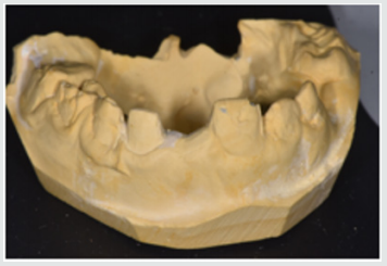
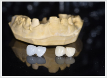
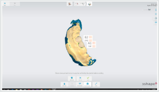
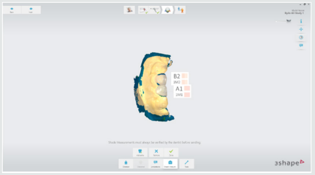
This study starting with collection of forty study casts of children aged 2-4 years old of both gender, with prematurely lost one of the maxillary central incisors and the adjacent lateral incisor was carious and considered to be abutment tooth were involved in this study for construction of cantilever bridges (Figure 1). For each cast, two bridges were constructed; 1st one is CAD/CAM acrylic bridge and the other one is manually performed acrylic bridge (Figure 2). Construction CAD/CAM bridge: The cast was 3D scanned by special scanner (710 3D) (smart optics Sensortechnik GmbH, Germany). The design of the bridge was carried out using Exocad Program (smart optics Sensortechnik GmbH, Germany). Acrylic block (Poly-methyl methacrylate) of classic shade A1 (Ivoclar vivadent, Switzerland) was used for fabrication of the bridge using CAD/ CAM machine (Charly dental, ZI Fonlabour, France). The bridge was finished and polished very well [4]. Construction of manually acrylic bridge: Wax pattern was fabricated on cast then followed the technique of typical wax loss; the heat-cure acrylic (Ivoclar vivadent, AG, FL-9494 Schaan/Liechtenstein) of classic shade A1 was used for bridge fabrication. Finally, surface finishing and polishing was done [7]. Saturated chocolate solution was prepared using 15g chocolate powder (MacChocolate TM, Malaysia) with 100ml distilled water. Baseline color readings for acrylic bridges were taken then immersed in chocolate solution for different time intervals (one week and two weeks) and maintained in incubator of 37 °C, Fresh chocolate solution was prepared every day. Before color measurements after one week and two weeks’ time intervals, the bridges were rinsed with distilled water for 30 seconds, cleaned with a soft bristle toothbrush and then dried with tissue paper [8]. Color measurement was carried out in the facial surfaces at the center third of the abutment and the center third of the pontic part of each bridge as shown in Figure 3. Color measurements of the bridges were measured using 3Shape scanner system (3 Shape A/S, Holmens Kanal 7.1060 Copenhagen K Denmark) and according to the software program of the system, Classic shade (Ivoclar vivadent, Switzerland) was depended.
Figure 1: One of the study casts involved in this study
lupinepublishers-openaccess-journal-pediatric-dentistry
Figure 2: One of the study casts involved in this study
lupinepublishers-openaccess-journal-pediatric-dentistry
Figure 3: Demonstrated the color shade measurement of the abutment and pontic portions of the acrylic bridge.
lupinepublishers-openaccess-journal-pediatric-dentistry

Results

Table 1 demonstrated the color shade of all samples at the baseline and after one-week and two weeks-time intervals. The results of the present study revealed that for all samples, the color measurement demonstrated that in CAD/CAM group even with using A1 shade acrylic block but at the baseline measurement the abutment revealed A0 shade while the pontic revealed A1 shade. Meanwhile, in manual group the abutment measured to be A1 shade and the pontic gave B1 shade. The results demonstrated that for all samples there were no changes in the color shade of CAD/CAM and manually fabricated acrylic bridges after one-week time interval, meanwhile, there were significantly color changes of all abutment and pontic portions of all samples of both bridge types after two weeks-time interval immersed in chocolate solution (Figure 4 & 5).
Figure 4: color shade measurement of CAD/CAM acrylic bridge after two weeks.
lupinepublishers-openaccess-journal-pediatric-dentistry
Figure 5: Color shade measurement of manual acrylic bridge after two weeks.
lupinepublishers-openaccess-journal-pediatric-dentistry
Table 1: Demonstrated the color shade of all samples at the baseline and after one-week and two weeks-time intervals.
lupinepublishers-openaccess-journal-pediatric-dentistry

Discussion

Restorations in the oral cavity are exposed to several factors that make them vulnerable to color changes, such as temperature, humidity, food and beverages. In the oral environment, restorative materials are also subjected to numerous other liquids, to temperature and load stress, and to tooth brushing. The success of restorations depends not only on mechanical and physical properties, but also on the esthetic appearance [9]. The color measurement in this study demonstrated that in CAD/CAM group even with using A1 shade acrylic block but at the baseline measurement the abutment revealed A0 shade while the pontic revealed A1 shade. These occurred because the thickness of the abutment was only about 0.5mm lead to that the color measured of the abutment was lighter than the pontic portion. Meanwhile, in manual group the abutment measured to be A1 shade and the pontic gave B1 shade. These results agreed with other studies those found the thickness of the material significantly affected the color shade of the prostheses [10,11].
The results demonstrated that the color changes demonstrated only after two weeks-time intervals immersed in chocolate solution. Even the color shades recorded in the CAD/CAM group considered to be lighter than in manual fabricated group, the discoloration from chocolate solution was probably due to adsorption of color colorant of chocolate solution at the surface of the prostheses.
The CAD/CAM bridges fabricated from blocks of pre-polymerized acrylic resin those had a hydrophobic surface that repels water [12]. As well as, perfect polishing surfaces of the bridges involved in this study revealed the limited discoloration that occurred agreed with other research [13]. As the duration of immersion increased, the color change values of both types of prostheses were recorded by 3Shape scanner system. Thus, the time is considered to be important factor in the staining of the dental prostheses and these results agreed with others [14,15]. Fabrication of dental prostheses with the help of CAD/ CAM technology is related to the advantages of high-density polymers based on highly cross linked polymethylmethacrylate [16]. Those advantages include; good esthetic, low water solubility and absorption, sufficient strength, low toxicity, easy repair with simple fabrication technique [17]. The using of hot cure acrylic for fabrication of dental prostheses even of some advantages but the main disadvantages include porosity with the presence of residual monomer which is a potential allergen, increased finishing time, brittle and uneven thickness [18]. A limitation of this study is that it was an in vitro study and need to be collected with in vivo study to measure the degree of color changes of the prostheses with presenting the effect of saliva and oral hygiene measures. Further clinical and in vitro studies are necessary to evaluate the susceptibility of CAD/CAM and manually acrylic bridges to discoloration by other beverages and nutrients.

Conclusion

Color considered as the most important factors for aesthetic appearance of dental restorations. In addition to the optimal chemo mechanical properties of acrylic resins, their availability in different color-shades has increased their application in fixed and removable prostheses. Acrylic resins can have acquired discoloration over time because of the process of adsorption and liquid molecules adhere to resin materials which was decreased their effect with using of CAD/CAM technology over conventional methods of acrylic resin prostheses fabrication.