Thursday, 12 December 2019

Lupine Publishers | Medical Procedure Guide

Lupine Publishers | Open access Journal of Complementary and Alternative Medicine

Opinion

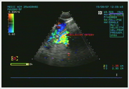
This book provides a comprehensive guide to the procedures to be followed when performing various techniques commonly practiced in hospitals and clinics. All these procedures are expected to assist students and medical practitioners to make a diagnosis and determine a reasonable treatment. The authors hope that with this guidebook, students and medical practitioners will better understand and understand these procedures and be able to do so properly and effectively. The contents of this book can be said to cover a broad scope and how the writing is in such a way as to facilitate the instruction being practiced. The writing and content of this book is based on the experience of the author as a medical practitioner and instructor and based on what the author usually does at their institution, Universiti Kebangsaan Malaysia.
This book is started with a cardiovascular system. Each electrocardiography has a positive pole and a negative pole. In essence, these canals can be concentrated on any angle and in relation to the heart. According to the convention, there are 12 electrocardiographys, ie, I, II, III, aVR, aVL, aVF, and VI to V6, from the perspective of the respiratory system, pleural effusion often requires inhalation of pleural fluid and pleural biopsy to be performed for diagnosis purposes. In most cases, both procedures are best carried out at the same time. The indicator is for diagnosis purposes, in particular to ensure whether or not there is effusion, infection, malignancy, empiema or hemothorax, the second is for treatment purposes, for example to relieve the congestion, to remove pus or blood and occasionally inject antibiotic or anticancer drugs. From the point of gastroenterology, the nasogastric tube will be used. The indications are lavage gaster to detect some diseases, distributing adequate food for body needs, detecting and treating poisoning, emptying gaster to treat acute abdominal pain or before or during an emergency surgery is to be performed. The equipment used is a large nasogastric tube so it is not easy to clog, but for the sake of nutrition it is preferable to use small size, lignocaine jelly, 50ml and litmus paper. The procedure is first, the patient is adequately informed about the procedure and the purpose. The second is the distance between the nose and the gaster measured first, usually between 40-50cm. Thirdly, there’s no need for a sterile way but keep the situation clean. The fourth is insert a little Lignocaine jelly into the nose groove and apply it to the end of the tube. Fifth is to advise the patient to sit if possible, then insert the tube according to the nasal floor (rather than the direction of the nose), preferably a little bow head. The sixth is the patient being swallowed when the tube is inserted (if there is no danger, give a little water to swallow, the seventh is the suction of the gaster with the siring.) If it does not work, enter the air by swirling while listening to the sound of bubbles in the gastric epi with the stethoscope. the test of liquid is inhaled with litmus paper The ninth is, if you are still in doubt, take the abdomen x-ray to ensure the end of the tubing Tenth is to use normal water first if the tube is used to feed the food. This condition occurs, pull the tube out and try again. If the tube is left too long, this can cause ulcer and graft penetration.
From the renal system, the urethral stenosis is a procedure that is often performed and should not be considered mild and should be performed by trained staff. From the point of view of the hematological system, venous punctures are the most frequent and important procedures in medicine. It has a very wide indication and there is no absolute contraindication, but it should be exercised with caution especially if it is carried out against patients with coagulation or bleeding problems. The equipment used is cotton, 5% spirit solution, 2I G-type disposable needle, binder rubber, 10ml or 20ml and specimen bottle.

For more Lupine Publishers Open Access Journals Please visit our website:


To Know More About Open Access Publishers Please Click on Lupine Publishers


Follow on Linkedin : https://www.linkedin.com/company/lupinepublishers
Follow on Twitter   :  https://twitter.com/lupine_online

Wednesday, 11 December 2019

Lupine Publishers | No Fact Check, Pharmaceutical Instituions from Reputed Pune University and Socially Situated and Socially Constituted Agency

Lupine Publishers | Journal Of Orthopaedics

Abstract

The behavioral view of corporate governance has derived from the behavioral view of the firm, which arguably rests on more realistic assumptions than the economics rooted positive agency theory with regard to the heuristics of managerial action. In addition to the bounded rationality condition, the principle of satisfying and the assumption on routinisation in the decision-making process, the behavioral view treats organizations as complex social systems. They constitute venues of power battles among the coalitions of corporate actors realizing often conflicting goal agendas. It is therefore probably the most explicit about the existence of phenomena of power and politics in the corporate settings among all theories of the firm.

Introduction

Recently published work, suggest enriching the under socialized agency perspective with the predictions of the behavioural theory of the firm. They refer to this cross theoretical hybridization as the behavioural theory of corporate governance. Within that framework, they distinguish two main mechanisms that impact on behaviour and actions of actors involved in governance processes in corporations, i.e. the socially situated and socially constituted agency [1,2]. PAT, as an under socialized and actor centric theory, concentrates on examining patterns, according to which individuals voluntarily, however rationally, realize their own goal agendas. They are motivated by self-interest and differential personal risk preferences, as well as are subject to informational and incentive constraints. In effect, PAT governance mechanisms tend to be formal in nature. They take a form of either incentive for managers as agents or means of monitoring/controlling them. They are construed to provide safeguards against such actions of managers, who driven by their self-interest may be potentially deviating from the desired organizational and/or societal outcomes. This unfolds by aligning managerial interests with those of shareholders or disciplining managers as agents. Infuse the agency relationships with the social context. They emphasise that corporate leaders do not operate in the social vacuum. On the contrary, they act in the socially constructed and interpreted reality. In the methodological sense, they enrich the of the individual human action in corporate governance, as posited in PAT, with the social fabric of norms, values and beliefs, and point towards the socio-cognitive processes as actual frames, within which particular board members enact their decision-making processes [3,4].
In other words, in this cross-theoretical framework and conceive the missing link between the macro-social explanations of well-functioning corporate governance practice, as offered by the economics-rooted PAT, and the micro-behaviour that is most likely to actually unfold in the boardroom reality. The term ‘socially situated’ is thought of in recognition of the fact that in any given situation individuals are enmeshed in a set of social relationships, networks, as well as institutions, which have influence on their perceived individual agency (e.g., a manager being accountable to non-executive directors directly, and to shareholders indirectly). Therefore, they represent crucial contingencies that ultimately shape the behaviour of individuals. The notion ‘socially constituted’, in turn, is conceived to capture a deeper kind of influence of the social context on the perception of the individual agency than it is the case with the socially situated agency. This concept emphasizes ways in which individuals’ socialization into performance of their particular roles (e.g., as a manager, a Chairman, a non-executive director), as well as their cumulative personal experiences to date, determine what they regard as possible or realistic in a given situation. The perceived individual agency, shaped through these processes, ultimately precipitates in a specific socio-cognitive orientation that particular board members adopt in their socially constructed boardroom reality. There have emerged entire streams of empirical research, which, even if it does not fully explain the theoretical rationale of the suggested behavioural theory of corporate governance, explicitly examines the socio-cognitive processes and behavioural tactics that are likely to unfold in the boardroom reality. They act as contingencies that shape decisionmaking processes by particular board members. For example, predicts the likely board outcomes as a result of competition and collaboration between the executive and non-executive directors in the boardroom. Research pluralistic ignorance on boards. Scrutinize favour rendering, ingratiation tactics and norms of reciprocity. Analyze the processes of symbolic and impression management, together with organizational/ institutional decoupling [4,5]. Finally, look at the social distancing tactics as a means of disciplining and/ or demonstrating ostracism towards those minority coalitions, which step out of the line dictated by the dominant board fraction.

Culturally determined agency

The notions of social situatedness and constitution fall close to concept of habitus. He coined it in elaborating on his view of power as internalized constraints. His perspective is methodologically akin to the conceptualizations of power, who regarded it as a ubiquitous abstract and subtle force that is impacting on individuals in such a way, that they actually act as their own over-seers. They discipline themselves and the existing social relationships thus arise as the natural order. These arguments suggest that the individual agency as perceived by particular social actors is de facto socially constructed, whereby this process is hugely influenced by the position of a given actor in the existing structure of social relationships. In corporate governance of domestic firms, the socio-cognitive processes that shape board members’ perception of their individual agency are described in the aforementioned contributions. However, such developments can also occur on Pharmaceutical Instituions’ boards [6-8]. There is one characteristic, though, which makes corporate governance in Pharmaceutical Instituions distinctively different from corporate governance in their domestic counterparts.
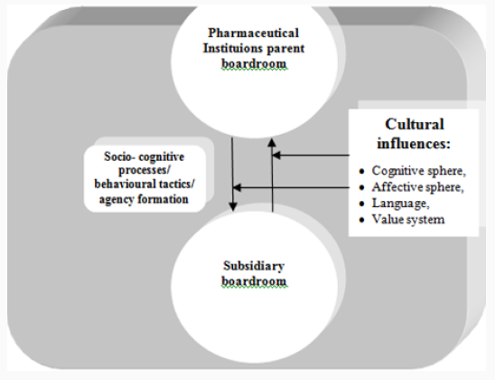
This is the phenomenon of culture and cultural differences between nation states. It is also the distinctive feature of the entire international business research, and hence we have it as a separate field of study in management science. Without drilling deeply into intricacies of accountability chains on boards within the Pharmaceutical Institutions headquarters and within its foreign subsidiaries separately, I therefore propose the view of culturally determined agency. The notion is to capture the socio-cognitive processes that particular board members in a given foreign subsidiary and their counterparts in the Pharmaceutical Institutions headquarters are exposed to, being located at the interface of Luo’s (2005a, 2005b) 1st- and 2nd-tier governance [9,10]. I suggest this view as a specific and distinctive feature of the corporate governance in Pharmaceutical Institutions. It incorporates cultural influences on the processes of social construction of the perceived individual agencies by particular board members at both governance levels and constitutes a significant portion of the overall variance of all types of impact factors on these processes [11,12].

Conclusion

Other corporate governance mechanisms typically distinguished in the academic literature comprise:
a) Minority investors’ protection rights.
b) Ownership concentratio
c) Incentive alignment (performance- related executive pay contingency).
d) Direct shareholders’ control (e.g. voting at the annual general meeting (AGM)).
e) Managerial labour market (reputation effects).
f) Market for corporate control (takeover activity).
g) Product market competition.

Controversies

Pharmaceutical Instituions has putting less effort to national economic systems. Principal of the Institutions has not report to college in time. They have alcohol dependency and smoker addict [13-15]. Self‐Report of such habit, Inconsistent alcohol use and Non-use are Poor Predictors of malpractice Prevalence among Pharmaceutical Instituions principal who have alcohol dependency with other Pharmaceutical Instituions Principal. Assessment of the population‐level effectiveness of the Avahan bad habit ‐prevention programme in Pharmaceutical Instituions in India: a preplanned, causal‐pathway‐based modelling analysis has been under way. This reasoning is summarised in Figure 1 A model of culturally determined agency.
Figure 1:A model of culturally determined agency.

Acknowledgment

This study has been guided by under supervision and guidance of Retd. Director’ National AIDS Research Institute India. I express my deep gratitude towards Respected Sir’ for motivation and being great knowledge source for this research.


For more Lupine Publishers Open Access Journals Please visit our website:
For more Open Access Orthopedics and Sports medicine articles Please Click Here:

To Know More About Open Access Publishers Please Click on Lupine Publishers


Follow on Twitter   :  https://twitter.com/lupine_online

Tuesday, 10 December 2019

Lupine Publishers | Caries Survey in 3-5 Year Old Children in Dubai Schools


Abstract

Introduction: Dental caries is considered the most prevalent chronic disease in the world, which affect individual from all ages. Host acceptability, cryogenic bacteria, fermentable carbohydrates and time are the four elements that are essential for the caries to start. dmft index is a measurement used to determine the degree of caries in any given population, in which it could help in setting aims and strategies to reduce and prevent this disease.
Aim: The aim of this study was to estimate the caries prevalence and the value of the dmft index of 3 to 5 year old children of Dubai private schools and its association with demographic factors as age and gender.
Methods and Materials: The study was a cross-sectional survey, which examined 3 to 5 year old children, for the presence of caries in their primary dentition. The survey was conducted by three calibrated dentist, who examined the children in the participating schools, in a school class setting using natural light and dental mirrors. Caries were recorded for each primary tooth, following WHO 1987 criteria. dmft and Significant Caries (SiC) Indices were calculated by age, gender and country of origin. Statistical analysis of the data obtained using parametric tests (t-test and ANOVA), to determine the presence of statistical significant differences.
Results: Two thousand nine hundred fifty-seven children were included in the study. 1527 males and 1430 females, in the male group 134 children were in the 3 year age group, while 630 and 763 children were in the four and five year old age group. The females group consisted of 139 children at 3 year of age, 575 in the four year age group and lastly 716 in the 5 year old age group. Caries prevalence was found to be 25.6%, 36.8% and 42.2% for the three age groups respectively. Dmft value was found to be 0.90, 1.50 and 1.72 for the 3, 4 and 5 year old age groups, with a dmft value of 1.55 for the total surveyed sample.
Conclusion: Caries prevalence in three, four and five year old children was 25.6%, 36.8% and 42.2%, respectively. Mean dmft score was 1.55 for the total sample, with a breakdown for each group to be: three year old group 0.90, four year old group 1.50 and five year old group 1.72. The SiC was found to be 2.07, 4.45 and 5.06; for the three age groups while the SiC of the total sample found to be 4.55. Comparison between the age groups showed that the caries prevalence and the SiC index increased as the age increased. The last national survey in 2001 had higher dmft for the 5 year old children in the state of Dubai

Introduction

Dental caries is the most prevalent chronic disease globally with multi-factorial etiology and pathogenesis WHO.int. Petersen [1,2].
It affects more than 5 billion people of total global population [24]. Children and adults could suffer dental caries at any time; once the oral hygiene measures are compromised, and the contributing factors are met together in specific condition and over a period of time [28,31]. Calling dental caries as a multifactorial, makes.com think about the elements responsible for such a disease formation. Tooth surface as a host, cryogenic bacteria, fermentable carbohydrates and time are the necessary elements. When they meet in the appropriate proportions formation of the disease will start. Characteristics of the tooth as a host such as, size, structure, anatomy and position of the tooth have an important impact in the progression of the caries and the rate of it. This could explain why the primary teeth are more susceptible to dental caries, having a wider proximal contact surface instead of contact point in permanent teeth [62,63,64,65,73].
Progression of the dental caries is faster which is sent back to the dental tubules arrangement, which is found as S-shaped course in permanent teeth and straight course in the primary teeth [27]. If dental caries are left untreated, then the general health of the individual is at risk. As a multifactorial disease, elimination of the cause will aid in the prevention of the disease. Alteration of a causative factor can be another way to prevent the disease. Dental caries is suggested to have big impacts on the person’s life, as it could impair the social function, self-esteem and psychological status Caries prevalence is another measurement, which represents the percentage of the affected population by dental caries at any given time [53]. One of the most important goals regarding global oral health was set by the World Health Organization (WHO) in 1979 is to reduce caries prevalence worldwide [2]. Repeated campaigns are designed to fulfil this goal. Most of these campaigns mainly focused on permanent teeth and few were exclusively targeted to the primary dentition using the dmft tool. Prevalence of dental caries among children from 0-5 year had been reported, as part of the WHO goal. Many studies have been conducted in UAE, the Gulf countries and the rest of the World to calculate the prevalence of the dental caries in primary dentition [45,16,33].
The caries prevalence’s for 5 years old children worldwide varied from 19.9% to 94%, while dmft score for the same age group varied from 0.87(1.16) to 9.8(5.5). In regard to the children aged 3 and 4 years old, little studies was conducted; which has results for the caries prevalence in 3 year old children between 19.4% and 85% and for the 4 year old it was between 23.8% and 90%. On the other hand the dmft scores worldwide was found to be 0.41(1.16) and 0.72(1.62) as lowest findings and 7.4(5.5) and 8.8 (5.6) as highest finding for the two age groups respectively [51,54,57,59,71,72].

Aim and Objectives

The aim of this study was to estimate the caries prevalence and the value of the dmft index of 3 to 5 year old children of Dubai private schools and the association of them with demographic factors as age and gender. The objectives were to compare the caries experience between the 3 age groups, to compare the caries experience between genders and to investigate possible association of the caries prevalence and demographic factors such as age and gender.

Methods and Materials

This study was part of European University College Oral Health Campaign in Dubai - United Arab Emirates. All the licenses needed for conducting this campaign were obtained from the Ministry of Higher Education and the Ethics committee of European University College that approved the project. Permission for participation in the study was granted from the public and private schools. Written explanation of the aim of the study and request for consent forms were given to the parents, in Arabic and English about the study. Sample From the list provided by the European University College Oral Health Campaign, three schools enrolled preschool and kindergartens children and were included in this study. Data were collected during the academic year 2012-2013. Recruitment was based on the age and the positive parental written informed consent. The age of children recruited was between 3 and 5 year old, starting at the day of their 3rd birthday up to the day before their 6th birthday date. The date of birth was obtained from the school files and entered into the computer program.

Procedure

Children of the chosen age groups were screened for dmft according to WHO 1987 criteria, for caries detection [1]. Examination was performed by three licensed dentist, who were calibrated based on the WHO 1987 criteria, for intra and inter liability score of k was 0.80 and 0.78 respectively, based on images of sound and carious teeth.
All children were examined at their school classroom under room lighting conditions. Gloves and sterile protocols were used, as well as tongue depressor, which aided in retraction of the tongue, lips and cheeks for better visibility of the teeth. Examination was done by dental mirror only, no dental probes were used. For moisture control and removal of plaque, cotton rolls and gauze were used. According to the WHO 1987 criteria, diagnosis of all primary teeth is based on the presence or absence of the caries. dmft was recorded for decayed, missing (due to caries) and filled teeth. The data from every child were recorded in individual charts. Each chart had an illustration of full primary dentition. The examiners were marking each tooth as decay (dt), missing (mt) or filled (ft). Mean dmft was calculated for the entire sample, as well as individually for each age and gender group separately. Standard deviation (SD) for the dmft was also calculated. Significant caries index (SiC) which represents the mean dmft of the one third of the sample with the highest caries score was also calculated for the total sample as well as for each age group and gender. SiC index gives an indication of the severity of the caries of the most affected part of the population and also illustrates in combination with dmft the distribution pattern of caries in this population [3].

Statistical Analysis

Data were statistically analyzed and compared between each age group as well as within each age group by gender. In order to statistically analyze the data of this study, t–test, ANOVA (Analysis of variance) and LSD (least square difference) tests were used at a level of significance p<0.05. ANOVA and t-test were used to identify differences between the means of dmft, dmft components and SiC index from different groups. When ANOVA detected a difference between groups then LSD was used to detect which groups were different.
Table 1:
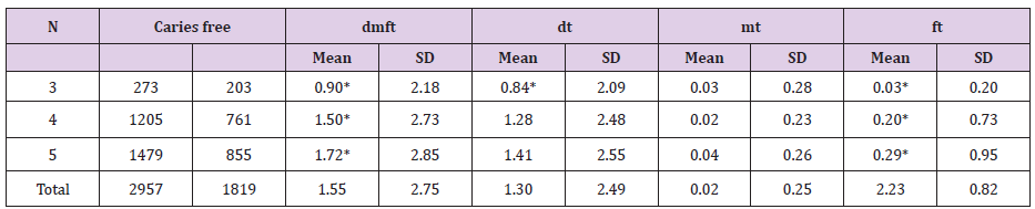
* Indicates statistical significant difference.

Results

The sample consisted of two thousands nine hundreds fiftyseven child (Table 1). 1527 males (51.6%) and 1430 females (48.4%), 273 (9.2%) in the 3 year old group and 1205 (40.8%) and 1479 (50.0%) in the 4 and 5 year old group, respectively. 3 year old children group consisted of 273 children, 134 (49.1%) males and 139 (50.9%) females, there were 203 (74.4%) caries free children. Of the male sample 104 children (77.6%) were caries free while of the females 99 children (71.2%) were free of caries for the 3 year old sample. Mean dmft (SD) was 0.90 (2.18) and dt = 0.83 (2.09), mt = 0.02 (0.27) and ft = 0.03 (0.19). Comparing the caries experience between males and females the result showed that males have lower dmft values, with a mean dmft (SD) of 0.69 (1.71), while for the females the mean dmft (SD) was 1.09 (2.53). Similarly, all of the dmft components in the male group presented lower values as compared to females; dt 0.67 (1.69), mt 0.02 (0.27) and ft 0.02 (0.14) for the males and dt 1.00 (2.41), mt 0.02 (0.27) and ft 0.04 (0.23) for the females. Differences in the means between the two genders were explored with ANOVA which did not show any statistically significant difference between the means of dmft (F= 1.52), dt (F= 1.30), mt (F= 1.51) and ft (F= 0.86). Moreover there were more males without caries (77.6%) compared to females (71.2%).
SiC value for the males was 2.07 (2.45) which are lower than the females 3.23 (3.50). 4 year old children group this age group included 2015 children, 630 (52.3%) were males and 575 (47.7%) females. There were 761 (63.2%) caries free children, 382 (60.6%) of the males and 379 (65.9%) of the females. The dmft values were found to be 1.50 (2.73) for the total sample, with dt = 1.28 (2.48), mt = 0.02 (0.23) and ft = 0.20 (0.72). It was also observed that dmft value for the males was 1.54 (2.69), with dt= 1.33 (2.69), mt= 0.03 (0.30) and ft= 0.19 (0.71), while for the females the respective values were, dmft = 1.45 (2.77), dt= 1.45 (2.77), mt= 0.01 (0.12) and ft= 0.21 (0.74). The mean dmft and dt was higher in the male group than the female group while females had higher dt and ft values. SiC value for the 4 year old sample was found to be 4.40 (3.11), while the SiC values for males and females were 4.44 (2.99) and 4.33 (3.25), respectively 5 year old children group The 5 year old group consisted of 1479 children, 763 (51.5%) males and 716 (48.5%) females. The mean dmft value of the total sample was 1.72 (2.99), with dt = 1.41 (2.55), mt = 0.03 (0.26) and ft = 0.29 (0.95). Comparing males to females in the caries experience within this group showed that the males had higher dmft value 1.85 (2.99) while the females had 1.59 (2.69). In regard to the dmft component, dt, mt and ft values; it was 1.53 (2.73), 0.04 (0.23) and 0.28 (0.87) for the males, and 1.28 (2.34), 0.04 (0.29) and 0.31 (1.04) for the females, respectively. Significant Caries index SiC for the total sample in this age group was 4.86 (2.97). Males presented statistically significant higher SiC 5.02 (3.05) than females 4.51 (2.82) using Student’s t-test (t= 5.11).
Comparing between the three age groups (3, 4 and 5 year old) The sample consisted of 1138 (38.5%) children with caries and 1819 (61.5%) caries free children. Caries prevalence for the 3 year old group was 25.6% (70 children) and 36.8% (444 children) and 42.2% (624 children) for the 4 and 5 year old group respectively. The mean dmft value was increasing as the age of the sample was increasing. The lowest dmft value was observed for the 3 year old group 0.90 (2.18) and then it increased for the 4 year old 1.50 (2.73) and the highest value was observed for the 5 year old children 1.72 (2.85). Regarding the dmft components, dt (decayed teeth) ranged between 0.84 (2.09) and 1.41 (2.55) and dt was the component with the higher contribution to the dmft index while the component with the lowest contribution was the ft which ranged between 0.03 (0.20) and 0.29 (0.95). ANOVA showed that among the three age groups there was statistically significant difference in the mean dmft (F= 10.81), dt (F= 6.16) and ft (F= 13.14) values, while there was no statistical significance difference in mt (F= 2.74) component. LSD test was used to explore the differences between the means that ANOVA suggested. LSD test showed that there was statistically significant increase of the mean dmft and dt values as the age of the children progressed. It was also found that only the three year old mean value of the dt component was different from the means of the other ages which had no significant difference.
Comparing dmft and its component between the three age groups showed that 5 year old children had higher value 0.9 (2.18), as compared with the 3 and 4 year old groups with 1.5 (2.73) and 1.72 (2.85), respectively. About the dmft components, dt was highest among the 5 year old group 1.41 (2.55) followed by the 4 year dmft 1.28 (2.48) and the 3 year old 0.84 (2.09). mt component was also highest among the 5 year old group 0.04 (0.26), the lowest was 4 year old children 0.02 (0.23), 3 year old group scored 0.03 (0.28). The values for ft was 0.03 (0.2) for the 3 year old group, 0.2 (0.72) for the 4 year old and 0.29 (0.95) for the 5 year old group. Dental caries formation in primary dentition increases with age, as the primate spaces and the physiological spaces reduces. Lack of hygiene and interproximal cleaning, aids in the increase of proximal caries as well the flat contact surfaces, which increase the progress of these kinds of caries. ANOVA was also used to test the difference of the dmft values between the males and females among the different age groups. Statistically significant difference was found between the different mean dmft male values (F=10.19) and LSD showed that the difference existed between all the three groups. Statistically significant difference was also observed in the dt and ft components of the dmft (F=6.71 and 7.10). LSD showed that males had statistically significant differences in dt and ft values among all three age groups. In female groups mean dmft did not have statistically significant difference, the only observation with statistically significant difference was seen in the mean dmft component, specifically ft value (F= 6.17). LSD test showed that all the three means in the ft females component were statistically significant different .

Discussion

United Arab Emirates has improved in many aspects regarding the dental health and the dental awareness of the population. Never the less through the few last year dental caries are still considered a national health problem among adult and children. Previously conducted National Survey in 2001 by El Nadeef et al. [4] studying dental caries among children, found dmft in 5 year old children 5.1, and specifically 6.6 for the state of Dubai. In this study 5 year old children dmft score was found to be 1.72. This is showing a decline of the dmft value as a result which could be attributed to the increase awareness in regard to dental health and the development of the dental care facilities. In this survey it is interesting to note the large drop of the dmft scores compared to previous studies. This big difference was primarily due to a substantial decrease of the dt components suggesting a drop in caries presence in the population. Despite this drop dt still remains the major contributing factor of the dmft index suggesting that even today caries are left untreated in children. Probably this reflects an attitude among the parents not identifying caries of primary teeth as a disease which needs treatment. In the past years there have been many awareness programs and educational campaigns conducted in the UAE trying to change this attitude. Towards the same direction this study shows also a slight increase in the ft component between the two studies. The awareness of the public regarding caries and caries prevention cannot therefore explain the substantial drop of the dmft values. Most likely this drop should be attributed to other confounding factors like wider use of fluoridated toothpaste and fluoridated products or healthier eating habits.
For the three and four year old group no studies were performed in Dubai, thus no data were available previously for the state of Dubai at the time of conducting this study. The results obtained from this study are the first data for this age group in Dubai, although the sample was not representative of the total population, the data may be considered as a pilot sample for an upcoming thorough investigation. AlHosani, Rugg-gunn 1998 conducted a study which included 4 and 5 year old children, in the western part of the UAE, AbuDhabi, AlAin and The West region. Prevalence of caries in the four year old children was ranging between 71-86%, while for the five year old it was between 82-94%. dmft was found to be 8.4 in AbuDhabi, 8.6 in AlAin and 5.7 in The West region. Comparing this result to the result obtained from this study, we can conclude that the dmft value in the state of Dubai is lower than the other UAE regions. Another study by Hashim R 2006 [6] in the city of Ajman found dmft in 5 year old children 4.0 (4.1), with caries prevalence of 72.9%, while in this study the prevalence was found to be 42.2%. Comparing the present findings to those conducted in the Middle East countries, findings suggest that this study is having lower dmft and caries prevalence than those conducted in the last decade. Paul, 2003 found in KSA caries prevalence of 83.5% and dmft of 7.1 (6.1) Paul 2003 [52]; Salem, Holm 1985 [7] had mean dmft 1.2 (0.08) for children age 3-5 year old in KSA, Morris et al. 1999 [8] had 19% dental prevalence in 18-48 months old children in Kuwait, also in Kuwait in 1993, Martomaa H found 22% prevalence in 3-7 year old children in Kuwait 1993 and Wyne, AH, 2008 found mean dmft 6.1 (3.9) for 3-5 year old children [70].
Among the 3 year old children the lowest dental caries (19.4%) was experienced in India 2001 [61], while the highest (85%) was in Philippines 2003 [26]. The result of this study falls in between this range, close to the lower end being 26%. There are no previous surveys conducted in the UAE for this age group in order to compare the result of this study. The only possible comparison can be done with surveys conducted in other countries of the world. We can only have an approximate comparison with other studies of the Gulf region since they report dmft and prevalence values for a range of ages that include the three year group making a direct comparison impossible. Viewing the previous studies conducted for the 3 year old children worldwide and comparing those findings with the finding of this study, we could conclude that the dmft score of this age group for the city of Dubai is being the lowest compared to other studies conducted in other countries in the world. The highest dmft value 7.4 (5.5) was reported in the Philippines 2003 [26], followed by 5.5 (4.5) in China 2002 [38]. dmft value in India varied between 0.41 (1.16) in 2001 [61] and 4.43 (2.33) in 2009 [65]. Brazil’s dmft values reported in 2009 were the lowest 1.13 (1.90) for 3 year old children [29], while in 2004 it was 2.10 [66].
For four years old children group the highest caries prevalence was found to be between 71% and 86% in AbuDhabi 1996 [5] which is very high compared to 37% in this study. In India (2001) [61], the reported caries prevalence was the lowest 28.2% [34], followed by Brazil 2007 with 23.8%. Philippines 2003 had the highest caries prevalence of 90% for this age group [26].
Comparing to the previous studies from different regions of the world, it could be noticed that dmft of this study falls in the lowest range of all dmft values of this age group. Australia 2002 [36] and Brazil 2009 [29] had the lowest dmft values 1.29 (2.74) and 1.42 (3.11), respectively. Next comes the dmft value of this study 1.50 (2.73).
Most of the dmft studies are conducted for 5 year old children. After the announcement of WHO goal for the year 2000, of having caries prevalence to be less that 50% for 5 year old children, many studies have been conducted all over the world to have this baseline and efforts have been put together afterwards to achieve the goal in that period. It worth mentioning that these efforts had been successfully noticed as dmft values had been significantly decreasing . In the UAE, significant decrease in the dmft value could be noticed between 1996 and 2001, and even further decrease could be noticed in dmft value of this study. Even though this decrease is obvious, the WHO target of having dmft score below 3 for the 5 year old children, haven’t been achieved yet. KSA 2000 [19], have achieved WHO target by having 0.95 (2.03) in a specific population. Other countries like India 2001 [61], Australia 2002 [36], India 2006 [44], Brazil 2007 [34], China 2008 [42], Brazil 2009 [29], Italy 2009 [14], Norway 2010 [74] and Greece 2011 [67] had also reach the target goal but after the set date. Highest dmft value could be observed for Philippines 5 year old children 2003 [26], followed by KSA 2008 [70] and FYROM 2014 [12] with same dmft value of 6.1. In regard to the caries prevalence in the 5 year old children, the highest prevalence could be observed in the Philippines 2003 [26] (94%) and in UAE 1996 [5] (94%).
The lowest prevalence was reported by Ferreira et al., Italy 2007 [34] (19.9%). It worth mentioning here that India 2001 [61], Kuwait 2002 [21], Brazil 2004+2007+2009 [66+34+29], UK 2006 [55], Italy 2009 [14], Norway 2010 [74] and Greece 2011 [50] all had caries prevalence that falls below 50%, which is the goal set by WHO in 2000 Khan 2014 [9] had reported a systemic review of the dmft in the Arab world for children aged between 3 and 10 year of age, for the period from 2000 to 2012. In this report Kingdom of Saudi Arabia in 2000 [19] reported the lowest dmft values 0.95 while in 2007 [52], dmft value was the highest 7.1. These studies were conducted in different places in KSA, follow up studies worth to be conducted in the same places to have a frank comparison between the new and old obtained dmft values. According to the finding results in this study, and due to the selection of the sample, which was drawn mainly from private schools and the fact that those children are from a medium socioeconomic status families, the result are not representative to the children population in the city of Dubai. Furthermore, the three examiners were calibrated through photographs, which may have contributed to some bias for caries underestimation. Also the caries were scored according the 1987 WHO caries criteria which does not account for caries with underlining dark shadow from dentin, lesions that are usually accounted in WHO 1997 criteria, used by most of the epidemiological studies [15,25,30,32,40,41,46,49,56, 58,60,68,69]. Since the introduction of dmft and SiC indices 1938 [35], it was considered as ideal indices, as they met a number of criteria, which made them useful epidemiological indicators. These criteria included simplicity, reliability versatility and statistical manageability [17,18,20,22,23,37,43,47]. dmf describes the mean experience of dental caries in specific population, without giving any information about the level of the dental caries in the most affected population. On the other hand significant caries index (SiC), does represent the mean dmft in the highest one third of the population with the highest dmft scores giving a picture of the severity of the disease in the most affected portion of the sample.
The SiC index indicated that the study sample had a score of 2.07 (2.45) for the 3 year old group, 4.45 (2.99) for the 4 year old group and 5.06 (3.13) for the 5 year old group. The SiC score for the 5 year old children is considered of lower score compared to the SiC indices from other part of the world, for example in SiC score of 5 year old children in Greece 2011 was 5.01 [50], while In Sri Lanka 2012 5.84 for 4 and 5 year old children [11]. In the Republic of FYROM, SiC index was found to be 8.83 [12] found SiC in Turkish children to be of 7.75 [13] and according to in Italy, SiC for 5 year children was 5.32 [14]. Recommendations since the National survey of dental caries in United Arab Emirates is almost fifteen year old, efforts have to be put together in a governmental and personal level to update this important data, which will aid in thorough prevention of the problem. Health education of the parents and the children has to start as early as possible, which will aid in prevention and detection of the problem at its initial stages. Dental visits have to be scheduled annually for routine checkups. Parent’s education regarding the importance of the primary teeth to the child’s overall health has to be emphasized; this important information will result in maintaining healthy children .

Conclusion

Caries prevalence in three, four and five year old children were found to be 25.6%, 36.8% and 42.2%, respectively. Mean dmft score for the total sample was 1.55 (2.75), with a breakdown for each group to be: three year group 0.90 (2.18), four year group 1.50 (2.73) and five year old group 1.72 (2.85). The SiC was found to be 2.07 (2.45), 4.45 (2.99) and 5.06 (3.13); for the three age groups, 3, 4 and 5 year old children, respectively [61-68]. The SiC for the total sample was found to be 4.55 (3.12). Comparison between the age groups showed that the caries prevalence and the SiC index increased as the age increased. The last national survey in 2001 had higher dmft for the 5 year old children in the city of Dubai. The prevalence of primary dental caries is greater in 5 year old children, especially in male children. Dental caries formation in primary dentition increases with age, as the teeth are exposed to the cariogenic bacteria for longer time. The sample consists of only private schools in Dubai; dmft scores are lower than the previous national survey which was conducted at public schools. Dental awareness is still minimum among the population new national survey has to be conducted to update the previous data [69-73].

For more Lupine Publishers Open Access Journals Please visit our website:
http://lupinepublishers.us/
For more Journal on Pediatric Dentistry articles Please Click Here:
https://lupinepublishers.com/pediatric-dentistry-journal/

To Know More About Open Access Publishers Please Click on Lupine Publishers

Follow on Linkedin : https://www.linkedin.com/company/lupinepublishers
Follow on Twitter   :  https://twitter.com/lupine_online

Monday, 9 December 2019

Lupine Publishers | Congenital Craniofacial and Cervical Cysts, Sinuses and Fistulas- A Review Article

Lupine Publishers | Journal of Otolaryngology Research Impact Factor


Abstract

Congenital cysts, sinuses and fistulas of the head and neck are anomalies that may be encountered in pediatric life. They represent abnormal development of the brachial apparatus or inclusion of the ectoderm at any site of the lines of fusion of the face. In the head, preauricular, salivary and median nasal fistulas as well as dermoid cysts and Van der Woude syndrome are examples. In the neck, median thyroglossal cyst and fistula, and lateral brachial cyst and fistula are well-known congenital anomalies. Proper diagnosis of these malformations is essential to decide the line of treatment. Standard and modified surgical procedures are adopted for excision and repair of these anomalies with the possibility of incidence of complications, recurrence and malignancy.
Keywords: Congenital; Cysts; Sinuses; Fistulas; Craniofacial; Cervical; Thyroglossal; Branchial
Abbreviations: CT: Computed Tomography; MRI: Magnetic Resonance Imaging; MULFs: Median Upper Lip Fistulas; TGDCs: Thyroglossal Duct Cysts; VWS: Vander Woude Syndrome

Introduction

A fistula is an abnormal tract communicating the cavities of two internal organs or the cavity of an organ with the body surface whereas a sinus is one-end blind tract that opens on the surface. Congenital cysts, sinuses and fistulas of the head and neck are uncommonly detected in children. They include congenital craniofacial and cervical anomalies; the latter may be mediocervical or latero-cervical in position. Surgical excision of these lesions is the only option of treatment.

Congenital Craniofacial Cysts and Fistulas

Congenital periauricular cysts, sinuses and fistulas occur commonly in children are developmental anomalies of the first branchial cleft and first pharyngeal arch [1,2]. Children with recurrent postauricular abscesses show associated congenital anomalies like postauricular sinuses, post auricular dermoid cysts, first branchial cysts, aural fistulas and aural atresia [3]. Congenital salivary fistulas of the main part of the parotid gland are rarely reported; surgical excision of the fistula and parotid gland is an effective line of treatment for this anomaly [4]. Odontogenic cutaneous fistula results from abnormal tooth canalization caused by chronic periapical infection; it may be wrongly diagnosed as a congenital fistula [5]. Abnormal protrusion of a part of the brain and its meninges through a defect in the skull bones is termed meningoencephalocele. A case of an anterior meningo-encephalocele presenting as a small swelling in the right lower eyelid was reported; if it was mistreated as a lachrymal swelling it might result in meningitis and cerebrospinal fluid leakage [6]. The nasal dermoid cyst and median nasal sinus are rare congenital anomalies that are attributed to early embryonic inclusion of the ectodermal germ layer during closure of the anterior neuropore of the neural tube [7]. Congenital nasofrontal fistulas (also termed nasofrontal dermal sinuses) are extremely rare malformations that may be complicated with serious meningitis [8]. Patients with congenital nasal fistulas and dermoid cysts are operated upon through vertical midline or horizontal glabellar, and nasofrontal osteotomy [9]. A craniofacial dermoid cyst at the lateral wall of the orbit was reported to be associated with a cutaneous frontotemporal fistula that extended to the dura of the temporal lobe of the brain through the temporal and sphenoidal bones; lateral orbitotomy was surgically performed in this case [10].
Congenital sinuses and fistulas of the lip are uncommon and median upper lip fistulas (MULFs) are extremely rare. A case of congenital MULF, at the philtrum of a girl infant, was surgically removed. The fistulous tract was histological lined with squamous epithelium that contained sebaceous and mucous glands as well as hair follicles. [11,12] Van der Woude syndrome (VWS) is a single gene autosomal dominant congenital craniofacial abnormality that is featured by labial cysts, accessory salivary glands, and lower lip pits, fistulas and paramedian sinuses; it is commonly associated with cleft lip or palate or both [13,14]. Excision of lower lip sinuses and accessory glands, reconstruction of the lip, nose, and associated cleft lip and palate are the components of VWS surgery [15]. Surgical excision of congenital lower lip sinuses has an increased rate of mucoceles formation and recurrence [16].

Congenital Cervical Cysts and Fistulas

Neck masses are frequently met with in children and they may be congenital or acquired in origin. Congenital neck swellings include branchial cysts, thyroglossal cysts, hemangiomas and cystic hygromas. They may be complicated into sinuses and fistulas [17,18]. Congenital cervical cysts, sinuses and fistulas are not common. Thyroglossal duct cysts (TGDCs) are the commonest malformations followed, in frequency, by branchial cleft anomalies and dermoid cysts [19]. Congenital cysts and fistulas of the neck are divided, according to their locations, into midline or mediocervical and latero-cervical anomalies [20,21]. In man during the 3rd to the 4th week of development, a pharyngeal (branchial) apparatus is formed in the wall of the pharyngeal gut. It consists of six mesodermal arches, four ectodermal clefts and five endodermal pouches. The derivatives of these branchial elements include essential structures in the head and neck while their remnants may develop into congenital neck anomalies like branchial cyst (and fistula) and thyroglossal cyst (and fistula) [22]. Laterocervical congenital cysts and fistulas are mentioned to be due to abnormal development of branchial clefts particularly the 2nd one; they are usually found at the anterior border of the lower third of the sternocleidomastoid muscle. The TGDCs (and fistulas) are the most common medio-cervical congenital anomalies; they are usually found in the median thyrohyoid region. Surgical resection of the cysts and fistulas should be complete to avoid recurrence and risk of malignancy [23]. Congenital cervical salivary fistulas result from ectopic non-branchial salivary tissue that has a sinus draining serous or mucous secretion to the anterior part of the base of the neck [24]. In congenital dermoid fistula of the anterior chest region, a skin orifice could be seen at the anterior border of the sternocleidomastoid muscle with subcutaneous extension of the fistulous tract till the sternoclavicular joint [25].

Median Neck Cysts and Fistulas

TGDCs are the commonest congenital swellings of the neck [26]. They represent 2% of cervical masses and 70% of congenital neck swellings [27,28]. TGDCs result from patency of a part of the embryonic thyroglossal duct that extends from the foramen cecum (on the dorsum of the tongue) till the thyroid primordium (pouches). They are painless and commonly diagnosed in children below the age of 5 years. A TGDC may ulcerate spontaneously leading to the formation of a thyroglossal fistula that rarely communicates with foramen cecum [28]. Thyroglossal fistulas may be suprahyoid, hyoid or infrahyoid in position [29]. It is agreed that congenital mento-sternal fistulas are embryologically and pathologically different from thyroglossal fistulas. Mento-sternal fistulas are most probably caused by midline cervical inclusion of ectodermal or endodermal tissue during fusion of the branchial elements [30]. Excision of the TGDC and fistula is performed through Sistrunk procedure which includes removal of the body of the hyoid bone. The lining epithelium of the excised cysts varies according to the sites of the cysts; it is cuboidal, columnar, pseudo stratified or stratified squamous in type [31]. In an adult female patient, during a Sistrunk procedure for a TGDC with recurrent purulent discharge, two fistulous tracts terminating at the hyoid bone were discovered; it was mentioned that similar cases were missing in the literature [32]. Moreover, a branched thyroglossal duct terminating in two separate cysts with a chronic fistula was diagnosed in a young woman with a history of an incomplete surgical excision of a TGDC [33].
Clinically, most of the thyroglossal fistulas are considered as the squeal of interventional procedures: incisions, punctures, enucleations or radiations of TGDCs. Radical Sistrunk procedure is an effective option for removal of a TGDC [34-37]. Standard or modified Sistrunk operation includes removal of the TGDC and/or fistula together with the middle part of the hyoid bone; the duct is microscopically or macroscopically dissected till the foramen cecum to avoid recurrence [38-43]. The recurrence rate of TGDCs removed by Sistrunk procedure is reported to be 4% [44]. In a group of patients with TGDCs and fistulas, the size of the cyst varied from 1 to 4 cm; Sistrunk procedure had the advantages of low rates of complications (9.08%) and recurrence(1.82%), avoidance of antibiotics usage and short hospital stay [45]. Preoperative neck ultrasonography is recommended as a diagnostic tool of a TGDC whereas postoperative histopathology of the excised cyst and fistula is mandatory to exclude rare occurrence of malignancy [46,47]. An unusual case of a TGDC at the base of the tongue and fistulizing to the anterior part of the tongue was described and successfully excised via a combined trans-hyoid and intra-oral surgical approach [48].
Piriform sinus fistulas are congenital anomalies of the third and fourth branchial arches. They are usually unilateral and associated with thyroiditis and recurrent abscesses in the lower part of the front of the neck. A case of rare bilateral congenital piriform sinus fistulas is mentioned in the literature [49].

Lateral Neck Cysts and Fistulas

Branchial anomalies of the 1st to 4th branchial clefts and arches represent 20% of all congenital head and neck swellings in children [50-52]. Branchial cleft anomalies can present as a neck swelling,sinus, or fistula. Second branchial cleft anomalies account for about 95% of all anomalies of the branchial apparatus. Repeated preoperative infections of these anomalies lead to a higher rate of postoperative recurrence [53]. Branchial cysts are supposed to result from incomplete developmental obliteration of the branchial cleft or trapping of cell nests in the branchial apparatus that can later form branchial cysts [54]. Second branchial cleft cysts and sinuses are the most common type of branchial cleft anomalies. Failure of obliteration of the cervical sinus of His leads to the formation of a branchial cyst and failure of fusion of the 2nd branchial arch with the 5th arch results in a branchial sinus (fistula) [55]. A rare coexistence of TGDC and branchial cyst with fistula in the same child is reported in the literature [56]. Unilateral first and bilateral second branchial cleft fistulas were concomitantly observed together with a preauricular sinus and moderate bilateral hearing loss in branchiootic and branchio-oto-renal syndromes [57]. A case of an ectopic tooth in a branchial cleft anomaly was described in a young girl who had Townes-Brocks syndrome [58]. Another case of second branchial cleft sinus remained asymptomatic till the sixth decade of life when it was surgically excised together with ipsilateral carotid endarterectomy [59]. Furthermore, a congenital branchial fistula may be complete with an internal orifice at the tonsillar fossa; complete surgical excision of the fistulous tract could be performed in one setting [60].

Conclusion

Congenital cysts, sinuses and fistulas of the head and neck must be properly diagnosed and managed to diminish the rate of complications and recurrence, and to avoid the risk of malignancy.

For more Lupine Publishers Open Access Journals Please visit our website: h
http://lupinepublishers.us/
For more Journal of Otolaryngology Impact Factor articles Please Click Here:
https://lupinepublishers.com/otolaryngology-journal/

To Know More About Open Access Publishers Please Click on Lupine Publishers

Follow on Linkedin : https://www.linkedin.com/company/lupinepublishers
Follow on Twitter   :  https://twitter.com/lupine_online

Friday, 6 December 2019

Lupine Publishers-Msct In Diagnosis of Congenital Heart Diseases in Viet Nam

Lupine Publishers | Advancements in Cardiovascular Research


Abstract


Background: Congenital heart diseases associated with more malformations, complex aortopulmonary collaterals and anomalous coronary artery. Echocardiography is the initial diagnostic method but this method can be limited in complex congenital heart diseases.
Purpose: To assess the role of MDCT in congenital heart diseases (CHD) diagnosis compare with operative result and interventional angiography.
Methods: 910 patients with congenital heart diseases of 31.000 patients underwent cardiac angiography with 64 and 320 section CT at Medic Medical Center since 09/09/2006 to 30/12/2015.
Results: There are 658 operated cases, most of operated cases demonstrated the exact diagnosis of MDCT in congenital heart diseases.
Conclusions: MDCT is the fast and non-invasive diagnostic method with the high accuracy, overcomes the limit of echocardiography in complex congenital heart diseases diagnosis and provides the panorama and useful information’s prior to the operation.
Keywords: Congenital heart diseases; Cardiac multi-detector computed tomography, Multi-detector computed tomography in congenital heart diseases; Congenital heart diseases computed tomography

Introduction

Congenital heart diseases effect ~ 1% of all live births in the general population. Complex congenital heart diseases associated with more malformations, complex aortopulmonary collaterals and anomalous coronary artery. Over the past few decades, the diagnosis and treatment of congenital heart diseases have greatly improved [1-6]. Diagnostic tools: X-ray, ECG, echocardiography, MRI and MDCT. ECG and X-Ray suggest the diagnosis but are not specific. Echocardiography is the initial diagnostic method for patients with suspected CHD but this method can be limited in complex CHD. The great capabilities of MRI for anatomic and functional assessment of the heart but MRI is time-consuming and may require patient sedation. Now enable CT to be used as an accurate noninvasive clinical instrument that is fast replacing invasive cine-angiography in the evaluation of CHD [1,2,5].
I. Improves both spatial and temporal resolution.
II. Increases scanning speed.
III. Improves diagnostic image quality by reducing respiratory artifacts

Purpose

To assess the role of MDCT in congenital heart diseases (CHD) diagnosis compare with operative result and interventional angiography.

Material and Methods

Subject: 910 patients with congenital heart diseases of 31.000 patients underwent cardiac angiography with 64 and 640 section CT at Medic Medical Center since 09/09/2006 to 30/12/2015.

Means and scanning techniques

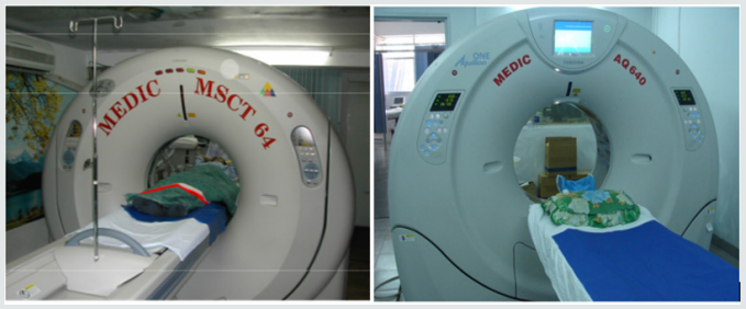
a) Medic Medical Center scanned cardiac CT by 64 MDCT Toshiba Aquilion machine and Toshiba Aquilion One (320 MDCT), 0.5mm slice thickness, 0.5mm imaging reconstruction.
b) Two phases scanning: Don’t inject phase and contrast media injection phase: +Phase doesn’t inject contrast which help locate and assess coronary artery calcification.
c) +Phase inject contrast media: Medicine chasing phase and water chasing phase.
d) Contrast pumping machine is double-barreled Stellant (Medrad).
e) To inject contrast by intravenous right hand.
f) Contrast dose used 1mL/ kg.
g) Drug pump speed depends on patient status and disease.
h) Vitrea software: Reconstructed images by MPR, MIP and VRT.
i) Effective radiation dose is low (320-MDCT is 3.69±061mSv; 64-MDCT is 12-14mSv) (Figures 1).
Figure 1.
Lupinepublishers-openaccess-cardiology

Data analysis

a. The prospective study and case series report compare with operative and interventional angiography.
b. Data collection at the HCM city Heart Institute, Tam Duc Heart Hospital and Medic medical center (Figures 2-17).
Figure 2: Atrial septal defects and Ventricular septal defects.
Figure 3: Patent ductus arteriosus.
Figure 4: Coarctation of aorta.
Figure 5: Double aortic arch.
Figure 6: Tetralogy of Fallot.
Figure 7: Pulmonary atresia with ventricular septal defect.
Figure 8: Transposition of great vessels.
Figure 9: Double outlet right ventricle.
Figure 10: Single ventricle.
Figure 11: Aortopulmonary window:
Figure 12: Truncus arteriosus.
Figure 13: Anomalous systemic venous return.
Figure 14: Anomalous pulmonary venous connection.
Figure 15: Single pulmonary artery.
Figure 16: Pulmonary artery trunk aneurysm:
Figure 17: Congenital pulmonary arteriovenous malformation.

Results

There are 658 operated cases, most of operated cases demonstrated the exact diagnosis of MDCT in congenital heart diseases.

Discussion

Congenital heart diseases associated with more malformations, complex aortopulmonary collaterals and anomalous coronary artery. Echocardiography is the initial evaluative method for preand post-operation congenital heart diseases but this method can be limited in complex cases. Multi-detector computed tomography overcomes the limit of Echocardiography by multiplanar reconstruction (MPR) and volume rendered techniques (VRT) reconstruction . Volume rendered techniques (VRT) reconstruction clearly demonstrates the relationship between the heart and great vessels.

Conclusion

Multi-detector computed tomography is the fast and noninvasive diagnostic method with the high accuracy. Overcomes the limit of Echocardiography in complex congenital heart diseases. Provides the panorama and useful information’s prior to the surgery.


For more Lupine Publishers Open Access Journals Please visit our website:

For more Advancements in Cardiovascular Research articles Please Click Here:

To Know More About Open Access Publishers Please Click on Lupine Publishers


Follow on Linkedin : https://www.linkedin.com/company/lupinepublishers
Follow on Twitter   :  https://twitter.com/lupine_online
Lupine Publishers-The Left Common Carotid Artery Rises from the Main Pulmonary Artery

Lupine Publishers | Advancements in Cardiovascular Research


Abstract


A young female patient of 15y.o presented at my hospital by dyspnea on effort and palpitation for one year. Mental deficiency was notified. Physical examination detected a 3/6 continuous murmur at the 2ndRICS. In the past history, PDA had been suspected by her physician, associated with recurrent bronchitis. Trans-thoracic Echocardiography showed an enlarged LV of 57mm with normal EF of 69% , LCA=5mm, RCA=3.5mm at origin, no suspected sign of PDA was seen. Only a continuous flow was visualized in the PA. CT-Angiography with IV contrast medium showed the Left Common Carotid Artery rising from the Pulmonary Artery trunk. PDA was not presented. The Left Common Carotid Artery then was re-implanted into the aortic arch normally with a favorable postoperative progress.

Keywords: Carotid Artery; Pulmonary Artery; Anomalous origin


Introduction


Anomalous origin of the left common carotid artery is very rare and has been reported previously. We present an operated case of this topic with clinical finding, cardiac ultrasound and MDCT imaging.

Case Report


Figure 1: Right aortic Arch.
Lupinepublishers-openaccess-cardiology
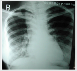
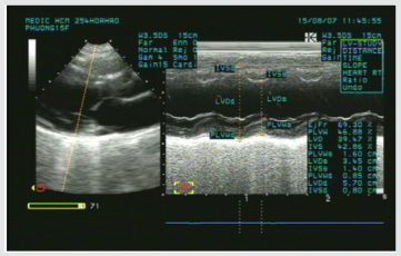
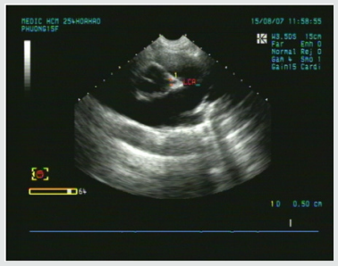
A young female patient of 15y.o presented at my hospital by dyspnea and palpitation when running and fast walking for one year. Mental deficiency was notified, she had some difficulties to learn at school. Physical examination detected a 3/6 continuous murmur at the 2ndRICS. In the past history, PDA has been suspected by her physician, associated with recurrent bronchitis. Her body state was normal with 1m60 of height and 48 kg of weight. She was evaluated immediately by a chest X ray that showed a right aortic arch (Figure 1). The trans-thoracic echocardiography that revealed an enlarged LV of 57mm with normal EF of 69% (Figure 2), LCA=5mm, RCA=3.5mm at origin (Figure 3). No suspected sign of PDA was detected except a continuous flow presented in the Pulmonary Artery (Figure 4).
Figure 2: Enlarged LV& normal systolic function.
Lupinepublishers-openaccess-cardiology
Figure 3: Normal LCA at origin.
Lupinepublishers-openaccess-cardiology
Figure 4: Continuous flow in the PA.
Lupinepublishers-openaccess-cardiology
Figure 5: Absence of aortic origin of the LCCA.
CT-Angiography (MDCT 64) with IV contrast medium Ultravist, slice thickness=1mm visualized a right aortic arch, aberrant origin of the left subclavian artery, dilatation of the branches rising from aortic arch with increased collateral vessels (Figure 5). Especially, MDCT 64 showed the Left Common Carotid Artery ( LCCA ) rose from the PA trunk (Figure 6) PDA was not detected. Patient underwent uncomplicated surgical repair: the Left Common Carotid Artery was re-implanted into the aortic arch normally with a favorable post-operative progress (Figure 7).
Figure 6: The LCCA rising from the Main PA roof.
Figure 7: Re-implantation of the LCCA.


Discussion


Anomalous origin of the Left Common Carotid Artery from the Pulmonary Artery Trunk has been previously reported as rare cases. Kagami Mijaji et al. [1] has reported a case of anomalous origin of the Artery from the Right Pulmonary Artery. Onyekachukwu et al. [2] has described a case of anomalous origin of the Left Common Carotid Artery from the Main Pulmonary Artery. In this article, my patient was not infant with CHARGES syndrome that includes multiple congenital anomalies like the patients in their reports. She was a teenage patient without other congenital disease. The role of ultrasound is orienting for the indication of Computed Tomography or DSA. In case of present turbulent flow in the PA, Coronary Fistula and other shunts from the head and neck vessels should be considered [3].


Conclusion


Anomalous origin of the Left Common Carotid Artery is very rare congenital defect that maybe isolated or associated with some syndromes. Noninvasive diagnostic methods as Ultrasound and CTA may confirm the diagnosis and inform the anatomical relation of the anomalous vessels prior to operate.


For more Lupine Publishers Open Access Journals Please visit our website:

For more Advancements in Cardiovascular Research articles Please Click Here:

To Know More About Open Access Publishers Please Click on Lupine Publishers


Follow on Linkedin : https://www.linkedin.com/company/lupinepublishers
Follow on Twitter   :  https://twitter.com/lupine_online

Thursday, 5 December 2019

Lupine Publishers | Risk Stratification at Patients with the Defect Vessels and High Blood Pressure: New Mathematical Model

Lupine Publishers | Open access Journal of Complementary and Alternative Medicine


Short Communication

It is noted, that today the population of Uzbekistan unprecedentedly increases consumption of salt and have low physical activity in general. Besides for the last 25 years, the population of Uzbekistan was enlarged twice. Today such tendency of population increase is followed by its aging. The positive aspect of this phenomenon is augmentation of average life expectancy. However, a negative side that the augmentation of life expectancy doesn’t correspond to its quality. Deterioration first of all is bound to augmentation of prevalence of age chronic diseases, such as a hypertension. For example, today in Uzbekistan, one of three adults has raised by the ABP, and excess weight occurs at every second (WHO/STEPS 2014). As a result it caused double increasing of visits of the doctor and total number of patients in general. The purpose of our research was in developing the prognostic model of the turnpike arteries biological age at hypertensive patients. To study the arterial blood pressure daily profile at sick with the arterial hypertension (AH) with the metabolic syndrome (MS) [1].

Material and Methods

The study included 96 healthy volunteers and 96 men with arterial hypertension AH in average age of 56.06 ± 7.86 years, diagnosed abdominal obesity (BMI 34.54 ± 3.83 kg/m2) and metabolic disorders. In order to estimate the clinical status, the following risk factors were studied: elevated arterial BP, smoking; clinical and biochemical parameters: 12-lead ECG; 24-hour Holter ECG monitoring; exercise stress test; echocardiography (EchoCG); carotid artery intima-media thickness (IMT) [2,3].

Prognostic Model

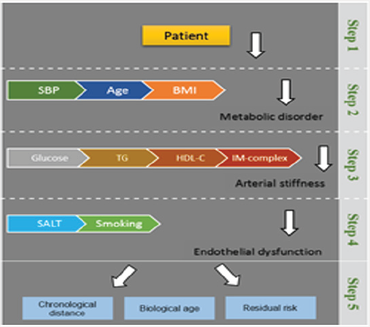
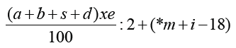
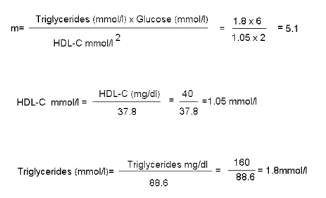
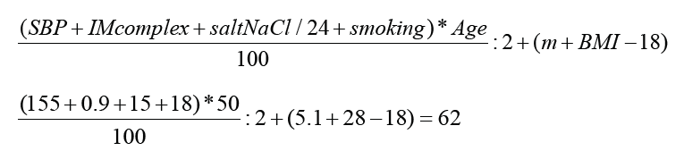
We selected clinical signs for creation of the prognostic table by means of the method of the consecutive diagnostic procedure based on a technique of the sequential analysis offered by A. Wald. For each informative sign gradation of this or that indicator were selected to equal the diagnostic value of each of indicators (Figure 1). On the basis of local research grant analysis the calculator of risk was developed to provide patients with AH I-II -degrees the important prognostic information. We also considered that population of modern Uzbekistan have tendencies of negative impact at vessels damage such as: salt and the increased body weight. As a result, nine best factors for stratification of risk at patients with the defect vessels and raised SBP, including easy available, clinical parameters were included in this calculator (Figure 1). We applied multistage stratification of risk, based on nine parameters which were included in the developed equation of nine factors [4]:
Figure 1: Prognostic model.
Lupinepublishers-openaccess-complementary-alternative-medicine-journal
(a-SBP; b- IMT intima-media thickness; s- Salt; d-smoking; e-age; *m- metabolic index *m= t x g / h2; i-BMI; t- Triglycerides, g- Glucose; h- HDL-C).
Lupinepublishers-openaccess-complementary-alternative-medicine-journal
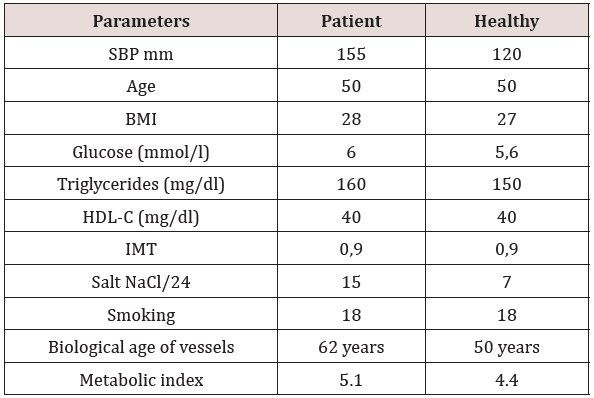
During this experiment, patients noted informational content of the calculator for the objective comprehension of their cardiovascular status (Sensitivity–85 %, Specificity–63%) (Table 1). However long-term preventive effect, in prevention of the recurrence was not confirmed [5-7].
Table 1: Example Clinical data.
Lupinepublishers-openaccess-complementary-alternative-medicine-journal

Conclusion

On completion of the therapy clinical tests indicated the high sensitivity at average specificity model that was acceptable in an opportunity to estimate medical effect. Unfortunately, the calculator could show the rate of damage vessels in the cases of actual disease, but not in cases of its prediction or probable emergence.


For more Lupine Publishers Open Access Journals Please visit our website:


To Know More About Open Access Publishers Please Click on Lupine Publishers


Follow on Linkedin : https://www.linkedin.com/company/lupinepublishers
Follow on Twitter   :  https://twitter.com/lupine_online