Lupine Publishers is a multidisciplinary, scholarly Open Access publisher focused on Genetic, Biomedical and Remedial missions in relation with Technical Knowledge as well. Our journals maintains a scrupulous, methodical, fair peer review System. Besides, quality control is riveted in each step of the publication process. https://in.linkedin.com/company/lupinepublishers
Objective: FB (foreign bodies) in ear, nose and throat
are often encountered by otolaryngologists in their daily practice and
is
commonly seen in both children and adults. Depending upon the
type and location of FB, it may have serious impact on individual’s
health if instant appropriate action is not taken. That’s why,
there’s frequent visits to ED (emergency department) on having FB in
ear and aerodigestive tracts. The objective of this study was
to evaluate the nature, common sites, modes of presentation, modes of
management of FB, age and gender distribution. Materials and Methods: A retrospective hospital-based study was done in Universal College of Medical Sciences, Bhairahawa,
Rupandehi, Nepal from March 2014 to September 2017. The information was obtained from hospital record books. Results: Out of 483 total patients, 287 (59.42%) were
male and 196 (40.57%) were female. Most of them were less than 10
years old. Of the 483 patients, 202 (41.82%) had FB in the
ear, 132 (27.32%) in the nose and 149 (30.84%) in the throat. Living FB
were found in 54 (26.73%) patients out of 202 in the ear, 10
(7.57%) patients out of 132 in the nose and none in the throat. Of the
total patients, 97 (20.08%) required general anesthesia (GA)
to remove FBs and the rest 427 (88.4%) patients were dealt with or
without local anesthesia. Most of the FBs were removed
promptly on presentation otherwise within 24 hours of presentation in
the
hospital. Conclusion: FB in ENT were found more commonly in the
children and the commonest site was ear. Timely presentation,
prompt diagnosis and needful management in a center with
otolaryngology practice reduces the morbidity and mortality. Most
of the FB in ENT can be removed in outpatient department (OPD)
or emergency room (ER) with or without local anesthesia (LA). Keywords: Ear; Nose; Throat; Foreign Bodies; Local Anesthesia
Introduction
A foreign body (FB) is any object or substance that is not
derived from the individual’s own body part and can cause harm by its
mere presence if prompt medical care is not provided [1,2]. They may be
found in Ear, Nose and Throat. They are very common in
otorhinolaryngological clinical practice. It can be introduced
spontaneously or accidentally by both children and adults. However,
children are common victims as they have habit of inserting nearby
objects in their nose, ear or mouth, imitation and also other
contributing factors are like boredom, playing, mental retardation,
insanity, and attention deficit hyperactivity disorder, along with
availability of the objects and absence of watchful caregivers.
Consequently, it may cause minor irritation to life threatening problem.
A proper technique, good light, appropriate instrument, a co-operative
or fully restrained patient and a gentle approach by the related doctor
or health worker are required for the removal of FB. One should have a
clear diagnosis before making attempts to remove the FB so as to lessen
the morbidity [3,4]. FB may be classified as animate (living) and
inanimate (nonliving). The inanimate FB can again be classified as
vegetative (organic) and non-vegetative (inorganic) FB, and hygroscopic
(hydrophilic) and non-hygroscopic (hydrophobic) [1,2]. The objective of
this study was to evaluate the nature, common sites, modes of
presentation, modes of management of FB, age and gender distribution.
Materials and Methods
A retrospective study was conducted in the Department of ENT –
Head and Neck Surgery, Universal College of Medical Sciences, from
March 2014 to September 2017. The data were obtained from the hospital
record books. Otoscopy and anterior rhinoscopy were performed to
diagnose FB of the ear and nose respectively. Instruments such as Jobson
Horne probe, FB hook, Tilley’s forceps, and crocodile forceps were used
in FB removal from the nose and ear. Syringing and suctioning were also
done for FB ear removal. Plain X-ray of the neck was done in patients
with a history of FB ingestion. Flexible nasopharyngo laryngoscopy (NPL)
and flexible upper gastrointestinal (UGI) endoscopy were done in cases
where the FB was not visible in X-ray to rule out presence of a FB or to
determine its site of impaction and in selected cases UGI endoscopy was
used for FB removal too. It was followed by removal of the FB from the
oropharynx/hypopharynx and esophagus with direct laryngoscopy or rigid
esophagoscopy, respectively under general anesthesia (GA). FB struck in
the oropharynx or parts of hypopharynx were confirmed with the help of
Lack’s tongue depressor and head light or indirect laryngoscopy and
removed with the Tilley’s forceps under local anesthesia (LA) in the OPD
with patient co-operation.
Results
There were 483 patients recorded, out of which 287 (59.42%)
were male and 196 (40.57%) were female with male to female ratio 1.46:1.
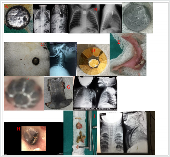
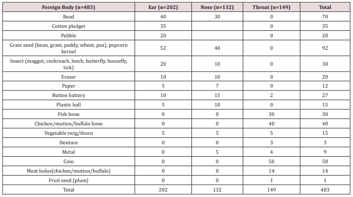
The number of FB (Table 1 & Figure 1) in ear was 202 (41.82%), 132
(27.32%) in nose and 149 (30.82%) in the throat. Out of 202 FB in ear,
54 (26.73%) was animate and 148 (73.26%) inanimate. Out of 132 nasal FB,
10 (7.57%) was animate and 122 (92.42%) was inanimate. The FB
encountered in throat was entirely of inanimate nature.
Figure 1. Few examples of ENT foreign bodies. A) FB toy battery in esophagus and removed with rigid esophagoscope, B) FB
coin in esophagus, C) FB button battery in esophagus, D) FB bead removed from ear, E) FB denture, F) FB bead removed from
nose, G) FB metal hook in esophagus, H) FB insect in ear and I) FB chicken bone with meat bolus in esophagus.
FB in Ear
Total of 202 patients were recorded with FBs in the ear. The
most common type of FB in the ear was grain seed 25.74% (52) followed by
FB bead 19.8% (40). Fifty-four (26.73%) patients had animate (living)
FBs. These were mostly insects in the form of maggots, cockroach,
grasshopper, butterfly, housefly and ticks. Hundred and forty-eight
(73.26%) patients had inanimate (nonliving) FBs. Out of 148 patients,
fifty-two (35.13%) had hygroscopic FBs in the form grain seed in the
form of bean, pea, wheat, paddy and gram as shown in Table 1. The rest
ninety-six (64.86%) patients had non-hygroscopic FBs in the form of
bead, cotton pledget, pebble, eraser, paper, button batteries, plastic
ball and vegetable twig or thorn. Majority of cases were seen in 0-10
years age group i.e. 72% (147). The most common site of FB lodgement was
found to be the external auditory canal. Most of these FBs were removed
in the OPD or in the ER with or without local anesthesia (LA). In 4
children, the FB were found impacted in the deeper part of EAC (3 button
batteries and 1 plastic ball) and had to remove under GA via post-aural
approach.
FB in the Nose
Hundred and thirty-two (27.32%) had FB lodgement in the nose.
The most common was grain seed 40 (Out of it, only 10 (7.57%) patients
had living FBs i.e., 9 had maggots and 1 had leech. The rest 122
(92.42%) patients had nonliving FBs as shown in Table 1. Forty FB
(30.3%) were of hygroscopic nature in the form of grain seed and the
rest sixty nine percent being non-hygroscopic as in Table 1. Of the
total number of 132 patients 122 patients (92.42%) were children and the
rest 10 (7.57%) patients were adults with animate type of FBs. Hundred
and five (86.06%) of the children presented with history of FB insertion
nose by their caretakers, while in 17 (12.87%) children neither the
patients nor the caretakers were certain of FB insertion. Unknowingly,
they were treated as a case of sinusitis due to complaints of nasal
blockage, headache and unilateral fetid discharge by the pediatricians
and primary care physicians, which was later, referred to our center and
revealed to be forgotten FB. Sometimes, even one has to depend on
imaging like x-rays /CT scans to rule out the FBs where the patients are
unable to recall the events. Otherwise, most of the times the typical
history provides clue for clinching the diagnosis. Most of the FB were
removed in the ER and OPD with or without LA and only 3 cases (2.27%)
required removal under GA i.e. 2 cases of beads and 1 case of grain seed
which on manipulation went posteriorly and also patient being
uncooperative.
Foreign Bodies in the Throat
A total of 149 patients presented with the complaint of
ingestion of FB. The most common type of FB was coin of one rupee, 2, 5
and 10 rupees and the common site of the impaction was cricopharyngeal
junction in all the 50 patients (33.55%). The sites of other types of FB
impaction were oral cavity, oropharynx, hypopharynx, thoracic esophagus
and lower gastro-esophageal sphincter region. All the ingested FB were
inanimate, with 84 (56.37%) being organic and 65 (43.62%) being
inorganic. Organic FBs were meat bolus and bone (fish, chicken, mutton,
and buffalo meat) and one of plum seed. The inorganic FBs included
button battery, thorn, denture, coin, and metallic objects as shown in
Table 1. Age less than 10 years old were the most common group with FB
coin. FBs fish bone and vegetable twig/thorn lodged in oral cavity and
oropharynx were removed under LA. Out of 40 FB coin, 10 were dislodged
spontaneously via gastrointestinal route, 5 FB meat bolus and 7 FB
chicken bone were removed by flexible endoscopy and the rest of the FBs
were removed under GA without postoperative complications.
Table 1. Different types of Foreign bodies (FB) in ENT.
Discussion
In our study, we’ve found higher incidence of FB in children
less than 10 years old in 66.04% (319) patients. This is consistent with
study by Iseh and Yahya [5], Ogunleye AOA and Sogebi R [6], Ahmad M, et
al. [7]. This may be consequent of children’s exploring habit and
lodging objects into the natural orifices of body, accidentally or
intentionally. We found 59.42% of patients to be male and 40.5% to be
female with male: female ratio 1.46:1. The male: female ratio was shown
to be 1:1.05 by Gregogri et al. [8] whereas it was 1:1.26 in the study
of Ogunleye AO et al. [9] and 1.35:1 by Agrawal S, Ranjit A study [10].
This suggests male are more susceptible than female to foreign body
insertion in the orifices. In this study we observed ears were the most
common site of lodgement of foreign bodies (41.82%) followed by throat
(27.32%) and nose (27.32%). Parajuli R [11] and Shrestha I, et al. [4]
also found in their study ears as the most common site for impaction of
foreign bodies followed by throat and nose. The most common foreign
body in the ear and nose was the variety of grain seeds like bean, pea,
paddy, wheat, gram, maize and foreign body coin was highest in throat.
Removal methods, most commonly used for ear, nose and throat FBs were
similar to those presented by Parajuli R [11], in order of preference
the alligator forceps, Jobson Horne probe, foreign body hook, Tilley’s
forceps and ear syringing. No patient required endoscopy or indirect
laryngoscopy to remove oropharyngeal FB. The need for general anesthesia
to remove FB varies in literature, with percentages varying from 8.6%
to 30% [12]. There were no complications reported post FB removal.
Conclusion
FB in ENT are common in both pediatric and adult population.
Comparatively the children are seen to be more vulnerable to have ENT FB
lodgement. Significant complications may arise if FB in ENT are not
taken care of immediately and skillful removal is must. Thus, proper
care and watch must be provided by care takers or the family members in
order to prevent the encounter of such objects, especially in pediatric
group.
Dental technology that depended on the standardized lost-wax
casting technology has been greatly improved with the
introduction of dental CAD ⁄ CAM systems. The aim of the present study
was to compare between the color changes of CAD/CAM
acrylic and manually performed acrylic bridges used for pediatric
patients. Forty study casts of children aged 2 - 4 years old of both
genders, with prematurely lost one of the maxillary central incisors and
the adjacent lateral incisor was carious and considered to
be abutment tooth were involved in this study for construction of
cantilever bridges. For each cast, two bridges were constructed;
1st one is CAD/CAM acrylic bridge and the other one is manually
performed acrylic bridge. After immersing the bridges in saturated
chocolate solution for different time intervals, color changes of the
bridges were measured using 3Shape scanner system.
Trauma and/or dental caries is the common causes those
result in premature loss of teeth in children. Cosmetic/aesthetic
restoration of such condition considers to be challenging in the
pediatric dental field. In case of premature tooth loss in anterior
incisal segment there will result in arch space loss and teeth’s
inclination that causing a collapse of the anterior teeth and
midline shifting [1], as well as may lead to parafunctional habits
[2]. Mahmoud (2009) found that anterior tooth loss had effect on
patient’s quality of life and gave negative effects on him/his [3]. Al
Rawi (2017) found that placement of cantilever acrylic bridges for
restoring the aesthetic dental appearance of preschool children
resulted in positive successes both to the child and parents [4].
Extrinsic discoloration of teeth and oral prostheses is stains caused
by foods or beverages. In pediatric patients such stain mostly
occurred due to colored foods such as beets or chocolate as well as
berries and candies [5, 6]. This study considered to be the first step
of our series studies deal with determining different physical and
mechanical properties of the prostheses used for pediatric patients
we planned to carry out (in vitro and in vivo studies). Starting
with the present study that aimed to compare between the color
changes of CAD/CAM and manually performed acrylic bridges used
for pediatric patients. After immersing the bridges in chocolate
solution for different time intervals, color changes of the bridges
were measured using 3Shape scanner system.
Material and Methods
This study starting with collection of forty study casts of children
aged 2-4 years old of both gender, with prematurely lost one of
the maxillary central incisors and the adjacent lateral incisor was
carious and considered to be abutment tooth were involved in this
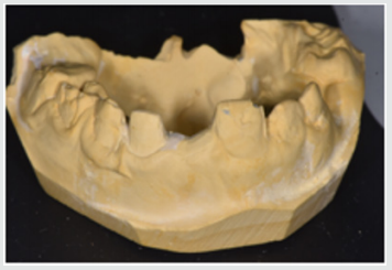
study for construction of cantilever bridges (Figure 1). For each cast,
two bridges were constructed; 1st one is CAD/CAM acrylic bridge
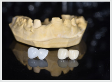
and the other one is manually performed acrylic bridge (Figure 2).
Construction CAD/CAM bridge: The cast was 3D scanned by special
scanner (710 3D) (smart optics Sensortechnik GmbH, Germany).
The design of the bridge was carried out using Exocad Program
(smart optics Sensortechnik GmbH, Germany). Acrylic block
(Poly-methyl methacrylate) of classic shade A1 (Ivoclar vivadent,
Switzerland) was used for fabrication of the bridge using CAD/
CAM machine (Charly dental, ZI Fonlabour, France). The bridge
was finished and polished very well [4]. Construction of manually
acrylic bridge: Wax pattern was fabricated on cast then followed
the technique of typical wax loss; the heat-cure acrylic (Ivoclar
vivadent, AG, FL-9494 Schaan/Liechtenstein) of classic shade A1 was used for bridge fabrication. Finally, surface finishing and
polishing was done [7]. Saturated chocolate solution was prepared
using 15g chocolate powder (MacChocolate TM, Malaysia) with
100ml distilled water. Baseline color readings for acrylic bridges
were taken then immersed in chocolate solution for different time
intervals (one week and two weeks) and maintained in incubator
of 37 °C, Fresh chocolate solution was prepared every day. Before
color measurements after one week and two weeks’ time intervals,
the bridges were rinsed with distilled water for 30 seconds, cleaned
with a soft bristle toothbrush and then dried with tissue paper [8].
Color measurement was carried out in the facial surfaces at the
center third of the abutment and the center third of the pontic part
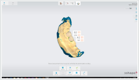
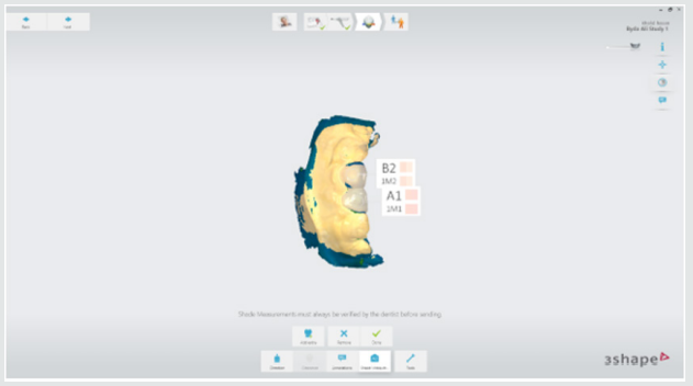
of each bridge as shown in Figure 3. Color measurements of the
bridges were measured using 3Shape scanner system (3 Shape A/S,
Holmens Kanal 7.1060 Copenhagen K Denmark) and according to
the software program of the system, Classic shade (Ivoclar vivadent,
Switzerland) was depended.
Figure 1: One of the study casts involved in this study
Figure 2: One of the study casts involved in this study
Figure 3: Demonstrated the color shade measurement of the abutment and pontic portions of the acrylic bridge.
Results
Table 1 demonstrated the color shade of all samples at the
baseline and after one-week and two weeks-time intervals. The
results of the present study revealed that for all samples, the color
measurement demonstrated that in CAD/CAM group even with
using A1 shade acrylic block but at the baseline measurement the
abutment revealed A0 shade while the pontic revealed A1 shade.
Meanwhile, in manual group the abutment measured to be A1 shade
and the pontic gave B1 shade. The results demonstrated that for all
samples there were no changes in the color shade of CAD/CAM and
manually fabricated acrylic bridges after one-week time interval,
meanwhile, there were significantly color changes of all abutment
and pontic portions of all samples of both bridge types after two
weeks-time interval immersed in chocolate solution (Figure 4 & 5).
Figure 4: color shade measurement of CAD/CAM acrylic bridge after two weeks.
Figure 5: Color shade measurement of manual acrylic bridge after two weeks.
Table 1: Demonstrated the color shade of all samples at the baseline and after one-week and two weeks-time intervals.
Discussion
Restorations in the oral cavity are exposed to several factors
that make them vulnerable to color changes, such as temperature,
humidity, food and beverages. In the oral environment, restorative
materials are also subjected to numerous other liquids, to
temperature and load stress, and to tooth brushing. The success
of restorations depends not only on mechanical and physical
properties, but also on the esthetic appearance [9]. The color
measurement in this study demonstrated that in CAD/CAM
group even with using A1 shade acrylic block but at the baseline
measurement the abutment revealed A0 shade while the pontic
revealed A1 shade. These occurred because the thickness of the
abutment was only about 0.5mm lead to that the color measured
of the abutment was lighter than the pontic portion. Meanwhile,
in manual group the abutment measured to be A1 shade and the
pontic gave B1 shade. These results agreed with other studies those
found the thickness of the material significantly affected the color
shade of the prostheses [10,11].
The results demonstrated that the color changes demonstrated
only after two weeks-time intervals immersed in chocolate solution.
Even the color shades recorded in the CAD/CAM group considered
to be lighter than in manual fabricated group, the discoloration
from chocolate solution was probably due to adsorption of color
colorant of chocolate solution at the surface of the prostheses.
The CAD/CAM bridges fabricated from blocks of pre-polymerized
acrylic resin those had a hydrophobic surface that repels water
[12]. As well as, perfect polishing surfaces of the bridges involved
in this study revealed the limited discoloration that occurred
agreed with other research [13]. As the duration of immersion
increased, the color change values of both types of prostheses were
recorded by 3Shape scanner system. Thus, the time is considered
to be important factor in the staining of the dental prostheses and
these results agreed with others [14,15]. Fabrication of dental
prostheses with the help of CAD/ CAM technology is related to the
advantages of high-density polymers based on highly cross linked
polymethylmethacrylate [16]. Those advantages include; good
esthetic, low water solubility and absorption, sufficient strength,
low toxicity, easy repair with simple fabrication technique [17]. The
using of hot cure acrylic for fabrication of dental prostheses even
of some advantages but the main disadvantages include porosity
with the presence of residual monomer which is a potential
allergen, increased finishing time, brittle and uneven thickness
[18]. A limitation of this study is that it was an in vitro study and
need to be collected with in vivo study to measure the degree of
color changes of the prostheses with presenting the effect of saliva
and oral hygiene measures. Further clinical and in vitro studies are
necessary to evaluate the susceptibility of CAD/CAM and manually
acrylic bridges to discoloration by other beverages and nutrients.
Conclusion
Color considered as the most important factors for aesthetic
appearance of dental restorations. In addition to the optimal chemo
mechanical properties of acrylic resins, their availability in different
color-shades has increased their application in fixed and removable
prostheses. Acrylic resins can have acquired discoloration over time
because of the process of adsorption and liquid molecules adhere
to resin materials which was decreased their effect with using of
CAD/CAM technology over conventional methods of acrylic resin
prostheses fabrication.
Lupine Publishers| Orthopedics and Sports Medicine: open access
journal
Abstract
Purpose:We are presenting this pattern of a rare variant of a clavicle malunion with an apex posterior-inferior deformity. This occurred in an elite major junior hockey player during his draft season. This illustrates that such a deformity will most likely result in shoulder weakness, altered shoulder mechanics and may cause brachial plexus neurological findings. In addition, this can cause associated sterno-clavicular deformity which can lead to sternoclavicular joint subluxation secondary to the increased strain placed on the sternoclavicular joint from an apex posterior inferior malunited clavicle. Deformity of > 20 degrees in any direction interferes with normal motion and normal cortical strength even in a young patient.
Introduction:Symptomatic malunion is fortunately less frequently observed (4) since the significant shift to operative treatment for displaced shortened mid shaft clavicle fractures. Symptomatic patients are typically those with marked displacement and significant shortening at the fracture site. Patient’s report weakness of the involved shoulder with rapid fatigability plus an increased deformity comes with an increased risk of recurrent fractures. Although not commonly described in the literature, clavicle malunion usually has a very consistent deformity pattern. As illustrated by McKee et al, the patient usually presents with a complex three dimensional deformity with shortening, an anterior apex at the fracture site and associated joint pain around the shoulder or sternum (6). The influence of the coraco-clavicular and a cromio-clavicular ligaments on the fracture fragments is hypothesized to cause an effect on the displacement of these fractures which involves the lateral segment of the clavicle being carried forward by virtue of its retained a cromio-clavicular and residual coraco-clavicular attachments. Angulations are more acute the closer the break is to these pivot points. This has had associated significant alteration in normal clavico-scapular motion.
Method:Case report and literature review.
Conclusion:Symptomatic clavicular malunion is rare but definitely higher with non-operative management and can cause discomfort and shoulder weakness. Neurological symptoms and signs are more likely to occur in inferior malunited clavicle, particularly with an inferior-posterior deformity. We illustrated the steps necessary to correct all deformities and lengthen the clavicle using a long working length precountored plate construct. This has improved the clinical symptoms of the patient and illuminated the risk of repeat fracture due to deformity. Plate removal is planned but is still an unanswered question.
An 18 year old elite Canadian Hockey player presented with a new fracture to his left clavicle and associated pain at the sternoclavicular joint with an obvious deformity. He had sustained a previous injury to his left mid shaft clavicle two years ago playing hockey. This was treated on operatively and went on to heal with a 25 degree posterior-inferior deformity. A review of his initial injury films, from two years ago, illustrated a moderately displaced mid shaft clavicle with a significant amount of shortening (2 cm)due to inferior apex deformity( 25 degrees).However, it was decided to treat him on operatively as it was a closed injury in a relatively young male and he was neurovascularlyintact. His fracture healed with 2.5 cm of shortening, slight scapular inward rotation and a 25-30 degree posterior-inferior deformity. The sternoclavicular joint deformity on the left side stopped him from playing hockey at an elite level for about two months but a steroid injection seemed to remove most of his symptoms and allowed him to compete. He also complained of an ongoing occasional shoulder weakness and an occasional fleeting numbness in his arm and hand. This was significant enough to warrant a CT of the chest to rule out thoracic outlet syndrome.n 2010, the Czech Republic participated in the World Health OrgAn 18 year old elite Canadian Hockey player presented with a new fracture to his left clavicle and associated pain at the sternoclavicular joint with an obvious deformity. He had sustained a previous injury to his left mid shaft clavicle two years ago playing hockey. This was treated on operatively and went on to heal with a 25 degree posterior-inferior deformity. A review of his initial injury films, from two years ago, illustrated a moderately displaced mid shaft clavicle with a significant amount of shortening (2 cm)due to inferior apex deformity( 25 degrees).However, it was decided to treat him on operatively as it was a closed injury in a relatively young male and he was neurovascularlyintact. His fracture healed with 2.5 cm of shortening, slight scapular inward rotation and a 25-30 degree posterior-inferior deformity. The sternoclavicular joint deformity on the left side stopped him from playing hockey at an elite level for about two months but a steroid injection seemed to remove most of his symptoms and allowed him to compete. He also complained of an ongoing occasional shoulder weakness and an occasional fleeting numbness in his arm and hand. This was significant enough to warrant a CT of the chest to rule out thoracic outlet syndrome.nization WHO Research to determine the quality of medical decAn 18 year old elite Canadian Hockey player presented with a new fracture to his left clavicle and associated pain at the sternoclavicular joint with an obvious deformity. He had sustained a previous injury to his left mid shaft clavicle two years ago playing hockey. This was treated on operatively and went on to heal with a 25 degree posterior-inferior deformity. A review of his initial injury films, from two years ago, illustrated a moderately displaced mid shaft clavicle with a significant amount of shortening (2 cm)due to inferior apex deformity( 25 degrees).However, it was decided to treat him on operatively as it was a closed injury in a relatively young male and he was neurovascularlyintact. His fracture healed with 2.5 cm of shortening, slight scapular inward rotation and a 25-30 degree posterior-inferior deformity. The sternoclavicular joint deformity on the left side stopped him from playing hockey at an elite level for about two months but a steroid injection seemed to remove most of his symptoms and allowed him to compete. He also complained of an ongoing occasional shoulder weakness and an occasional fleeting numbness in his arm and hand. This was significant enough to warrant a CT of the chest to rule out thoracic outlet syndrome.
This 18 year old male continued to play elite major junior hockey (prime pathway to the NHL in Canada) then unfortunately sustained another injury where he was checked into the boards during an elite hockey game. He felt immediate pain and tenderness along his clavicle and therefore presented to the hospital emergency. Interestingly, since his initial incident, he had never been free of symptoms and he subsequently fractured his clavicle with relatively low trauma within 18 months of his last fracture. Plus he had significant sterno-clavicular associated symptoms with pain and anterior subluxation of the ipsilateral sterno-clavicular joint
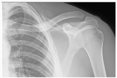
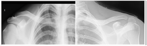
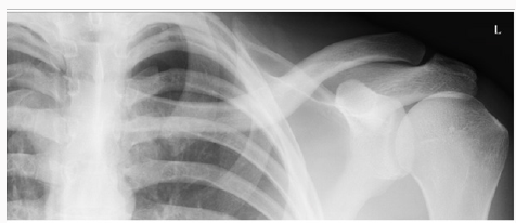
In the Emergency Department he was evaluated by the ER physician and the orthopaedic on call team. He had normal vital signs and good air entry bilateral chest, his neurological exam of both motor and sensory nerves of his left upper extremity showed no deficit, no signs of thoracic outlet syndrome and he illustrated a normal vascular exam. His investigation included x ray of his left clavicle with a contra lateral clavicle x ray for comparison. Both clavicles had an AP and orthogonal clavicular views (see images below). His clavicle demonstrated a more pronounced posterior-inferiorapex deformity (30-35 degrees), shortening and malrotation plus a significantly deformed (anterior subluxation) sternoclavicular joint as noted over the last year.
A detailed discussion with the patient about the findings was complete along with the possible operative and non operative treatment modalities available. Given the latest research and paper by McKee et al on the increased fracture rate in significantly deformed clavicles, an operative approach was chosen. This choice was also enhanced by the history of increased discomfort generally around the shoulder girdle discomfort plus the significant shoulder weakness, sterno-clavicular pain, neurological symptoms and reduced maximal function. We, therefore, elected to book him for a corrective osteotomy to restore length, alignment, rotation and angulations to augment the mechanics of his shoulder and the biomechanical ability of this clavicle to absorb an impact without re-fracturing.
Operative Procedure
The patient underwent general anaesthesia and was placed in a beach chair position in a 45 degree semi sitting position with a small pad behind the left shoulder blade and the involved upper extremity was draped freely with the distal arm placed in a sterile extremity drape. An oblique incision was made along the superior surface of the clavicle to expose the nonunion site. The skin and subcutaneous tissue was raised as a flap, and the underlying myofascial planes identified. This layer was raised as contiguous flaps and was preserved so that a two-layered closure could subsequently be achieved. Next, the malunion site was identified, and a long oblique, superior to inferior, osteotomy was performed. This provided a long osteotomy surface to correct the inferior apex deformity while allowing for the three dimensional correction with excellent bone to bone contact.
The osteotomy was performed with a, well irrigated, cooled, micro sagital saw. After careful dissection a small blunt Haworth elevator was placed underneath the clavicle to protect the neurovascular structures during the osteotomy and elevation of the deformity. Very importantly, the medullar canal was re-established, on both sides of the osteotomy, with a 3.5-mm drill-bit plus very aggressive curettage of the sclerotic bone in order to obtain an excellent opening in the medullar canal in the proximal and distal segments.
However, we have a very novel solution in the Czech Republic - whetSmall reduction clamps were then utilized to perform a reduction that would allow lengthening of the clavicle along with rotational and ambulatory correction utilizing the precountored plate as a reduction tool. First, shortening was corrected and held by translating the medial lateral fragment over the large surface osteotomy area to gain the planned length of 2.5 cm based on our preoperative planning. This was accomplished almost entirely by deformity correction. Secondly, rotation was corrected by rotating the lateral fragment about forty degrees clockwise until the flat surface of the lateral fragment was facing superior as desired. We then placed a long 10 whole precountored clavicle plate on the superior surface of the clavicle using the construct, with its long working length, to help gradually realign the bone back to the plate. This was and should be done very slowly and carefully as the underlying neurovascular structures can be tethered to the deformed bone. This was then held using absolute stability fixation with non locking screws on each side of the osteotomy. The screws were then gradually tightening of screws on either side of the deformity.r you are an individual patient crippled and dying for legal or iSmall reduction clamps were then utilized to perform a reduction that would allow lengthening of the clavicle along with rotational and ambulatory correction utilizing the precountored plate as a reduction tool. First, shortening was corrected and held by translating the medial lateral fragment over the large surface osteotomy area to gain the planned length of 2.5 cm based on our preoperative planning. This was accomplished almost entirely by deformity correction. Secondly, rotation was corrected by rotating the lateral fragment about forty degrees clockwise until the flat surface of the lateral fragment was facing superior as desired. We then placed a long 10 whole precountored clavicle plate on the superior surface of the clavicle using the construct, with its long working length, to help gradually realign the bone back to the plate. This was and should be done very slowly and carefully as the underlying neurovascular structures can be tethered to the deformed bone. This was then held using absolute stability fixation with non locking screws on each side of the osteotomy. The screws were then gradually tightening of screws on either side of the deformity.
Intra operatively, significant improvement in the shoulder contour was obvious as well as a noticeable reduction in the anterior subluxation of the sternoclavicular joint. Screw length was checked with an image at the end of the procedure. Deformity correction usually necessitates some screw changes as the initial screws can be long once the deformity is reduced. Wound closure was done in layers closing the myofascial flap over the plate and subsequently the subcutaneous tissue and the skin was re approximated with narrow skin staples.
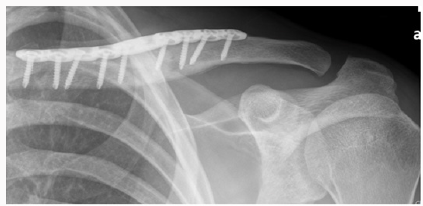
Post operatively the patient was placed in a shoulder sling for comfort and scheduled for early physio to initiate shoulder and elbow function. His post op exam confirmed intact neurovascular status of his left upper extremity. Chest x ray taken in recovery room confirmed we had not created a pneumothorax. The operative procedure was performed as an outpatient. The patient went home on the same day and returned at 10 days for wound examination and staple removal. Aggressive physio was initiated that day following the initial gentle ROM and pendulum exercises which were initiated immediately post op (Figures 1-9).
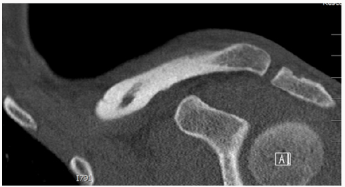
Figure 1:Axial CAT scan of the chest delineating the sternoclavicular deformity related to the clavicle malunion.
Figure 2:Coronal CT showing the direction of malunited clavicle.
Figure 3:
Figure 4:(a) Comparison right (normal)(b) Left (Malunited) clavicle
Figure 5:
Figure 6:
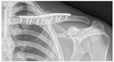
Figure 7:Early post operative.
Figure 8:
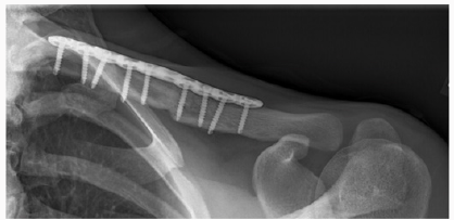
Figure 9:Three months post-operative (signs of radiographic healing).
Discussion
Clavicles fractures are common injuries and are reported to represent 2% to 5% of all adult fractures [1]. More recent evidence suggests that specific subsets of patients may be at higher risk for nonunion, symptomatic malunion, or suboptimal functional outcomes [2]. A recent meta-analysis suggests that the incidence of clavicle nonunion after nonsurgical treatment is approximately 5.9%, but can be as high as 15%for some fracture subtypes [3]. Nonsurgical treatment universally results in some degree of malunion; however, symptomatic malunion is fortunately quite low and is usually used particularly in very young patients [4]. Symptomatic patients are typically those with marked displacement at the fracture site, with shortening of >2 cm. Patients that are symptomatic may report weakness of the involved shoulder, rapid fatigability, numbness and paresthesia of the hand and forearm with elevation of the limb, and an asymmetric, “droopy,” “ptotic,” or “driven in”shoulder [5].
McKee et al performed a review of a cohort of patients to analyze the functional results of corrective osteotomy of a mal united clavicular fracture in patients with chronic pain, weakness, neurologic symptoms, and dissatisfaction with the appearance of the shoulder. Fifteen patients (nine men and six women with a mean age of thirty-seven years) who had amalunion following non operative treatment of a displaced mid shaft fracture of the clavicle were reviewed both preoperatively and postoperatively. The mean time from the injury to presentation was three years (range, one to fifteen years).Follow-up, at a mean of twenty months (range, twelve to forty-two months) postoperatively, illustrated that the osteotomy site had united in fourteen of the fifteen patients. All fourteen patients expressed satisfaction with the result. There was one nonunion, and two patients had elective removal of their plates. With regards to the patho anatomy of the deformed clavicle, McKee et al. noted that the deformity of the clavicle was a complex three-dimensional problem with all their patients illustrating a superior-anterior apex deformity. In his series there were certain consistent features seen in patients who presented with symptoms following non operative treatment and a healed clavicle. The hall mark characteristic is shortening in the medial-lateral dimension, with inferior displacement of the distal fragment and superior displacement of the proximal fragment. They, therefore, concluded that the shortening in the medial-lateral plane had a negative effect on muscle-tendon tension, and muscle balance. The anatomic boundaries of neurovascular structures were of paramount importance in the development of symptoms [6].
In a study by Edelson et al, he studied the bony anatomic details in 73 cadaver specimens which had clavicle malunions in different regions of the clavicle. According to the Allman classification. Edelson found that in the middle-third fractures, similar anterior angulations to the lateral third fracture malunion was indeed present. The most consistent finding at the middle-third level was that the lateral shaft fragment was almost invariably displaced posterior to the medial shaft fragment. The author also commented that initial anterior-posterior radiographs of clavicle fractures are often dominated by inferior displacement or ptosis of the lateral fragment. However, in the cadaveric specimens, anterior angulations rather than drooping of the lateral fragment were the predominant deformity. Although often initially displaced in a down ward direction, the lateral fragment does not usually heal in this position, unless it is a greenstick fracture as occurred in our patient.
Therefore the literature concludes that the principle deformity in a healed malunionis anterior, superior angulations. In this series there were only 4 cases in which the lateral clavicle healed with downward angulations of 20° or more at the fracture site as occurred in our young patient with his greenstick type of fracture. The author hypothesized that inferior displacement of the lateral fragment, which predominates on the initial radiographs, is most likely due to post-traumatic muscle a tony, principally of the deltoid and trapezius, similar to that which can cause the glenohumeral joint to appear subluxed after fractures of the humeral head and claimed that as soon as the muscle tonus returns, the clavicle resumes a horizontal orientation, and fracture position is then dominated by the pronators and internal rotators of the scapula and upper arm, which reposition the fragments into the anteriorsuperior apex position [7].
We believe that corrective osteotomy can lead to predictably good results (> 95%), however one should be careful with the inferior dissection as it can and has produced neurological and vascular issues in the past. So which fracture requires surgical correction? In general principles, according to the Canadian Orthopedic Trauma Society (COTS)and the McKee et al papers, “symptomatic deformity” with significant shortening of 2-3 cm , angulations deformity >30 degree or translation of >1cm . This has been supported in numerous repeated studies since 2008. In addition softer indications would be symptoms of thoracic outlet syndrome, weakness or rapid fatigability with overhead activity, a relatively weak arm at over a year from the fracture or more commonly a combination of all of these symptoms, should be considered for an operative correction [6].
Another area of controversy between surgeons who treat this type of injury is the need for hardware removal to decrease the risk of re-fracture. Some surgeons prefer to remove the implant in all patients after clavicle fracture union, whereas others plan for additional surgery only if the patient complains of symptomatic hardware. In either case, adolescent patients undergoing surgical fixation for clavicle fracture must be warned of the possibility of return to the operating room to remove the implant.
Conclusion
Malunion of the clavicle with > 20 to 30 degrees of deformity and symptoms of weakness and malfunction should be considered for corrective osteotomy. The success rate is very high (.95%) and results in excellent patient satisfaction. This again supports McKee’s initial study that highlighted the clinical impact of mid shaft clavicle deformity and the importance of surgical reconstruction with an absolute stability. We also believe that if a surgeon carefully follows the steps of the surgical technique described in this case report; the incidence of vascular and neurological injuries can be mitigated although not entirely illuminated as a risk.
Lupine Publishers | Journal of Otolaryngology Research Impact Factor
Opinion
pinion
This editorial is based on my personal professional experiences
of some four decades work in health and business applications
of statistics. I am fully aware that these experiences cannot be
interpreted as a random sample and there is no warranty of any
kind for the absence of possible biases. You might well be familiar
with the US-FDA view that there is no unbiased data in the scientific
universe available. Historically, the use of statistics in medicine is
a well-documented fact since about two centuries. My personal
experiences cover the transition period from electronic desktop
calculators with paper and pencil, to the omnipresence of cheap
computing power and sophisticated statistical software of today.
I remember my early professional study design activities as a
statistician as heavily impacted by cost considerations: Human
work time was and still is quite expensive. During the last period
of about some two decades I got the strong impression that the
medical profession, especially those doctors who are working
scientifically – showed a quite strong trend to higher statistical
understanding and knowledge as compared to my early working
times. This positive trend is highly appreciated by me and I see it
as an enormous economic advantage that computers took over
the tedious workload of numbers crunching nowadays. There
is a negative trend involved over the last two decades as well: I
observed a growing trend of numbers of “so called” statisticians
who actually are experts in using available statistical software
packages only but have little or no statistical expertise. I see a
professional statistician as a human who understands the primary
objective of the study’s objective and to assess the medical question
under consideration and to decide the statistical model selection
for the very question based on the scope of the scientific question
and all of the constraints and limitations in practice. I know that
this is an “ideal world” assumption and we all are but humans with
our limitations. My experience that it pays to strive for perfection
is illustrated by some examples which I consider to be potentially
useful and beneficial for your work as doctor:
Project Planning Phase
a) Overall considerations of project management and quality
control, legal requirements.
b) Definition of research question(s).
c) Definition of data selection criteria, sampling variables
and observation time(s) schedule.
d) Aspects of data collection and documentation, ongoing
project quality controls.
e) Feasibility considerations of various project designs.
f) Administrative aspects: financials, selection of partners,
estimation of required realization times.
g) Statistical methods for data analysis, feasibility of pilot-
(sub)-projects.
h) Final definition of data selection criteria.
i) Aspects of results publication.
j) Aspects of possibly necessary actions in case of
emergencies.
Project Realization Phase
a) Ongoing control of “plan vs actual” progress.
b) Ongoing communication between all project partners.
c) Maintenance of open minds for early signs of project’s
critical developments.
d) Ongoing monitoring for possible emergency actions.
e) The KISS principle: Keep everything as simple and stupid
as possible.
I’d like to recommend using the items in the above project
management as guidance for your project but under no condition
as a comprehensive cookbook! As ENT scientist you should
permanently remember that you are doing research in humans and
not in bolts and nuts!
How you could easily detect the difference between a
“user of statistical software” or a “real” statistician
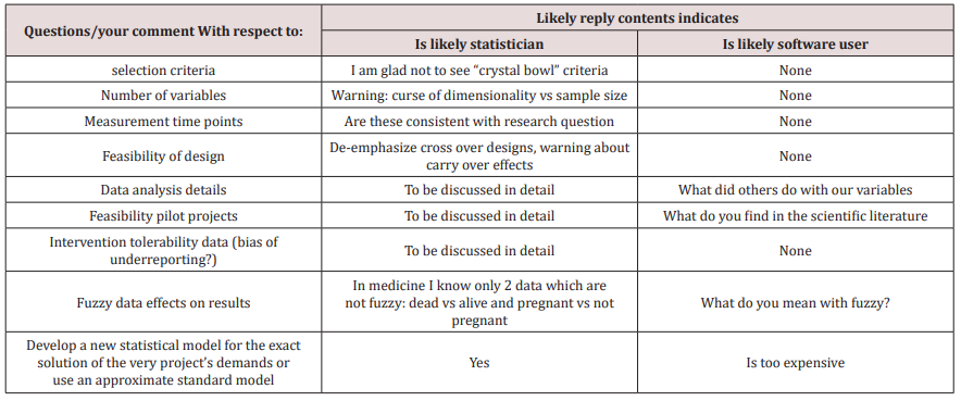
The next table provides you with selected questions/topics to
distinguish between professional statistician and software user
(Table 1).