Wednesday, 15 September 2021

Lupine Publishers| Comparison between the Second Zagreb Eccentricity Index and Eccentric Connectivity Index of Graphs

 Lupine Publishers| Current Trends in Computer Sciences & Applications (CTCSA)


Abstract

For a graph G, the second Zagreb eccentricity index E2(G) and eccentric connectivity index ∈c(G) are two eccentricity-based invariants of graph G. In this paper we prove some results on the comparison between and of connected graphs G of order n and with m edges.

The authors demonstrated how a combination of both techniques and human interventions enhances control, decision-making and data analysis systems.

Keywords: Graph; Eccentricity (of vertex); Second Zagreb eccentricity index; Eccentric connectivity index

Introduction

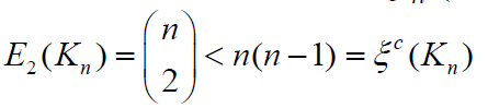
Throughout this paper we only consider the note, undirected, simple and connected graphs. The degree of v∈ V(G), denoted by degG(v), is the number of vertices in G adjacent to v. For any two vertices u; v in a graph G, the distance between them, denoted by dG(u; v), is the length of a shortest path connecting them in G. As usual, let Sn, Pn, Cn, Kn be the star graph, path graph, cycle graph and complete graph, respectively, on n vertices. Other undefined notations and terminology on the graph theory can be found in [1]. For any vertex of graph G, the eccentricity ∈G (v) (or ∈(v) for short) is the maximum distance from v to other vertices of G, i.e., ∈G (v)= maxu≠v dG(u,v). The eccentricity of a vertex is an important parameter in pure graph theory. The radius of a graph G is denoted by r(G) and defined by . Also, the diameter of G, denoted by d(G), is the maximum distance between vertices of a graph G and hence . A vertex v with ∈G(v)= r(G) is called a central vertex in G. A graph G with d(G) = r(G) is called a self-centered graph. A graph which contains only two non-central vertices is called almost self-centered graph [2] (ASC graph for short). Moreover, the eccentricity is also applied in chemical graph theory. There are several eccentricity-based topological indices, including the second Zagreb eccentricity index E2(G) [3] and eccentric connectivity index ∈c (G) [4], of graphs G where



In particular, we have or any graph G. In this paper we prove some comparison results between and of connected graphs G of order n with m edges. Main results In this we prove several results on the comparison between and of graphs G. Firstly we present two useful lemmas.

Lemma 2.1: [5] Let G be a connected graph of order n with maximum degree Ξ” . If Ξ”= n −1 then E2(G) =ΞΎc(G) .Otherwise, E2(G) ≥ΞΎc(G)with equality holds if and only if G is a 2-SC graph.

Lemma 2.1: [6] If u and v are two adjacent vertices of a connected graph G, then ∈(𝒰)−∈(𝒱) |≤1.

Denote by Gn(m; d) the set of connected graphs of order n with m edges and diameter d.

Theorem 2.3. Let G∈ΞΆ(𝓂,𝒹) with n>5 and 𝒹≤2. Then <. Proof. If d = 1, G∈ΞΆ(𝓂,𝒹) contains a single graph Kn with and . Then our result follows. Next it suffices to consider the case when d = 2. If G has maximum degree Ξ” = n −1by Lemma 2.1, we have E2(G) <ΞΎc(G) for any graph G∈ΞΆ(𝓂,𝒹) .Moreover, we have 𝓂≥𝓃−1 If 𝓂=n-1, then G≅Sn with for any n ≥ 5. Moreover,<holds clearly form ≥ n. If Ξ” ≤ n − 2 then G is a 2-SC graph. By Lemma 2.2, G is never a tree. Therefore m ≥ n with equality holding if and only if G ≅ C4 or G ≅ C5 . Consider that n > 5, m > n holds immediately. It follows that . This completes the proof of the theorem.

In the following we consider the graphs G∈ΞΆ(𝓂,𝒹) with diameter d ≥ 3.

Theorem 2.4: Let G∈ΞΆ(𝓂,𝒹). with d ≥ 3, n > 5 be a tree or a unicyclic graph. Then >Proof. If d ≥ 3, then Ξ”(G) ≤ n − 2 . From Lemma 2:1, we have E2(G) <ΞΎc(G). Note that m ≥ n for any tree or unicyclic graph G. Thus, it follows . finishing the proof of the theorem. Next we consider the case
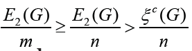
m > n. In the following theorem we give a sufficient condition for the graph G of order n with ≥ .

Theorem 2.5. Let G∈ΞΆ(𝓂,𝒹) with d ≥ 3, m = n + t and . If r(G) ≥ 3, then ≥ ,

Proof. Making a difference, we have

Set Ξ”1= nE2(G)−(n + t)ΞΎc(G) . From Lemma 2.2, we have

Since r(G) ≥3 and , we have

Therefore, Ξ”1 ≥ 0 with equality holding if and only if ∈(π“Š) = 3for each vertex π“Š∈V(G) that is, G is a self-centered graph with radius 3. This completes the proof of the theorem.

F or, G∈ΞΆ(𝓂,𝒹) with d ≥ 3, r = 2 and considering that r(G)≤d(G)≤2r(G) we have d(G) = 3 or d(G) = 4. In this case, the value of Ξ”1 may be negative, zero or positive. Let

Denote by mi the cardinality of i∈{1, 2,3, 4,5}. Then

In the following result we present some comparison results for ASC graphs.

Theorem 2.6: Let G∈ΞΆ(𝓂,𝒹) with d = 3, r = 2, m = n + t, t ≥ 1 where .

If G is an ASC graph, then < Proof. If G is an ASC graph with d = 3, r = 2, from the structure of ASC graph, we have 𝓂3≤ 𝓃 − 2,𝓂3=0, that is, 1 𝓂 ≥ t + 2 . If 𝓃 ≤ 5t, clearly, we have Ξ”1 ≤ 0 .For n >

5t, we have

holds if and only if Note thatThus Ξ”1 < 0 is equivalent that with t ≥1. Therefore the result holds immediately. It is much interesting to search more generalized graphs G with different comparison results between and which can be a topic for further research in the future. 

Read More About  Lupine Publishers Current Trends in Computer Sciences & Applications (CTCSA) Please Click on Below Link: https://computer-sciences-lupine-publishers.blogspot.com/ 

Monday, 13 September 2021

Lupine Publishers| Anti Ulcer Activity of Leea Indica in Wistar Albino Rats

 Lupine Publishers| Archives of Nanomedicine Open Access Journal (ANOAJ)


Abstract

Objective: Leea indica is a well known plant with numerous pharmacological activities owing to the presence of the active constituents. Inspite of numerous therapeutic uses, efficacy of the plant in treating ulcers is not yet evaluated scientifically. Hence an attempt has been made to evaluate the Antiulcer activity of Leea indica.

Methods: The effect of Leea indica methanolic extract (LIME) on gastric ulcer in pylorus ligation-induced and aspirin induced models was studied by employing LIME at 200 mg/kg, 400 mg/kg. In both the models Ranitidine (40mg/kg) was employed as the standard. Depending on the model, parameters evaluated were total acidity, free acidity, volume and pH of gastric fluid, ulcer score and percent inhibition of ulcer index.

Results: Data were analyzed following graph pad instat version 3.0 followed by Dunnets multiple comparision test. LIME (400mg/kg) significantly (P<0.01) reduced gastric ulcer index in pylorus ligation-induced and Aspirin induced ulcer models comparable to that of Standard drug. Histopathological studies confirmed that LIME (400mg/kg) possess anti ulcer activity in both models. The elicited activity might be owing to the existence of secondary metabolites such as flavonoids, tannins, and saponins.

Conclusion: Obtained results elucidate the Antiulcer activity of Leea indica methanolic extract. Further investigations on isolation of specific phyto chemicals and elucidating mechanisms of action are needed.

Keywords:Antiulcer, Pylorus Ligation Model, Aspirin Induced Model, Histopathology, Leea Indica, Peptic Ulceration, Vitaceae, Anti Hyper Lipidaemic, Antitumor, Hydrocarbons

Mini Review

Peptic ulceration is one of the common modern age epidemics affecting nearly 10% of world population [1]. Even though numerous potent anti-ulcer drugs are marketed, most of them were associated with toxicities, thus emphasizing the essentiality in search for new alternatives. Approximately 80% of the world’s population trust on herbal-derived medicines reinforcing herbs was advantageous sources for new drugs. Leea indica a traditional Chinese medicine found to be distributed in India, Srilanka, Bangladesh, Burma, Nepal, Thailand, Laos, Camboida, China, Vietnam, New Guinea, Malasia, Solomon islands, North Australia, Santa Cruz island, New Hebrides and Fiji [2-4]. Leea indica, an evergreen large shrub of family vitaceae [5,6] grows up to 8mt. Leaflets are ovate-lanceolate with crenate to serrate margins. Leaves are 1-3 pinnate bearing 7 leaflets, with petioles 7-20cm long. Fruits are purplish black, bearing six seeds with 1 cm in diameter. Flowers are greenish white with 5mm across. Stems are glabrous to pubescent.

Leea indica leaves were scientifically evidenced for wide pharmacological activities like antitumor [7], antiviral [8], analgesic [9], sedative and anxiolytic [10], Nitric oxide inhibitory [11], phosphodiestrase inhibitory [12], antidiabetic [13]. Anti hyper lipidaemic [14]. Leaf decoction is employed in pregnancy and delivery for birth control and body pain [4,15]. Dried leaves ingested were effective against cancer [16]. Roasted leaves relieve vertigo. Leaves (31.4%) constitute to be the most widely used parts in medicine preparation then fruits and roots (16.05%) [17]. Therefore the current investigation was designed to screen the leaves for the antiulcer activity. Twenty three chemical constituents were detected in Leea indica leaves by Gc-Ms analysis, Spectroscopic techniques, and Co-TLC. Compounds identified were eleven hydrocarbons, palmitic acid, pthalic acid, 1-eicosanol,farnesol, soalnesol, gallic acid, three pthalic acid esters, lupeol, Ξ²-sitosterol and finally ursolic acid [18]. Though the plant was reported with numerous chemical constituents antiulcer activity was not yet screened. Hence the present study was taken up to investigate antiulcer activity of Leea indica.

Materials and Methods

Chemicals

Ranitidine(Lee Pharmaceuticals, Hyderabad.), Aspirin (Shalg pharmaceuticals, Goregaon, Mumbai.)

Plant

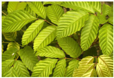
(Figure 1) Leea indica leaves were procured from Karthikavanam forest, Dhulapally, Hyderabad. These were authenticated by Dr. Madhavachetty, Professor, Sri Venkateshwara University, Tirupathi. The plant was placed in the college herbarium with a vocher specimen no. 438.

Figure 1: Leea indica leaves.

Lupinepublishers-openaccessjournals-Nanotechnology-nanomedicine

Animals

Male Wistar albino rats (200–250g) were purchased at Albino Research Center, Bachupally, Hyderabad. They were accomodatedin polypropylene cages with standard diet and water ad libitum. Experiment was designed as per ethical norms of CPCESA and IAEC Malla Reddy Institute of Pharmaceutical Sciences (1662/PO/a/12/ CPCSEA).

Extraction

Fresh leaves were shade dried, coarsely powdered and sieved with sieve no.40. Powdered material (500gm) was packed in soxhlet apparatus and subjected to extraction with methanol and water for about 48h. Solvents were evaporated under reduced pressure by rotary vacuum evaporator. Obtained methanolic extract (MELI) and aqueous extracts of Leea indica (AQLI) were subjected to phyto chemical screening and acute toxicity study [19].

Acute Toxicity Study

This study was performed as per OECD guideline 425 using limit test dose 2000mg/kg [20]. Rats were fasted overnight and a limit dose of 2000mg/kg was administered and observed for autonomic profiles (defecation and urination), behavioural profile (alertness, restlessness, irritability and fearfulness), neurologic profile (spontaneous activity, reactivity, touch response, pain response and gait), physical states such as lacrimation, loss of appetite, tremors, hair erection, salivation, diarrhoea and for morbidity or mortality continuously for 2 h, periodically during the first 24 h and daily thereafter, for a total of 14d.

Antiulcer Activity

Pylorus Ligation Method: Male Wistar albino rats were segregated into four groups comprising five each [21]. Group I negative control, received distilled water, Group II positive control, received ranitidine 50mg/kg; Group III LIME (200mg/kg, p.o); Group IV LIME (400mg/ kg, p.o). Plant extracts were administered for 10 d. At the end of experimental period rats were fasted overnight with water ad libitum. Standard, Ranitidine (40mg/kg) and LIME (200, 400mg/ kg) were administered. Under light ether anaesthesia, the abdomen was cut opened by incision below the xiphoid process. The Pylorus end of stomach was slightly lifted up and ligated avoiding damage to blood vessels. After completing ligation stomach was replaced and the abdominal wall is sutured and closed. Rats were deprived of water during post operative period for 3 to 4h [22]. Four hours later, stomach was dissected out. Stomach contents were drained into tubes and were subjected to centrifugation at 2000 r/m for 10 min. Supernatant obtained was analysed for gastric volume, pH, free acidity and total acidity. The stomach was incised along the greater curvature, and observed for ulcers and the ulcer index (UI) was calculated. Ulcers were scored and severity was assessed microscopically (10 X) employing hand lens (10 X). The scores were given as mentioned.

Read More About Lupine Publishers Archives of Nanomedicine Open Access Journal (ANOAJ) Please Click on Below Link: https://lupine-publishers-nano-science.blogspot.com

Saturday, 11 September 2021

Lupine Publishers | The Fetal Alcohol Spectrum Disorders: A Mini-Review

 Lupine Publishers | Journal of Gynaecology


Abstract

Since the first description of the fetal alcohol syndrome in 1973 the fetal alcohol spectrum disorders (FASDs) are still the leading preventable cause of birth defects,intellectual and neurodevelopmental disabilities. A recent WHO study estimates the global prevalence 7,7 per 1000 population,with large differences between countries.Renewed awareness results in new prevention and screening campaigning for this completely preventable global public health problem. In this mini-review, the various parts of FASD, as FAS, ARND and ARBD are discussed.

Introduction

The term fetal alcohol syndrome was first used in 1973 [1,2]. Fetal Alcohol spectrum disorders are caused by drinking alcoho during pregnancy [3,4]. Prenatal exposure to alcohol can damage the developing fetus and is the leading preventable cause of birth defects and intellectual and neurodevelopmental disabilities [1,3,4]. It presented as a cluster of birth defects[1]. Fetal alcohol spectrum disorders (FASDs) encompasses a range of possible diagnoses, including fetal alcohol syndrome (FAS), partial fetal alcohol syndrome (pFAS), alcohol-related neurodevelopmental disorder (ARND), alcohol related birth defects (ARBD) and neurobehavioral disorder associated with prenatal alcohol exposure (ND-PAE). FAS is a distinct clinical entity refering to a specific constellation of physical behavioral and cognitive abnormalities, resulting from prenatal alcohol exposure (PAE)-[1].

The lack of uniformly accepted diagnostic criteria for FAS and other related disorders has limited efforts to determine accurate prevalence figures. The World Health Organization (WHO) estimates the global prevalence to be 7,7 per 1000 population (95% CI; 4,9-11,7 per 1000 population). The WHO European Region had the highest prevalence (19,8 per 1000 population) and the WHO Eastern Meditarranean Region had the lowest (0,1 per 1000 population).South Africa was estimated to have the highest prevalence of FASD at 111,1 per 1000 population, followed by Croatia (53,3/1000 population)-[5].

Alcohol-related birth defects and developmental disabilities are completely preventable,when pregnant women abstain from alcohol use.Neurocognitive and behavioral problems resulting from prenatal exposure are lifelong.Early recognition,diagnosis, and therapy along the FASD spectrum can result in improved outcomes. There is no amount of alcohol intake to consider safe.There is no safe trimester to drink alcohol. All forms of alcohol pose similar risk and binge drinking poses dose-related related risk to the developing fetus [6]. There is a global need to establish an universal public health message about the potential harm of prenatal alcohol exposure and a routine screening protocol,according to the WHO [5]. Globally, nearly 10 percent of women drink alcohol during pregnancy [7]. In this mini-review the various parts of the fetal alcohol spectrum disorders will be discussed. The pathogenesis,pathophysiology and treatment are beyond the scope of this mini-review.

Fetal Alcohol Syndrome (FAS)

For the diagnosis FAS, patients must have at least one growth abnormality eg short stature,as well as all three characterisic facial abnormalities;short palpebral fissure length,a thin upper lip and a smooth philtrum.They must also have at least one diagnosed structural or functional abnormality of the central nervous system, eg microcephaly or impaired executive function. Confirmation of intrauterine exposure to alcohol is not obligatory for the diagnosis [8].

Growth abnormalities in FAS: The percentiles for weight are significantly lower in children with FAS at birth and in following outpatient consultations,,compared with children without FAS in a retrospective study of 322 FAS children. Moreover 22% had a body mass index below the third percentile compared with 3% of those withoit FAS[9]. Day et al. (cohort study,n=580) found, that 14 year old children,whose mothers had drunk alcohol in the first and second trimester of pregnancy showed reduced body weight, and maternal alcohol consumption in the first trimester led to smaller body length [10]. Explanation of the growth disturbance by other causes as eg prenatal deficiency states, hormonal disorders, malabsorption, malnutrition, neglect, and genetic syndromes should be excluded [11].

Facial anomalies in FAS: Several studies described the characteristic facial abnormalities in children with FAS [12-16]. Regardless of ethnicity and sex, the most powerful discriminating features for FAS proved to be smoothing of the philtrum, a thin upper lip and short palpabral length. These facial screening criteria for FAS showed a sensitivity of 100% and a specificity of 89,4%. To add quantitative assessment of upper lip thickness and philtrum smoothness Astley and Clarren developed a lip-philtrum guide with five photographs comparable to a five-point Likert scale. Upper lip and philtrum scores of 4 or 5 are considered pathological in the context of suspected FAS [17,18].

Central nervous system (CNS) abnormalities in FAS: Early injury of the brain,due to alcohol toxicity may be manifested by microcephaly. Affected children and adolescents show behavioral phenotypes of toxic damage to brain structures. As most studies are exploratory case-control studies no specific neuropsychological profile of children with FAS can currently be defined. Most of these children show below-average performance [19-27]. Functional abnormalities of the CNS should be evaluated by means of standardized neuropsychological tests together with behavioral assessment by a psychologist or physician [27]. Bell et al. found in a cohort from 2 FAS centers, that 5,9% of children with FASD showed epilepsy. THhs is much higher than the 0,6% prevalence found in the normal population [28,29]. THere is no agreement in the literature regarding a recorded cut-off value for the presence of microcephaly in children with FAS.

Studies about the head circumference in children with FAS yielded conflicting results. Therefore,a head circumference

Importance of confirmation of maternal alcohol consumption in FAS: Burd et al. [37] investigated the importance of confirmation of alcohol consumption of the mother for the certainty of the diagnosis of FAS. In cases where maternal alcohol consumption could not be confirmed sensitivity for the diagnosis FAS was higher (unconfirmed 89%,confirmed 85%), while specificity was lower (71,1% versus 82,4%). In other words,more children with FAS actually have FAS diagnosed,when alcohol consumption by their mother is not confirmed. Documentation of maternal alcohol intake is difficult. Many mothers are not questioned about their alcohol consumption during pregnancy, because carers are worried about loss of trust in the caregiving relationship. Otherwise mothers frequently deny alcohol use during pregnancy for reasons of social acceptability. The diagnosis of FAS remains difficult, because the characteristic abnormalities in children with FAS change with age. Facial abnormalities and growth deficiencies are obvious in childhood,but less distinct in adolescence and adulthood [38].

ARND

Unfortunately children with FAS represent only the tip of the iceberg of affected children, as numerous children exposed to alcohol in utero have significant physical or neuro developmental abnormalities, without all the features of FAS [39]. Alcohol related neurobehavior disorder (ANRD) refers to a constellation of neurobehavioral and central nervous system effects, occuring in the absence of the characteristic facial and growth abnormalities associated with FAS. These abnormalities include; head circumference< or equal to the 10th percentile,learning disabilities, poor impulse control, seizures, deficits in higher level receptive and expressive language, and problems with mathematical skills, memory, attention and judgement [40].

ARBD

Individuals that exhibit the typical FAS facies along with specific structural anomalies, that are known to be associated with alcohol exposure, such as low set ears,micrognathia, epicanthal folds, low nasal bridge, short upturned nose,strabismus, clinodactyly, ”hockey stick” palmar crease, radioulnar synostosis, renal anomalies and cardiac defects, but have normal growth and development, are classified as having alcohol-related birth defects ( ARBD)-[41]. The prevalence of ARND and ARBD is estimated to be at least 4 times more common than FAS [42,43]. Combining prevalence rates for FAS, ARND And ARBD indicates that 1% to 3% of all children born in the United States are affected by alcohol. This is probably an underestimation,because primary care providers and others, who care for children, do not routinely screen for FASD [44].

Conclusion

FASDs are still the leading cause of birth defects,intellectual and neurodevelopmental disabilities. There is a global need to establish an universal public health message about the potential harm of prenatal alcohol exposure and a routine screening protocol, according to the WHO [8,10]. Ten percent of pregnant women use alcohol and 1% to3% of all children born in the United States are affected by alcohol.

Read More About Lupine Publishers Journal of Gynecology Please Click on Below Link:
https://lupinepublishers-gynecology.blogspot.com/

Friday, 10 September 2021

Lupine Publishers | September 21: World Day of Alzheimer’s Disease

 Lupine Publishers | Journal of Psychology


Opinion

Before talking about Alzheimer’s disease, we must understand the concept of dementia, which is conceptualized as the progressive loss of cognitive functions due to brain damage or disorders. Characteristically, this cognitive alteration causes inability to perform the activities of daily life mainly seen in memory, calculation, way of relating and living together, as well as in decision making. Many times, dementia is caused by other types of ailments, such as brain tumors, hydrocephalus, multiple cerebral infarcts, metabolic diseases such as hypothyroidism, chronic infections; or it can also be simulated by anxiety and chronic depression. Being the most frequent cause of dementia (although we already stressed that it is not the only one), Alzheimer’s disease was discovered and reported in 1907 by the German Alois Alzheimer, and it is when until a few years ago it was of unusual characteristics due to the poor expectation of life, but by increasing this, a series of chronic degenerative diseases are derived at the same time, with an increase in their frequency of appearance, among them the condition that welcomes us today.

This degenerative dementia is characterized by a gradual onset, but irreversible at the same time, consisting of depression, anxiety, loss of memory, failure of judgment and calculation, view of the handling of money, unexpected exits of the patient from his home, repetition of ideas, as well as altered visions of reality and persecutory thoughts; this series of symptoms are increasingly frequent and intense, leading the patient to prostration, and being prey to respiratory or urinary tract infections, but not before altering the mental state of relatives and caregivers surrounding the patient. There are 40 million Alzheimer’s patients in the world, and it will continue to rise, due to the investment of the age pyramid, and the expected increase in the duration and quality of life in people, through the new medical interventions They are discovering day by day. The drugs are often symptomatic and fail to effectively stop the clinical picture, often the basis of treatment is the patient’s previous struggle, is the “store for the winter”; we refer to that we would face the eventual possibility of this disease through what we do in previous stages, and more specifically in a healthy lifestyle. A healthy lifestyle is always to have proper sleep habits, avoid smoking and drinking, have the habit of reading and always fortify our thoughts with education and culture, not live situations too stress, care and anticipate our economy with an adequate pension system and try to belong to a health system that has adequate coverage for problems of adults of the third age.

Read More About Lupine Publishers Journal of Psychology Please Click on Below Link:
https://lupine-publishers-psychology-journal.blogspot.com/

Thursday, 9 September 2021

Lupine Publishers | Role of Combinatorial, Medicinal & Biological Chemistry in Drug Discovery Development: An Introduction

 Lupine Publishers | Journal of Organic and Inorganic Sciences

Opinion

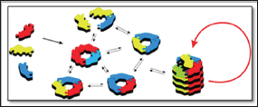
Figure 1: Example of Combinatorial Chemistry.

Lupinepublishers-openaccessjournals-chemical-science

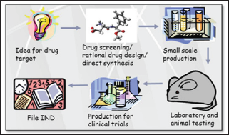
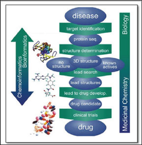
Combinatorial chemistry, a new approach for greatly reducing the time for the discovery of newer organic motifs, has been developed over the last few years. Combinatorial chemistry (Figure 1) is a technique for creating huge numbers of biologically potent motif and their hybrids, then rapidly screening them for desirable properties 1. Almost pharmaceutical and agrochemical industries uses this strategy because of the potential for immense reducing in time and cost, and several drug candidates presently in clinical trials are products of this methodology. Drug discovery process (Figure 2 & 3) is long, time and money intensive process [1]. This science of has found application in the production of molecules of commercial interest in the construction of newer pharmacological active therapeutic agents derived from rational drug design, into synthesize complex natural molecules, in the finding innovative approaches to render this chemical science more efficient [2,3]. The development of newer pharmaceuticals is currently a critical and challenging task to the pharmaceutical industry. The vital interest of the medicinal and agrochemical industries in organic synthesis is often related with their natural occurrence. Similarly, medicinal and pharmaceutical field, there has always been and continue to be a need for newer chemical entities with diverse pharmacophoric properties. Many works are still needed to minimize the time, expenditure, and attrition rate in the drug discovery process simultaneously addressing the huge unmet medical need across the world.

Figure 2: Drug development cycle.

Lupinepublishers-openaccessjournals-chemical-science

Figure 3: Role of Medicinal Chemistry and Biology in Drug Discovery.

Lupinepublishers-openaccessjournals-chemical-science

Referencing the study report, poor pharmacokinetic and preclinical toxicity were the main reasons for the failure in the drug development, in addition to the lack of efficiency and adverse effects [4,5]. New drugs are necessitated to cure new diseases, to find less hazardous drug and to cure diseases whose drugs have become ineffective due to resistant strains of microorganisms. Besides these causes, new drug discovery and researches are required to recognize pharmacophore present in the effective drugs [6-8]. We must always continue to search for drugs which exhibit clear advantages over the already existing respective drugs. Such advantages may be improvement in bioactivity, partial or total absence of adverse effects, minor toxicity, more nutritive value, improved stability and decrease in production cost. In present, research development department (R&D) of many pharmaceutical laboratories are working for synthesis of newer entities having improved drug activity and increasing yields of existing drugs [9-11]. Combinatorial, medicinal and biological chemistry have contributed to life processes and to the efforts to advance the quality of life as well as to the development of society from synthetic, biopharmaceutical, medical and industrial point of view. Finally, this communication opens the door to the combinatorial design, synthesis of biological active of a variety of newer hybrid molecules.

Acknowledgement

I thankful to chemistry staff members of the Mehesana Urban Institute of Sciences, Ganpat University, Gujarat for support.

Read More About Lupine Publishers Journal of Organic and Inorganic Sciences Please Click on Below Link:
https://lupinepublishers-chemicalsciences.blogspot.com/


Wednesday, 8 September 2021

Lupine Publishers| Beyond The Local Anesthesia

 Lupine Publishers| LOJ Pharmacology & Clinical Research (LOJPCR)


Editorial

One of the most important events in dentistry history was the discovery of local anesthetics in 1859 by Albert Niemann (1834 – 1861). In fact, this event represents the beginning of a new era in dentistry. Despite the adverse and lethal effects of cocaine, the first local anesthetic used in dentistry, this finding permitted the develop of other local anesthetics with less adverse effects and better safety index such as procaine, lidocaine, mepivacaine, bupivacaine, prilocaine and more recently ropivacaine and articaine. Local anesthetics constitute a group of elementary drugs in the professional arsenal of dentistry and are the most used drugs in this area. They act by preventing the conduction of the nociceptive electrical impulses in a reversible way, generating a loss of sensitivity to pain. Local anesthetics internally block Na+ voltage gated channels on neuronal fibers, thus decreasing nerve conduction. Na+ voltage gated channels are complex structures formed by two Ξ² subunits (Ξ²1 and Ξ²2) and a large Ξ± subunit in whose central part is located the entry pore for this ion.

In the last 100 years, the reversible local anesthetic effect added to the wide therapeutic index of these agents have allowed to perform invasive procedures in the oral cavity in different areas such as endodontics, oral and maxillofacial surgery, implantology, restorative dentistry and periodontics without pain during the performance of these treatments. This fact has meant an unprecedented advance in the world history of dentistry.

Several research have been conducted and published with the aim of improving the pharmacokinetic profile of these drugs such as inclusion complexes with cyclodextrins and encapsulation in liposomes and nanocapsules. These studies are aimed at increasing the liposolubility of these drugs through the neuronal cell membrane, as well as avoiding the use of vasoconstrictors that are present in most local anesthetic formulations to counteract the vasodilator effect of these drugs. Moreover, this new formulations could reduce the side effects of this drugs, because these cell carriers act as reservoir and from there they release (in a sustained manner) the local anesthetics molecules. These latest advances could allow in the near future the topical use of these agents and limit the use of dental needles for their administration.

Recently, there has been special attention in relation to the cytotoxic effects produced by local anesthetics. It has been shown, for example, that these agents in low tissue concentrations (as used in clinical practice) produce an induction of cellular apoptosis. This fact could mean a possible coadjuvant treatment to the base treatment of some types of cancer. Could it be that we are facing a group of old drugs with a new target?

Read More About Lupine Publishers LOJ Pharmacology & Clinical Research (LOJPCR) Please Click on Below Link:  https://lupine-publishers-pharmacology.blogspot.com/


Tuesday, 7 September 2021

Lupine Publishers| Utilization of Direct Acting Oral Anticoagulants in Patients with Liver Cirrhosis: Is It Safe?

 Lupine Publishers| Current Trends in Gastroenterology and Hepatology


Abstract

Patients with liver cirrhosis are known to have increased risk bleeding particularly from gastrointestinal tract. However, recent literature has shown that patients with liver cirrhosis are also at increased risk of thrombotic complications. Therefore, it is important to consider anticoagulation in cirrhotic patients. The purpose of this article is to review the epidemiological studies available in scientific literature comparing the risk of bleeding in cirrhotic patients utilizing DOACs vs traditional anticoagulation.

Abbreviations: INR: International Normalized Ratio; VTE: Venous Thromboembolism; DVT: Deep Venous Thrombosis; PE: Pulmonary Embolism; PTT: Partial Thromboplastin Time; LMWH : Low-Molecular-Weight Heparin; NO: Nitric Oxide; PTT: Partial Thromboplastin Time; DOACs: Direct Oral Anticoagulants; AT: Antithrombin; ULN: Upper Limit Of Normal; AUC: Area Under The Plasma Concentration-Time Curve; PD pharmacodynamics; PK: pharmacokinetics

Introduction

Liver plays central physiologic role in hemostasis as it synthesizes the majority of the procoagulant and anticoagulant factors. The levels of these factors are markedly affected by decrease function of liver associated with cirrhosis resulting in abnormal hemostatic mechanism. Generally, the impression in the clinical world is that liver cirrhosis is associated with decrease synthesis of procoagulant factors resulting in increased risk of bleeding. This phenomenon is known as auto-anticoagulation and is supported by elevated international normalized ratio (INR) and low platelet count usually observed in cirrhotic patients. In this regard, gastrointestinal bleeding and more specifically variceal bleed is of major concern since they contribute significantly to the mortality of patients with liver cirrhosis. At least, 30% mortality has been reported at the first episode with a 70% recurrence rate in this patient population and a one year survival estimate ranging from 32% to 80% [1]. However, decrease function of cirrhotic liver also results in reduce level of anticoagulant factors including anti thrombin III, protein S, and C which may result in increased tendency to form clots. Interestingly, recent data also indicates cases of venous thromboembolism (VTE) including both deep venous thrombosis (DVT) and pulmonary embolism (PE) in cirrhotic patients ranging between 0.5% to 6.3% [2-10]. Dabbagh et al. [4] found that even an elevated INR > 2.2 was not protective against VTE in this patient population [4]. Gulley D et al. [10] noted that hospitalized cirrhotic patients without predisposing co-morbidities (e.g. neoplasm, congestive heart disease and chronic renal failure) had similar risks for VTE as compared to non cirrhotic patients [10]. Thus, the myth of auto-anticoagulation seems to be only partially true. Therefore, the abnormal routine blood tests (like elevated INR, Partial Thromboplastin Time [PTT], high MELD score and low platelet count) may indicate increased hemorrhage risk in this patient population which may not be completely protective against risk of VTE as these tests do not accurately reflect the activity of aforementioned anticoagulant factors in the serum. As a result, utilization of anticoagulation is now being increasingly encouraged in cirrhotic patient population to avoid thrombotic complications.

Direct oral anticoagulants (DOACs) are relatively newer class of anticoagulants which selectively inhibit factor Xa (for e.g. Apixaban, Rivaroxaban, and Edoxaban) and factor IIa (for e.g. Dabigatran) of the coagulation cascade. Being able to be administered orally, rapid onset of action, lack of heparin induced thrombocytopenia, fewer interactions, and non requirement of laboratory monitoring are some of the advantages that DOACs carry over the traditional anticoagulant agents. DOACs may also be helpful in management of portal vein thrombosis (PVT) and portal hypertension (pHTN) in patient with cirrhosis as case reports have been reported about PVT controlled by Rivaroxaban treatment [11-13]. Vilaseca M et al. [14] investigated the effect of Rivaroxaban on various mediators of portal hypertension in CCl4 and thio acetamide-cirrhotic rats. Rivaroxaban significantly decreased portal pressure in both models of cirrhosis by reducing oxidative stress, improving nitric oxide (NO) bioavailability, and ameliorating endothelial dysfunction. Rivaroxaban also markedly reduced intrahepatic microthrmbosis by reduced fibrin deposition and deactivated hepatic stellate cells which plays major role in increasing intrahepatic vascular resistance by promoting fibrogenesis [14]. The purpose of this article is to review the epidemiological studies available in scientific literature comparing the risk of bleeding in cirrhotic patients utilizing DOACs vs traditional anticoagulation.

Methods

An electronic Medline search was conducted using the key terms anticoagulation, oral anticoagulant, direct acting oral anticoagulant, novel oral anticoagulant, direct thrombin inhibitors, direct factor Xa inhibitors, Apixaban, Rivaroxaban, Dabigatran, Edoxaban, liver cirrhosis, chronic liver disease, and decompensated liver disease. Studies written in the English from January 2000 to March 2018 were considered for this review article. All search results were reviewed.

Results

Hum J et al. [15] conducted a retrospective cohort study to compare the efficacy and safety of direct oral anticoagulants vs traditional anticoagulants in 45 patients with cirrhosis who were prescribed therapeutic anticoagulation over a 3-year period for thrombosis or prevention of stroke in patients with atrial fibrillation. 27 patients were prescribed one of the DOACs and 18 were prescribed vitamin K antagonist or low molecular weight heparin (LMWH). Similar total bleeding events (8 with DOACs vs 10 with other, P=0.12) were observed in the two groups but DOACs group had significantly less major bleeding events (1 [4%] vs 5 [28%], P = 0.03) [15]. In another retrospective study, Intagliata N et al. [16] compared the rates of bleeding in cirrhotic patients treated with DOACs (Rivaroxaban and Apixaban) to the cirrhotic patients treated with traditional anticoagulation (coumadin and LMWH) using a research database. The sample size consisted of 39 patients who received anticoagulation therapy over a 3-year period. 20 patients received DOACS and 19 received traditional anticoagulation. No significant difference in bleeding was observed in the two groups (three in the traditional anticoagulation group and four in the DOACS group, p = 0.9). Three major bleeding events were noted including two in the traditional anticoagulation group and one in the DOACS group [16]. Nagaoki Y et al. [17] also conducted a retrospective cohort study in fifty cirrhotic patients comparing the efficacy and safety of Edoxaban and warfarin for treatment of portal vein thrombosis (PVT). After treating for two weeks with danaparoid sodium, patients were switched to either Edoxaban (n = 20) or coumadin (n = 30). The efficacy and safety of Edoxaban and warfarin was compared for up to 6 months. Clinically significant gastrointestinal bleeding was encountered in 3 of 20 (15%) patients of the Edoxaban group and 2 of 30 (7%) of the warfarin group but the difference was not statistically significant (P = 0.335) [17]. In a relatively larger retrospective cohort study, Goriacko P et al. [18] compared the rate of bleeding in chronic liver disease patients with atrial fibrillation treated with oral anticoagulants (coumadin vs DOACs). No significant difference in all-cause bleeding (HR 0.9, 95% CI 0.4-1.8) and major bleeding were observed between the two groups [18].

Conclusion

The data on the safety of DOACs in patients with liver cirrhosis is in very initial stages. Based on our Medline literature search, we were able to find four studies comparing the risk of bleeding in cirrhotic patients utilizing DOACs vs traditional anticoagulation. All studies reported either decrease bleeding events in patients with liver disease treated with DOACs as compared to patients treated with traditional anticoagulation or no significant difference in bleeding risk. However, these studies were limited by retrospective nature, small sample size, and lack of randomization. Due to retrospective nature, underreporting of the bleeding events may have resulted in the underestimation of the risk of hemorrhage in these studies. Lack of randomization may have resulted in the underutilization of DOACs in cirrhotic patients at higher risk of bleeding such as those with high INR, low platelet count and presence of esophageal varices. This may have also confounded the results of these studies.

One of the major concerns regarding utilization of DOACs in cirrhotic patient population is the lack of specific antidotes in face of life-threatening gastrointestinal bleeding or urgent invasive procedure. Since DOACs have long half-life, drug discontinuation is insufficient in these circumstances. Recently, three agents including Idarucizumab, andexanet alfa, and ciraparantag have been introduced with promising antidotal effect against the DOACs [19]. Idarucizumab is the only agent to date which has been approved for use and is specific to Dabigatran [19]. Andexanet alfa is specific to factor Xa inhibitors and is still under investigation [19]. Ciraparantag is a universal antidote and is in earlier stages of development [19]. In a study on healthy volunteers, Prothrmobin complex concentrate has been demonstrated to reverse the anticoagulant effect of Rivaroxaban and Dabigatran [20]. In another study, prothrombin concentrates and recombinant factor VIIA were added in vitro to plasma from healthy volunteers receiving Rivaroxaban and Dabigatran with (partial) reversal of these agents [21].

Another concern regarding the utilization of DOACs in patients with liver disease is that the abnormal functioning of liver may affect the pharmacodynamics (PD) and pharmacokinetics (PK) of DOACs resulting in altered half-life and serum concentration of these agents in this patient population. As a result, caution and dose adjustment may be required when using DOACs in patients with abnormal liver function. Graff J et al. [22] observed that in patients with moderately impaired liver function (i.e. Child-Pugh classification B), the area under the plasma concentration-time curve (AUC) of Rivaroxaban after a single dose of 10 mg increased by 2.27-fold along with increase in factor Xa inhibition [22]. Since, Rivaroxaban is also excreted mainly by the kidneys (66%) and liver (34%), caution and dose adjustment of this agent is recommended in cirrhotic patients with cirrhosis with or without concomitant renal failure [22]. Rivaroxaban is also contraindicated in patients with liver cirrhosis associated with coagulopathy, increased bleeding risk, and patients classified as Child-Pugh B and C [22]. In contrast, the AUC of Dabigatran after a single dose of 150 mg decreased by 5.6 % in patients with moderately impaired liver function (i.e. Child-Pugh classification B) [22]. Also, Dabigatran is mainly (80%) eliminated via the kidneys and is likely the more safer choice in patients with liver cirrhosis [23]. Stangier J et al. [23] observed slower conversion of Dabigatran intermediate to active Dabigatran [24]. However, total drug exposure was comparable between healthy volunteers (n = 12) and patients with hepatic impairment (Child Pugh classification B, n = 12) [24]. Moreover, the parameters of coagulation, including activated partial thrmboplastin time, clotting time, and thrombin time relationships were basically similar in both groups [24]. Therefore, Dabigatran can be used in patients with moderate hepatic impairment without the need for dose adjustment [24]. Dabigatran should be avoided in patients with elevated hepatic enzymes (>2× ULN) and is contraindicated in patients with hepatic impairment expected to have any impact on survival [22]. Increased AUC by 1.09-fold was observed for Apixaban after a single dose administration of 5 mg whereas AUC of Edoxaban decreased by 5.6 % after single dose administration of 15 mg [22]. In patients with mild (Child-Pugh A) or moderate (Child- Pugh B) hepatic dysfunction or transaminase levels >2× upper limit of normal (ULN), Apixaban can be used with caution. Apixaban should be avoided in patients with severe hepatic impairment and in those with hepatic impairment with increased bleeding risk [22].

It will be helpful to monitor activity of DOACs in cirrhotic patients particularly at increased risk of bleeding for example patients with esophageal varices, elevated INR, and low platelet count. Novel coagulation assays need to be developed to monitor the activity of DOACs in serum from patients with liver disease. In one study, Potze W et al. [25] noticed substantial reduction in anti- Xa levels when antithrombin (AT) dependent anticoagulant drugs (Unfractioned heparin, LMWH, and fondaparinux) were added to the plasma of patients with cirrhosis as compared to plasma from healthy controls. Therefore, they concluded that anti-Xa assay cannot be used to monitor AT-dependent anticoagulant drugs in patients with cirrhosis, as it may result in underestimation of drug levels and increase risk of bleeding. However, this was not the case with Rivaroxaban and Dabigatran and they recommended that direct factor Xa and IIa inhibitors may be monitored through the respective anti-Xa and anti-IIa assays in patients with cirrhosis.

Read More About Lupine Publishers Current Trends in Gastroenterology and Hepatology Please Click on Below Link: https://currenttrendsingastroenterology.blogspot.com/


Friday, 3 September 2021

Lupine Publishers | Evaluation of the Anti-Inflammatory Activity of Semi-Purified Glycoside Extracted from the Leaves of Plumeriarubra (Kalachuchi)

 Lupine Publishers | Journal of Research & Reviews


Introduction

The researchers focused on skin inflammation. An inflammation is a biological response of the body aiming to remove the harmful stimuli that affects a part of our body. Based on visual observation, the ancients characterized inflammation by five cardinal signs; heat (calor) is caused by the increased movement of blood through dilated vessels into the environmentally cooled extremities, also resulting on the increased redness (rubor), swelling (tumor) is the result of increased passage of fluid from dilated and permeable blood vessels into the surrounding tissues, infiltration of cells into the damaged area, and in prolonged inflammatory responses deposition of connective tissue, pain (dolor) is due to the direct effects of mediators, either from initial damage or that resulting from the inflammatory response itself, and the stretching of sensory nerves due to oedema and loss of function (functiolaesa) refers to either simple loss of mobility in a joint. (Punchard N.A., Whelan C.J., Adcock I., 2004)[1-3].

Methodology

Studies were conducted using Sprague-dawley male rats having an average weight of 100- 160 grams. They were obtained from the animal house to be acclimatized to laboratory condition for three weeks before starting the experiment. Three rats were used for each step. The initial dose used was from 5mg/kg, 50mg/kg, 300mg/kg, and 2000 mg/kg body weight. The starting dose level should be that which is most likely to produce mortality in some of the dosed rats. A limit test should be conducted when the available information suggests that mortality is unlikely at the highest starting dose. It is recommended to use the starting dose of 300mg/kg when there is no information on a substance to be tested [4].

Result and Discussion

Carrageenan induced paw edema was the method used in the induction of inflammation due to the reason that, carrageenan is the phlogistic agent of choice. It is considered to be antigenic and has no systemic effect. As described by winter, the inflammation produced by carrageenan is acute, non-immune, well-researched and highly reproducible. The result of the Biological Testing using Carrageenan Induced Paw Edema shows that the 750 mg concentration of the extract exhibits the highest anti-inflammatory effect among the three concentrations [5].

Conclusion

After the phytochemical screening of the semi-purified glycoside extract the leaves of P.rubra constitute Alkaloids, Flavonoids, Glycosides, and Tannins. Based on the results obtained from the Fourier Transport Infrared Spectroscopy high amount of polyphenolic compound is found which indicates the presence of Glycosides. Therefore the researchers conclude that the Glycoside is the one that exhibits anti-inflammatory activity. After the Acute Oral Toxicity Testing, the researchers conclude that the extract of P. rubra administered is safe. There is significant difference between the anti-inflammatory activity of the 250 mg/kg and 500 mg/ kg concentration and the positive control. There is no significant difference between the anti-inflammatory activities of the 750 mg/ kg concentration derived from the leaves of P. rubraand the positive control.

Recommendations

After evaluating the anti-inflammatory activity of the semipurified glycoside extracted from the leaves of Plumeriarubra the researchers comes up with the following recommendation to widen the study regarding the activity of the plant especially on its antiinflammatory.

The following were recommended for future studies:

    a) To conduct further studies about the anti-inflammatory activity of P. rubra

    b) To isolate the pure compound that elicits the antiinflammatory effect of P. rubra

    c) To formulate a dosage form that is befit for the isolated compound.

    d) 7.4. To further develop another study using the other parts of P. rubra.

    e) 7.5. To accentuate the other pharmacological uses of P rubra using other solvent.

Thursday, 2 September 2021

Lupine Publishers | Double Malignancies: A Rare Entity

 Lupine Publishers | Journal of Surgery & Case Studies


Abbreviations: MPM: Multiple Primary Malignancies, SEER: Surveillance Epidemiology and End Results Programme, SPT: Second Primary Tumor, TT: Third Tumor, QT: Quadrant Tumor, SPM: Second Primary Malignancies, NAACCR: North American Association of Central Cancer Registries

Introduction

Patients which have diagnosed with a cancer, have a life time risk for developing another de novo malignancy depending on various inherited environmental and iatrogenic risk factors. Cancer victims could survive longer due to settling treatment modalities, and then would likely develop a new metachronous malignancy [1]. The incidence of multiple primary malignancies has not been rare at all. Screening procedures have especially been useful for the early detection of associated tumors, whereas careful monitoring of patients has treated for primary cancer, and then a good communication between patients and medical care team would certify not only an early detection for secondary tumors, but only finally & subsequently, an appropriate management [2]. Differentiation between multiple primary and multicentric cancers was addressed in the classification by Moertel CG [3]:

I. MPMNs of Multicentric Origin

a) The same tissue and organ.

b) A common, contiguous tissue shared by different organs.

c) The same tissue in bilaterally paired organs.

II. MPMNs of Different Tissues or Organs

III. MPMNs of Multicentric Origin Plus a Lesion (s) of a Different Tissue or Organ

In 2002, However in ‘A review of the definition for multiple primary cancers in the United States’ classified the association of different cancers in two categories depending on the timing of their discovery [4];

a) Synchronous in which the cancers occur at the same time or within two months and

b) Metachronous in which the cancers follow in sequence of more than two months apart.

Vaamonde [5]. reckoned the time factor as six months. In 2005, International Agency for Research on Cancer working Group has come out with International Rules for Multiple Primary Cancers [6].

Although the mechanism involved in the development of multiple primary cancers has not been clarified, some factors such as heredity, constitution, environmental and immunological factors, oncogenic viruses, radiological and chemical treatments have been implicated. Hereditary susceptibility explains only a small proportion of all second cancers though many hereditary cancer syndromes have been described. MPMNs can occur at any age. However, from the reviewed series, patients with MPMNs tend to be older than those with a single primary malignant neoplasm. In many autopsy series and clinical reports, the median age of 50- 94% of MPMN patients was over 50 years [7]. Multiple Primary Malignancies (MPM) were first described in 1879 by Billroth [8]. The neoplasms may be limited to a single organ or may involve multiple separate anatomical organs. The North American Association of Central Cancer Registries (NAACCR) classifies MPM into two categories:

a) Synchronous, in which the cancers occur at the same time (The Surveillance Epidemiology and End Results Programme (SEER) definition is within two months) and

b) Metachronous, in which the cancers follow in sequence, that is, more than six months apart [9].

According to Warren Gates criteria a diagnosis of MPM require the following criterias to be fulfilled

a) Each tumor should present a definite picture of malignancy

b) Each tumor should be histologically distinct

c) The possibility that one is metastasis of the other must be excluded [10].

Meta-analyses show the frequency of Second Primary Tumor (SPT) as 3-5%, Third Tumor (TT) as 0.5%, and fourth tumor, that is, Quadrant Tumor (QT), as 0.3%, in different organs and of different histogenesis. Metachronous primary malignancies are becoming increasingly common because of an increase in the number of elderly cancer survivors, greater awareness and improved diagnostic modalities [1]. The exact pathophysiology for MPM remains unknown. However certain factors have been postulated which includes the common carcinogen induced multiple cancers in a exposed epithelial surface, called as “Field-Cancerization” as seen in head-neck tumors. In addition other causative factors includes ionizing radiation, increased use of organ transplant, and the increasing use of newer treatment modalities like hormonal manipulation, target therapies, genetic manipulation, and immunomodulators [2]. In a study conducted by Chakrabarti [11] it has been reported that the over a period of 2 years, 12 cases of MPM were detected against a total of 1255 cases. Of these, five cases were synchronous malignancies and seven cases were metachronous. Head and neck was the commonest site of index malignancies with seven cases followed by the breast cancer with three cases and next gynaecological malignancies with two cases. Most common sites for Second Primary Malignancies (SPM) were head and neck with (four cases).

Male to Female ratio was 1:1.5 in the synchronous primary group and 1:1.3 in the metachronous group. Median age of presentation of the primary tumour was 52 years and 6 months. The age range for the SPM was 17-72 years with the highest incidence in the 6th decade of life. Studies have reported that that the relative risks of SPM ranges from 1.08 to 1.3. SPM are often missed during follow-up and are detected accidentally. According to various series, the onset of SPTs decreases the 5-year survival by 18-30% as compared to those with only a single tumor. The controversy between the lateral spread of clones vs multiple foci of independent alterations does not currently affect the surgical and medical management of theses premalignant and malignant lesions. In the future, the presence of altered clones at mucosal margins may be an indication for aggressive therapy, including chemoprevention or radiotherapy to treat altered clonal patches that are unable to be detected grossly and are beyond the initial scope of surgical excision [3]. The issue of whether those with an extensive visible mucosal field defect is more likely to benefit from chemotherapy, radiotherapy or chemoprevention is a complex one. Current management is often site specific: Recurrent oral premalignant disease is often treated by surgical excision, whereas diffuse high grade premalignant changes in the laryngeal mucosa are frequently treated with radiotherapy. Determination of the role for these and other treatment modalities for clinically occult, clonally altered patches of epithelium is likely to be a difficult issue, since treatment of mucosa with widespread visible alteration is already challenging [4]. The paucity of awareness about SPM has also prevented the formulation of population-based screening protocol. Multiple tumors that have been pathologically confirmed at the time of presentation should be evaluated and staged as independent tumors. The treatment plan should be decided after staging of both the primary and secondary tumors in view to attain maximum clinical response. Proper counselling and patient’s understanding of magnitude of the disease is paramount. Operable synchronous SPM can be operated in a single setting with minimal morbidity with better survival and is less taxing on the patient and his/her relative both psychologically and financially. A regular follow-up on the patient by the clinician increases the chances of early detection of metachronous SPM and the formulation of the treatment plan at the earliest with better overall survival

Conclusion

MPMNs are still elusive for want of proper guidelines regarding correct terminology and classification encompassing varying presentations of chronological, aetiological, clinical and histopathological combinations. Our case adds up to literature for further research. The possibility of occurrence of synchronous multiple primary malignancies should be considered during workup for any malignant condition to institute early intervention to achieve good outcome [12]. The possibility of multiple primary malignancies existence should always be considered during pretreatment evaluation. Screening procedures were especially useful for the early detection of associated tumors, preferably before clinical manifestations occurrence [12]. There were some evidences that screening would improve outcomes among patients who might develop second malignancies, although the data were limited. The optimal screening modalities and strategies for reducing mortality from second malignancies remained to be defined for most tumor sites [13]. The early diagnosis of secondary malignancies should not be neglected in patients treated for a primary malignancy, especially when the long clinical period before the diagnosis of subsequent tumors has been taken for management. With careful monitoring, secondary tumors could be detected earlier, and, with appropriate intervention, might be better managed, without compromising survival. Our data could guide oncologists towards a closer follow-up strategy in the management of patients treated for common tumors. Availability of data regarding incidence of MPM, particularly those from developing countries is very limited and hence further studies are needed. SEER is working in this direction with an aim to define and develop appropriate and reliable criteria’s for synchronous and metachronous cancers. It is imperative that patient with a primary malignant tumors should be thoroughly closely, and regularly followed. Genetic counselling, risk estimation, cancer screening and chemoprevention must be emphasized. Appropriate cancer prevention strategy in with proper emphasis on synchronous and metachronous cancer needs to be designed and incorporated in the National Control Programme as multiple primary cancers have unique, biological behaviour requiring specific diagnostic and therapeutic interpretation [14].

Read More About Lupine Publishers Journal of Surgery & Case Studies Please Click on Below Link:
https://surgery-casestudies-lupine-publishers.blogspot.com/