Showing posts with label journal of Nanomedicine. Show all posts

Monday, 17 July 2023

Lupine Publishers | Current Approaches in Nanomedicine

 Lupine Publishers | Archives of Nanomedicine: Open Access Journal


Introduction

Over the last years, nanotechnology has been introduced in our daily life. Nanotechnology may be able to part in a very range of applications, such as in nanomedicine, nanoelectronics, biomaterials energy production, and consumer product, etc. The application of nanotechnology for medical purposes has been termed nanomedicine and is defined as nanoscale tools (e.g., 1–1000 nm sized) for the diagnosis, prevention, and treatment of diseases. The term nanomedicine appeared in the 1990s, and since then, has the potential to significantly improve some current treatments. Commonly, nanomedicines consist of active pharmaceutical ingredients such as small molecules or biologics packaged into nano-sized carriers made of excipients like lipids and polymers. By packaging drug in the particles, drug concentration in the target is maximize by passive or active targeting and pharmacokinetic-pharmacodynamics profiles are improved. But due to their size related physicochemical properties, nanomaterials can require additional quality and safety testing compared to products with standard size.
In nanomedicine, nanomedical devices can be used for analytical, imaging, detection, diagnostic and therapeutic purposes and procedures, such as targeted cancer therapy, drug and gene delivery, improving cell-material interactions, scaffolds for tissue engineering, etc. The application areas of nanomedicine are shown in Figure 1.
With the developing technology and increasing life expectancy, new problems are faced in human life. Science is actively working to offer solutions to these problems. The advancing technology and increasing knowledge, the reliability of the products and techniques used for years, started to be questioned and the problems identified led to the development of new solutions. Over than 160 years, dentistry has been used silver amalgam for tooth filling material which contains approximately 50% Hg metal. During the past decay, science demonstrated the released Hg from the filling material covalently bound to cell proteins and shows toxic effect. So, the presence of toxic and trace elements in dental powders has led to the discover better dental filling materials and tests. In this context, Joseph published a research article about Al detection in dental powder by using handheld X-Ray Fluorescence (HHXRF) (Joseph, 2019). On the contrary of conventional XRF, HHXRF has an advantage of showing Al which is a prominent element in dental powders.

Figure 1: Schematic representation of the application areas in nanomedicine.

lupinepublishers-openaccess-journal-nanomedicine

Mycobacterium tuberculosis infection is one of the most common and deadliest infection for many years. During past decades, many efforts have been made to reduce the level of the diseases. Gupta et al. reviewed the mycobacteriophage to control tuberculosis infection (Gupta et al., 2019). Mycobacteriophages are the member of a group of bacteriophages that infects Mycobacterium and have two killing mechanisms such as lytic and lysogenic. Endolysin and lysB proteins have a major role to disrupt cell wall envelope of the bacterium. Mycobacteriophage therapy is a novel study for Tuberculosis which is a common and deadliest infection disease. Both in nuclear and thermonuclear power engineering, hydrogen could be the first reason of equipment damage. Because of that, reactor pressure vessels (RPV) was manufactured without stainless cladding as a product of RPV wall corrosion. In the RPV steel, hydrogen concentration was determined with gas chromatography. The hydrogen content in the irradiated steel were found less than 0,1 ppm. This was attributed to the increase in hydrogen content as fast neutron fluency increased (Krasikov, 2019).
“If one day, my words are against science, choose science” Mustafa Kemal Atatürk.


Read more about Lupine Publishers Archives of Nanomedicine: Open Access Journal Click on the below link: https://lupine-publishers-nano-science.blogspot.com/


Monday, 17 January 2022

Lupine Publishers| Degradation of Malachite Green by Green Synthesized Copper Nanoparticles by Using Aloe Barbadensis Leaf Extracts

 Lupine Publishers| Journal of Nanomedicine


Abstract

The present study was aimed to account a green synthesis of copper nanoparticle is by interaction of leaf extract and copper salt. The bio-synthesis of nanoparticles put forward a cost free and eco-friendly method of nanoparticle synthesis. Copper nanoparticles were synthesized by using aqueous solution of copper sulphate and extract of Aloe barbadensis. The prepared leaf extract was observed when 1mM copper sulphate solution is added in it. Color change of the reaction mixture was observed from deep blue to colorless and then to brick red and dark red indicating the formation of copper nanoparticles. The synthesized CuO NPs was characterized by using different technique such as UV, IR, XRD, and SEM. ward a cost-free and environmentally suitable method of nanoparticle synthesis. Synthesized CuO nanoparticles with average particle size of 60n. Shape of copper nanoparticles was spherical and cubic and their range was 80-120nm Different functional group in synthesized nanoparticles are examined by FTIR. UV spectrophotometer confirm peak of copper nanoparticle experiments Copper oxide nanoparticles shows maximum absorbance at 272nm. Catalytic activity of synthesized nano particles is also examined on the degradation of malachite green. This catalytic effect of copper oxide nanoparticles can be contributed to its small size.

Keywords: Aloe Barbadensis, SEM, Copper Oxide Nanoparticles, Green Synthesis, XRD, Green Synthesis, Highly Stabilized Nanoparticles , Ecofriendly, Phenolic Content, Degradation of Malachite Green

Introduction

Nanotechnology deals with manipulation of matter at low size normally lesser then that of the 100nm. Metallic nanoparticles can be prepared by chemical and physical method. These methods have certain flaws like toxic chemicals and also dangerous to environment [1]. Developing research in green chemistry employed prominent part in nanotechnology to attain benefit to society surface area and mass ratios increase adsorption property [2]. Green synthesis has been concerned in synthesis of highly stabilized nanoparticles. Synthesis of nanoparticles taking assistance of ecofriendly methods has achieved huge attention in the modern era. The particles produced by green synthesis differ from those using physio–chemical approaches. [3] Green synthesis, a bottom up approach, is similar to chemical reduction where an expensive chemical reducing agent is replaced by extract of a natural product such as leaves of trees/crops or fruits for the synthesis of metal or metal oxide NPs. Biological entities possess a huge potential for the production of NPs. Biogenic reduction of 
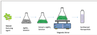
metal precursors to corresponding NPs is eco-friendly [4] (Figure 1). Copper nanoparticles were synthesized by leaf extract of Aloe barbadensis plant. The plant is also known as “Aloe vera”. The green synthesis of copper nanoparticle by Aloe vera plant extract is fast, easy and (Figure 2). environmentally suitable method [5].

Figure 1: Green synthesis of NPs.

Lupinepublishers-openaccessjournals-Nanotechnology-nanomedicine

Phenolic content in plant extract dissolved in water, degradable and catalyzed synthesis of nanoparticle as capping and reducing agent [6]. This old plant is well known for deeper healing effects. It is majorly present in cosmetics and skin creams. It has the ability to clean skin and anti-aging effects of it is also famous [7]. Aloe vera contains antioxidant vitamins A, C, and plus vitamin B12, folic acid, and choline [8]. Its gel juice is taken as power drink. Minerals such as calcium, copper, selenium, chromium, manganese, magnesium, potassium and zinc are present in aloe vera. Leaves of aloe vera provide anthraquinones [9]. Copper nanoparticles synthesis by using electron microscopy represented that their range is upto 50 to 130nm [10]. Copper nanoparticles important as compares to other nanoparticles due to their properties that are found at less cost than that of expensive metal such as gold and silver such as examination of catalytic activity of copper nanoparticles by degradation of malachite green [11]. Copper oxide particles show effective catalytic removal of organic dyes such as malachite green, when particles were added into it [12]. The use 0fgreen method increased so much because of its easy preparation and low manufacturing cost. Moreover, less to toxic starting materials and ease of handling make it more favorable [13]. Malachite green is extensively used in many industries as a dye for leather, textiles and also in aquaculture industry to control fish parasites and disease [14]. Malachite green is classified as a class II health hazard and they pose toxicity (mutagenicity, genotoxicity) to the aquatic organisms like fish, algae, bacteria etc. and it’s proved to be highly carcinogenic and is banned by many countries [15] (Figure 3). The removal of organic pollutants and dyes from industries remain as a challenge as these dye molecules are difficult to decompose. Varieties of organic and heavy metal pollutants were removed by nano adsorbents by various research groups [16].

Figure 2: Aloe veraplant.

Lupinepublishers-openaccessjournals-Nanotechnology-nanomedicine

Figure 3: Malachite Green

Lupinepublishers-openaccessjournals-Nanotechnology-nanomedicine

Materials and Methods

Material

Copper sulphate, Aloe barbadensis leaves, sodium borohydride (NaBH4), organic dyes such as Malachite green

Preparation of Plant Leaf Extract

50g of the Aloe vera is taken from the nearby garden. The leaves of aloe vera are first separated from the gel part o0f aloe vera. The leaves are then washed thoroughly with distilled water to remove soil and dust particles. After washing leaves were dried and finely chopped. These finely chopped leaves were allowed to boil for 15min at 100 °C with 100mL of de-ionized water in a250-mL flask and then allow to cooled down to come at least at room temperature. The resulting solution is passed through a filter paper to remove any solid particles and then again filtered throughaWhatmanfilterpaperofporesize0.2μm.Thefiltrate is stored at 6 °C as a stock for the synthesis of CuO NPs.

Green Synthesis of Cuo NPs: A copper sulphate solution of fifty milliliters was added to 15ml aloe vera leaves extract. The solution was stirred on a magnetic stirrer at 120degrees. The color change was observed. The color changes from deeply blue to colorless and then dark red at saturation. Brick red color confirms the nanoparticles formation. The resultant solution was centrifuged for ten mints at speed of 50,000rpm. After discarding supernatant copper oxide nanoparticles were dried in a watch glass. After drying, black color particles (Figure 4). were assemble for further characterization.

Figure 4: copper NPs synthesis.

Lupinepublishers-openaccessjournals-Nanotechnology-nanomedicine

Characterization of Green Synthesis Copper Nanoparticles

The morphological, structural and chemical composition of CuO NPs were analysed by using SEM (jsm-6480) and XRD (XPERTPRO) equipment. Optical properties of synthesized particles are investigated by UV spectrophotometer (DB-20). Size and shape of copper oxide nanoparticles were observed by SEM (jsm-6480). The crystal structure of synthesized nanoparticle is examined by XRD (XPERT-PRO), FTIR analysis is performed for the collection of the functional groups, present in this synthesis of CuONps.

Colour change observation: Color changes indicate the formation of nanoparticles of copper oxide. Blue color solution was turned into red or brick red indicated for formation of copper nanoparticles synthesis.

Result

X-Rays Diffraction Studies

Copper oxide nanoparticles were examined by X-ray diffractometer (XPERT-PRO). Copper oxide powder was put in cubes of XRD for calculation of intensity. The resultant pattern of synthesized nanoparticles was analyzed. The peaks at 2θ correspond to intensity as the peaks at28, 29.8, 32.1, 35.8, 36, 43.3, 47.5, 51.1, and have 112, 200, 103, 202, 004, 111, 301 and 200, pattern which is compare to JCPDS card no (049-1832). The pattern of Cu nanoparticles compared to JCPDS card no (01-085-1326), the peaks at 2θ. XRD pattern confirmed that CuO nanoparticles are highly crystalline with cubic crystal structure. The average size of the particle calculated by Scherrer equation was 60-100nm (Figure 5).

Figure 5: XRD pattern of Cuo NPs.
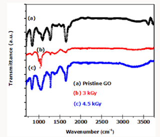
Lupinepublishers-openaccessjournals-Nanotechnology-nanomedicine

FTIR Analysis

In this analysis, FTIR (IPRrestige-21) spectrum was analyzed. The analyzation confirms the presence of copper nanoparticles. Different peaks were observed at 1100cm-1 confirm formation of Copper oxide nano particle speaks was observe in range of 400-4000cm-1. The FTIR spectrum of Copper oxide nanoparticle exhibits that the broad absorption band at 32cm-1 corresponds to the hydroxyl (OH) functional group in alcohols and phenolic compounds. The peak at 1601.2cm-1 is due C=C aromatic bendindg. Absorption peak at 1038.0 cm-1 stretching vibration of C–O group of primary and secondary alcohols (C–O), while smaller peaks at 900- 700cm-1 were also (Figure 6). Assigned to the aromatic bending vibration of C–H group (Table 1).

Table 1: Absorption peak at 1038.0cm−1 stretching vibration of C–O group of primary and secondary alcohols (C–O), while smaller peaks at 900–700 cm−1 were also assigned to the aromatic bending vibration of C–H group.

Lupinepublishers-openaccess-Nanomedicine

Figure 6: FTIR spectra of copper oxide nanoparticles.

Lupinepublishers-openaccessjournals-Nanotechnology-nanomedicine

Ultra violet spectroscopy: The presence of copper oxide nanoparticles is confirmed at the range of 200-1100nm. The ecofriendly method for the synthesis of copper oxide nanoparticles using Aloe vera leaves extract proved feasible, coast free and successful method. UV-Vis spectra analysis has apparently shown the formation of copper oxide nanoparticles. Nanoparticles synthesized have variety of application in the different field. The maximum absorption peak is between 265-285nm.The peak at about 280nm was achieved (Figure 7).This peak confirmed formation of the copper oxide nanoparticles.

Figure 7: Nanoparticles synthesized have variety of application in the different field.

Lupinepublishers-openaccessjournals-Nanotechnology-nanomedicine

SEM Analysis

The average particle size of copper nanoparticle was analyzed by SEM model (JSM-6480). The range of grain of copper oxide nanoparticle was calculated about 50.5-130nm by SEM micrograph. It was observed that particles were smooth with a spherical shape (Figure 8). The catalytic activity of the CuO NPs analyzed by the degradation of malachite green dye. The catalytic activity of the CuO NPs analyzed by the degradation of malachite green dye. Preparation of 1000mg/l dye S.S. 1000ppm solution of Malachite green dye was prepared by dissolving dye in 1-liter distilled water.

Different concentration of dyes was prepared from stock solution. 100ppm solution was prepared from 1000ppm solution after dilution. After that 150,200,250-ppm solution was prepared. 18g of NaBH4 is made up to 10mL and kept aside. Different concentrations of NaBH4 and catalyst are tested on the methylene blue dye. The catalytic degradation of organic dyes was observed by measuring UV-Visible spectra at regular time intervals.

Figure 8: SEM micrograph.

Lupinepublishers-openaccessjournals-Nanotechnology-nanomedicine

Malachite Green: Malachite green is extensively used in many industries as a dye for leather, textiles and also in aquaculture industry to control fish parasites and disease. The use has increased so much because of its easy preparation and low manufacturing cost (Table 2) (Figure 9).

Figure 9: Stucture of dye (malachite green).

Lupinepublishers-openaccessjournals-Nanotechnology-nanomedicine

Table 2: The use has increased so much because of its easy preparation and low manufacturing cost.

Lupinepublishers-openaccess-Nanomedicine

Structure

Dye Degradation: The degradation of malachite green in the absence and presence of CuO NPs were studied spectrotometrically by using DB-20 UV-Vis spectrophotometer determining the decrease in the absorbance at 631nm.The reaction was study spectrophotometrically at room temperature (25 0C). The colour of the reaction mixtures faded, indicating that degradation had occurred. The same procedure was followed for uncatalyzed reactions, in absence of CuO NPs.

1-Time Effect on Dye Removal: Decolorization of dye Malachite Green at room temperature was analyzed. Initially 20ml dye solution was taken and 1mg of greenly synthesized copper nanoparticles using aloe vera leaves extract dissolved in it. 0.1mg of NaBH4 was dissolved as a reducing agent. The solution was heated for 10- 20 mint at 100 degrees. The time interval was taken in consider gradually during reaction. The removal percentage of decolorization was calculated and draws graphically. The maximum time was 120 mints with70% color removal. This confirms the rapid reaction of copper oxide nanoparticles (CuO NPS) (Figure 10).

Figure 10: The time interval was taken in consider gradually during reaction.

Lupinepublishers-openaccessjournals-Nanotechnology-nanomedicine

Figure 11: Aloe vera synthesized copper oxide nanoparticles showed maximum percentage decolorization as pH was increased at a certain limit after more increase has a negative effect.

Lupinepublishers-openaccessjournals-Nanotechnology-nanomedicine

2-pH effect on dye removal: pH of the solution also majorly affected the de-colorization of dye. pH effect on the decolorization of copper oxide nanoparticles was analyzed in this research. Aloe vera synthesized copper oxide nanoparticles showed maximum percentage de-colorization as pH was increased at a certain limit after more increase has a negative effect. This may be happened due to the formation of more positive ion competition. Maximum de-colorization 70% was at pH 5 (Figure 11).

3-Concentration of dye effect on decolorization of dye: The increase or decrease in the concentration of Malachite green MG dye is also considerable in decolorization efficiency. The graph was obtained after experimenting various concentration of dyes. The maximum amount of dye taken was 20mg/l. After increasing concentration no effect on 70 decolorizatioof dye was observed (Figure 12).

Figure 12: After increasing concentration no effect on70 decolorizatioof dye was observed.

Lupinepublishers-openaccessjournals-Nanotechnology-nanomedicine

4-Effect of Copper Oxide Nanoparticles Amount on Dye Removal: The number of copper oxide nanoparticles exhibits positive results on decolorization. The number of nanoparticles 1 gram was taken showed maximum de-colorization power. This confirmed from the experiments that increasing of nanoparticle showed no effect on de-colorization. This concentration of nanoparticles was used in further experimentation of research (Figure 13).

Figure 13: This concentration of nanoparticles was used in further experimentation of research.

Lupinepublishers-openaccessjournals-Nanotechnology-nanomedicine

Conclusion

Here in conclusion, we concluded a method of green synthesis of Cu nanoparticles by leaf extract of Aloverabarbadensis plant. This eco-friendly way of synthesis of nanoparticles is more recommended over other methods as green synthesized CuO NPs are cost-effective, biogenic molecules with the capability to serve as dye absorbent. From vast of analyzation on nanotechnology for synthesis of nanoparticles it is declared that it is safer and best by using natural plants. With the huge plant variety much more plants are still not known for the synthesis of nanoparticles. Nanoparticles synthesized can be applicable in the different field of biochemistry, Pharma, agriculture and industry. Copper oxide nanoparticles have the ability to remove carcinogenic dyes. In the present study, Malachite green dye was removed by nanoparticles and its time, pH, contact time was observed. The maximum contact time was 120min, pH was observed 5, nanoparticle amount 1mg which proved green synthesized copper nanoparticles, as best removal of carcinogenic dye like Malachite green.

Read More About Lupine Publishers Journal of Nanomedicine Please Click on Below Link https://lupine-publishers-nano-science.blogspot.com

Tuesday, 19 October 2021

Lupine Publishers| Reduced Graphene Oxide Via Green Route Exposure of Gamma (γ) Ray for Surface Morphological Investigation

 Lupine Publishers| Journal of Nanomedicine


Abstract

γ-ray radiation was used to reduce graphene oxide thin films to partially reduced graphene oxide films at ambient conditions. Micro-Raman spectroscopy showed that irradiation of GO with γ-rays did not change the structure of graphene. The γ-rays are only active on the oxygen functional groups bonds because they are short and highly energized wavelengths. γ-rays penetrate the C-C lattice isotropically hence sustaining the graphene layer structure. This results in transparent and intact r-GO thin films with no structural defects. The defects on the structure would result to the opaqueness of the C-C lattice hence compromising the transparency of the material. The Scanning electron microscopy, Transmission Scanning electron, Fourier transform infrared and Raman spectrometry are presented.

Abbrevations: ITO: Indium Tin oxide, TCOs: Transparent Conducting Oxide, GFs: Graphene flakes, FE-SEM: Field Emission Scanning Electron Microscope, TEM: Transmission Electron Microscope, GO: Graphene oxide, FTIR: Fourier Transform Infrared

Introduction

Indium Tin oxide (ITO) has been for so many years used as a transparent and conductive material on our mobile phones and television screens. However, since ITO is expensive and environmentally harsh it is therefore, critically important to come up with an alternative material that will replace it. This material should be environmentally friendly, cost effective and much more efficient and accessible. Graphene which is a competitive candidate is atomically thick, two-dimensional platelets featuring carbon atoms in a honeycomb arrangement. These sp2-hybridized sheets of carbon possess a host of desirable properties, including high mechanical stiffness (1 TPa) [1-2], good thermal conductivity (5000Wm_1 K_1) [3-6], and excellent charge carrier mobility (250 000cm2 V_1 s_1) [3]. As such, graphene has been studied for use in a variety of applications that exploit these properties [7-10] ranging from gas sensors [6] to lithium-ion battery electrodes [11- 14] as well as transparent conducting oxide (TCOs) type electrodes. Current reduction methods for producing colloidal graphene for graphene based electrodes from graphene oxide typically afford Nano sheets with high C/O ratios (∼10) [15] that exhibit good electrical conductivity and optical transparency.

Graphene flakes (GFs) and carbon nanotubes are very promising for molecular sensors, single-electron transistors, super capacitors, non-volatile memory devices, integrated circuits, atomic scale switches and other carbon based electronic and magneto-electronic devices [16-18]. Graphene in particular exhibits outstanding properties in various novel applications [19-23]. Specifically, sensing is arousing continuous interests due to the low background noise and the excellent surface activity [24,25], targeting to employ them in practical application. Various graphene Nano sensors have been fabricated in previous works [26-28]. The industrialization and commercialization of graphene Nano sensors require the preparation of graphene in quantity. Several methods have been used to synthesize graphene, among which, there is the reduction of graphene oxide which is a promising path toward high-yield production of graphene. It is however, known that the negative influence rendered by this method, that is the defective graphene structure, hinders the application in the field requiring excellent conductivity such as transparent conductive film [2].

Chemical reagents are fundamental to the reduction of GO. For instances, hydrazine hydrate (H2O4) which is used to reduce GO to r-GO is poisonous and toxic to human life. Taking sensing into consideration, the introduction of chemicals is a serious disadvantage because the intrinsic response of graphene to trace analyte may be masked by the signals caused by the impurities [29]. Therefore, a reduction method without impurities involved should be developed. Herein, we report on facile and environmentally friendly reduction method to synthesis a well-dispersed graphene from graphene oxide as a conductive and transparent dye solar cell electrode. This contribution reports on the synthesis of reduced graphene coatings for TCOs type applications in Gratzel dye solar cells and the description of some fundamental reactions taking place during the reduction phase.

Characterization

The surface measurements were recorded with a JEOL 7500F field emission Scanning electron microscope (FE-SEM). Transmission electron microscope (TEM) images were obtained on Fei Tecnai G2-20 operated at 200kV using an energy filter of 20eV. The Chemical and crystalline nature on the wings was analysed with the Perkin Elmer spectrum 100 (FT-IR) in the transmission mode operating with the wave number range of 400-4000 cm-1 at a scan speed of 0.20cm/s and a resolution of 4cm-1. Raman spectra were carried out using Horiba Jobin Yvon HR 800 model DU 420AOE with power of 20m Watts at an excitation wavelength of 514.5 nm with an argon ion laser from 100 to 4000 cm-1. Gamma ray with Co60 source type GIK-9-4, S/N 08398 and 56 TBq was used.

Experimental Methods & Results

Raw Materials

Natural graphite flakes (Bay carbon) were bought from Germany. 98 % Sulphuric acid (H2SO4), Potassium permanganate (KMnO4), Sodium nitrate (NaNO3), 30 % Hydrogen peroxide (H2O2) were locally purchased from Sigma Aldrich. All chemicals were of analytical grade reagents and were used without any further purification

One-Step Synthesis Of Graphene Oxide (Go)

5g of commercial graphite flakes (Bay Carbon) was added to 3.8g of sodium nitrate while 169 ml of sulphuric acid (H2SO4) was slowly added in agitated ice bath. 22.5g Potassium Permanganate (KMnO4) was also added while agitating for an hour maintaining the temperature below 20 oC. The solution was cooled for 2hrs, then removed from ice bath and allowed to stand for five days in a gentle stir at temperature below 20 oC. A highly viscous liquid was produced. As prepared solution was added with 498 ml distilled water and 1.4ml sulphuric acid was slowly added in this viscous liquid while agitating for 1 hour. Stirring continued for two hours and a liquid turned brown. 15ml of 30% Hydrogen Peroxide (H2O2) was slowly added and stirring continued for another two hours while effervescence was observed. This resulted in brownish solution of Graphene oxide (GO).

GO Reduction

The GO thin films on SiO2/Si glass substrates were directly irradiated using a Co60 γ-ray source at dose rate of 6 Gy min-1 at ambient conditions.

Results and Discussion

The short wavelength and high energy γ-ray interaction with deionized water (d-H2O) produces excited electrons, ions and molecules which reduce GO. The number of layers of exfoliated graphene oxide and the concentration of graphene sheets is not dependent on the γ-rays induced but rather on the quality of graphene oxide synthesized and the fabrication of the thin film. The GO is not totally reduced as this is a very difficult task hence reduced graphene oxide (r-GO).

Scanning Electron Microscope

The morphological properties of the fabricated GO thin films were investigated by Scanning electron microscopy. Figure 1 shows SEM images of a typical large area GO and r-GO film transferred onto SiO2/Si substrates. The SEM images show that the vacuum filtration method produces GO and r-GO thin films which are of uniform thickness and large surface area. When there is chemical conversion of GO to r-GO, holes and defects are easily produced on the carbon grid. This could be attributed to the fact that these holes are a result of removal of oxygen functional group species during the reduction process [30]. But for the γ-ray reduction, that does not seem to be the case because the γ-rays are short wavelengths that are only active and penetrate on the carbon bonds without causing any structural damage. This is demonstrated by ultrathin and homogenous graphene film. However, r-GO image shows rippled graphene layer instead of completely flat layer. This could be attributed to the removal of water from the graphitic gallery.

Figure 1: SEM images (with 80 000xmag) of a large surface area GO (a) and r-GO (b) thin films on a SiO2/Si substrate fabricated using vacuum filtration method.

Lupinepublishers-openaccessjournals-Nanotechnology-nanomedicine

Transmission Electron Microscope

The morphology and structure of the graphene oxide and reduced graphene oxide were also studied by transmission electron microscope (TEM) analysis. The transmission electron microscope spectra of graphene oxide and reduced graphene oxide are presented in Figure 2a- 2c shows large GO sheets which were observed on the top of the copper grid. The most transparent and featureless regions are likely to be layer of GO. Different from bulk GO sheets, the reduced graphene oxide, r-GO sheets, are no longer totally flat and smooth but always exhibit some corrugation, where they resemble crumpled silk veil waves. Especially, any particular area of the particle shows considerable folding Figure 2b.

Figure 2: Transmission electron micrograph GO (a) and r-GO (b) thin films on a SiO2/Si substrate fabricated using vacuum filtration method (c) SEM image of graphene scrolled layers.

Lupinepublishers-openaccessjournals-Nanotechnology-nanomedicine

Fourier Transform Infrared (FTIR)

As reported in Figure 3, the FTIR spectra of the pristine GO shows the presence of oxygen species represented by the hydroxyl groups (3050-3800cm-1), ketones groups (1600-1650cm-1), carboxyl groups (1650–1750 cm-1) and contribution from C-O and C=O groups (1100-1280cm-1). The presence of the sp2-hybridized C=C (1500-1600cm-1) in Pristine GO shows that some parts of the GO material is not completely oxidized. Reduction of GO to r-GO is shown by a decrease or complete disappearance of the following oxygen species (peaks) represented by hydroxyl groups (3050- 3800cm-1), ketones groups (1600-1650cm-1, 1750-1850cm-1). Ketones groups (1600-1650cm-1), carboxyl groups (1650-1750 cm- 1) in the 3.0kGy irradiated GO films.

Figure 3: Typical ATR-FTIR spectra of of GO and r-GO irradiated at different γ-ray doses.

Lupinepublishers-openaccessjournals-Nanotechnology-nanomedicine

The γ-ray treatment was carried at ambient conditions. Continued irradiation of GO films resulted in the reappearance of the following oxygen species represented by the presence of hydroxyl groups (3050-3800 cm-1), ketones groups (1600-1650cm-1), carboxyl groups (1650-1750c) and contribution from C-O and C=O (1100-1280cm-1). This crystal clearly indicates that -radiation can be used to partially reduce GO thin films at ambient conditions but cannot be used to completely reduce GO films to r-GO because -radiation can also break C-C bonds. The breaking of C-C and C=C bonds at ambient conditions results in the formation of C-OH, C-O-C and, C=O bonds. This then explains why graphene undergoes what is termed self-healing process (reorganizing itself to the sp3 hybridized electronic configuration).

Micro-Raman Spectroscopy Investigation

Raman spectroscopy is a non-destructive technique used to obtain structural information about carbon-based materials [31]. Raman spectra have two characteristic peaks of graphitic carbon material that is the first order D and G peaks and their overtones. This first order peaks arise from the vibration of sp2-carbon corresponding to 1355cm-1 and 1585cm-1 respectively. The D peak which represents the breathing mode of aromatic ring arising from the defect in the sample [32] has intensity always used to measure the magnitude of the disorder [32]. The G peak corresponds to the optical E2g photons at the Brillouin zone center resulting from both stretching of sp2 and carbon atoms in both rings and chains while the D peak is due to the breathing modes of the six-atom rings and requires a defect for its activation [33,34]. Figure 4 reports the room temperature Raman spectra at various gamma irradiation doses. The changes in the G band peak positions and the ID/IG ratio of the GO films upon irradiation in Table 1. A slight downshift of the G Peak positions from 1591cm-1 in GO to 1592cm-1 in GO irradiated with 300Gy/min gamma radiation showing that indeed GO was reduced to partially reduced graphene oxide.

Figure 4: Raman spectra of GO and r-GO irradiated at different γ-ray doses.

Lupinepublishers-openaccessjournals-Nanotechnology-nanomedicine

Table 1: The Raman D, G, ratio intensities of samples at different γ-ray doses.

Lupinepublishers-openaccess-Nanomedicine

Conclusion

We have therefore, shown for the first time that γ-radiation can be used to reduce graphene oxide films using distilled water as aqueous media. We propose that the γ-rays break C-O bonds of the carbonyl, carboxylic and the hydroxyl group of the graphene oxide matrix leaving the graphene film intact. The SEM images demonstrate ultrathin and homogenous GO and r-GO films. TEM shows the thermodynamic stability of the 2D membrane as a result from microscopic crumbling via bending or buckling. This phenomenon also supports that the coarse aggregates have been exfoliated completely. FTIR analysis show reduction at 3kGy and reappearance of oxygen functional groups at 6kGy when carbon lattice undergoes self-healing process. The Raman shows the reduction of GO with a more ordered carbon structure at 3 and 4.5kGy irradiated samples.

Read More About Lupine Publishers Journal of Nanomedicine Please Click on Below Link: https://lupine-publishers-nano-science.blogspot.com/


Monday, 13 September 2021

Lupine Publishers| Anti Ulcer Activity of Leea Indica in Wistar Albino Rats

 Lupine Publishers| Archives of Nanomedicine Open Access Journal (ANOAJ)


Abstract

Objective: Leea indica is a well known plant with numerous pharmacological activities owing to the presence of the active constituents. Inspite of numerous therapeutic uses, efficacy of the plant in treating ulcers is not yet evaluated scientifically. Hence an attempt has been made to evaluate the Antiulcer activity of Leea indica.

Methods: The effect of Leea indica methanolic extract (LIME) on gastric ulcer in pylorus ligation-induced and aspirin induced models was studied by employing LIME at 200 mg/kg, 400 mg/kg. In both the models Ranitidine (40mg/kg) was employed as the standard. Depending on the model, parameters evaluated were total acidity, free acidity, volume and pH of gastric fluid, ulcer score and percent inhibition of ulcer index.

Results: Data were analyzed following graph pad instat version 3.0 followed by Dunnets multiple comparision test. LIME (400mg/kg) significantly (P<0.01) reduced gastric ulcer index in pylorus ligation-induced and Aspirin induced ulcer models comparable to that of Standard drug. Histopathological studies confirmed that LIME (400mg/kg) possess anti ulcer activity in both models. The elicited activity might be owing to the existence of secondary metabolites such as flavonoids, tannins, and saponins.

Conclusion: Obtained results elucidate the Antiulcer activity of Leea indica methanolic extract. Further investigations on isolation of specific phyto chemicals and elucidating mechanisms of action are needed.

Keywords:Antiulcer, Pylorus Ligation Model, Aspirin Induced Model, Histopathology, Leea Indica, Peptic Ulceration, Vitaceae, Anti Hyper Lipidaemic, Antitumor, Hydrocarbons

Mini Review

Peptic ulceration is one of the common modern age epidemics affecting nearly 10% of world population [1]. Even though numerous potent anti-ulcer drugs are marketed, most of them were associated with toxicities, thus emphasizing the essentiality in search for new alternatives. Approximately 80% of the world’s population trust on herbal-derived medicines reinforcing herbs was advantageous sources for new drugs. Leea indica a traditional Chinese medicine found to be distributed in India, Srilanka, Bangladesh, Burma, Nepal, Thailand, Laos, Camboida, China, Vietnam, New Guinea, Malasia, Solomon islands, North Australia, Santa Cruz island, New Hebrides and Fiji [2-4]. Leea indica, an evergreen large shrub of family vitaceae [5,6] grows up to 8mt. Leaflets are ovate-lanceolate with crenate to serrate margins. Leaves are 1-3 pinnate bearing 7 leaflets, with petioles 7-20cm long. Fruits are purplish black, bearing six seeds with 1 cm in diameter. Flowers are greenish white with 5mm across. Stems are glabrous to pubescent.

Leea indica leaves were scientifically evidenced for wide pharmacological activities like antitumor [7], antiviral [8], analgesic [9], sedative and anxiolytic [10], Nitric oxide inhibitory [11], phosphodiestrase inhibitory [12], antidiabetic [13]. Anti hyper lipidaemic [14]. Leaf decoction is employed in pregnancy and delivery for birth control and body pain [4,15]. Dried leaves ingested were effective against cancer [16]. Roasted leaves relieve vertigo. Leaves (31.4%) constitute to be the most widely used parts in medicine preparation then fruits and roots (16.05%) [17]. Therefore the current investigation was designed to screen the leaves for the antiulcer activity. Twenty three chemical constituents were detected in Leea indica leaves by Gc-Ms analysis, Spectroscopic techniques, and Co-TLC. Compounds identified were eleven hydrocarbons, palmitic acid, pthalic acid, 1-eicosanol,farnesol, soalnesol, gallic acid, three pthalic acid esters, lupeol, β-sitosterol and finally ursolic acid [18]. Though the plant was reported with numerous chemical constituents antiulcer activity was not yet screened. Hence the present study was taken up to investigate antiulcer activity of Leea indica.

Materials and Methods

Chemicals

Ranitidine(Lee Pharmaceuticals, Hyderabad.), Aspirin (Shalg pharmaceuticals, Goregaon, Mumbai.)

Plant

(Figure 1) Leea indica leaves were procured from Karthikavanam forest, Dhulapally, Hyderabad. These were authenticated by Dr. Madhavachetty, Professor, Sri Venkateshwara University, Tirupathi. The plant was placed in the college herbarium with a vocher specimen no. 438.

Figure 1: Leea indica leaves.

Lupinepublishers-openaccessjournals-Nanotechnology-nanomedicine

Animals

Male Wistar albino rats (200–250g) were purchased at Albino Research Center, Bachupally, Hyderabad. They were accomodatedin polypropylene cages with standard diet and water ad libitum. Experiment was designed as per ethical norms of CPCESA and IAEC Malla Reddy Institute of Pharmaceutical Sciences (1662/PO/a/12/ CPCSEA).

Extraction

Fresh leaves were shade dried, coarsely powdered and sieved with sieve no.40. Powdered material (500gm) was packed in soxhlet apparatus and subjected to extraction with methanol and water for about 48h. Solvents were evaporated under reduced pressure by rotary vacuum evaporator. Obtained methanolic extract (MELI) and aqueous extracts of Leea indica (AQLI) were subjected to phyto chemical screening and acute toxicity study [19].

Acute Toxicity Study

This study was performed as per OECD guideline 425 using limit test dose 2000mg/kg [20]. Rats were fasted overnight and a limit dose of 2000mg/kg was administered and observed for autonomic profiles (defecation and urination), behavioural profile (alertness, restlessness, irritability and fearfulness), neurologic profile (spontaneous activity, reactivity, touch response, pain response and gait), physical states such as lacrimation, loss of appetite, tremors, hair erection, salivation, diarrhoea and for morbidity or mortality continuously for 2 h, periodically during the first 24 h and daily thereafter, for a total of 14d.

Antiulcer Activity

Pylorus Ligation Method: Male Wistar albino rats were segregated into four groups comprising five each [21]. Group I negative control, received distilled water, Group II positive control, received ranitidine 50mg/kg; Group III LIME (200mg/kg, p.o); Group IV LIME (400mg/ kg, p.o). Plant extracts were administered for 10 d. At the end of experimental period rats were fasted overnight with water ad libitum. Standard, Ranitidine (40mg/kg) and LIME (200, 400mg/ kg) were administered. Under light ether anaesthesia, the abdomen was cut opened by incision below the xiphoid process. The Pylorus end of stomach was slightly lifted up and ligated avoiding damage to blood vessels. After completing ligation stomach was replaced and the abdominal wall is sutured and closed. Rats were deprived of water during post operative period for 3 to 4h [22]. Four hours later, stomach was dissected out. Stomach contents were drained into tubes and were subjected to centrifugation at 2000 r/m for 10 min. Supernatant obtained was analysed for gastric volume, pH, free acidity and total acidity. The stomach was incised along the greater curvature, and observed for ulcers and the ulcer index (UI) was calculated. Ulcers were scored and severity was assessed microscopically (10 X) employing hand lens (10 X). The scores were given as mentioned.

Read More About Lupine Publishers Archives of Nanomedicine Open Access Journal (ANOAJ) Please Click on Below Link: https://lupine-publishers-nano-science.blogspot.com

Tuesday, 20 July 2021

Lupine Publishers| Nanoparticles and the Building Industry-A Short Review

 Lupine Publishers| Archives of Nanomedicine: Open Access Journal (ANOAJ)


Mini Review

Nanoparticles have been used to protect the exteriors of built structures for many years, with nTiO2 having a major role in the production of self-cleaning surfaces. Photo catalysis leads to the liberation of substances such as Reactive Oxygen Species, which can effectively remove organic contaminants, including the disfiguring microbial growths, from the surfaces. Light exposure is not essential for this activity, however some engineered nanoparticles have been shown to have inherent antimicrobial properties. Other nanometals have been employed, sometimes together with TiO2, or with materials such as stone consolidants. A brief review of some recent research in the area, including ecological problems that can arise when the particles are released into the environment, is presented. It is essential that standard testing methods, both for nanoparticle efficacy and for ecotoxicological effects, be developed. Nanoparticles (NPs) of metal oxides have been used to protect building surfaces against microbial bio film formation for many years. NPs of TiO2 (n-TiO2), especially, have been used to produce surfaces that are self-cleaning on exposure to light, when photo catalytic activity destroys organic materials, including microorganisms. The antimicrobial effects are mainly due to interactions with DNA and proteins and penetration of the cell membranes Whang [1], and Reactive Oxygen Species (ROS), produced by photo catalysis, are responsible for much of the antimicrobial activity. However, engineered nanoparticles have also been reported to show nonphoto catalytic antimicrobial action Ortega-Morales [2] and Loh [3] demonstrated this for n-ZnO in cement. The latter authors also suggested that ZnO, either as nano- or micro- particles, could be a better option than TiO2 for use as an antimicrobial agent in cement-based materials, a suggestion that had previously been made for paint Lin [4] Particles are generally classified as “nano” sized if they measure less than 100nm and Folli [5] suggested that TiO2 particles of 154±48nm should be designated ‘‘microsized” (m-TiO2), as opposed to the ‘‘nanosized” (n-TiO2) particles of 18±5 nm. It is important to note that commercially available nanoparticle suspensions may include micro particles Ohtani [6] Loh [3] and this may alter the homogeneity of the application, since differently sized particles will show different degrees of aggregation. The total surface area of the particles is an important functional parameter and aggregation obviously affects this nanoparticles based on Ti, Ag, Zn and Si are those produced in greatest amounts worldwide, with Si>Ti>Zn>Ag, followed by Cu Nowack [7]. This is also the order of preferred use in the building industry, although Si NPs are not employed for an antimicrobial function. Suspensions of mixed NPs or NPs mixed with other chemicals have also been tested for their efficacy against bio fouling of building envelopes. Frequently the mixtures are found to be more effective than a single NP. Substances that have been mixed with nanoparticles to improve function include biocides, heavy metals, and water repellants/ stone consolidants. Examples can be found in Fonseca [8] Pinna [9] Banach [10] La Russa [11] Graziani [12] and Batista [13].

Ortega-Morales [2] have reviewed some of the literature on the use and testing of Ti, Ag, and Zn and Cu nanoparticles for protection of cultural heritage and have emphasized the importance of appropriate procedures for testing efficacy. Ag, Zn, and Cu, as well as the more commonly used Ti, NPs have been shown to be effective in control of bio film formation on built structures, either singly or in mixed formulations. However, it is difficult to compare the treatments because of the various methods employed intesting.

The use of standard test methods and organisms is greatly to be desired. After application, release of NPs may occur when the coatings are not adequately fixed to the stone or when they are not sufficiently effective to prevent stone degradation and crumbling Shandilya [14] Carmona-Quiroga [15]. Those NPs that end up in the water systems can adversely affect aquatic and marine life and in the soil essential microbial interactions may be interfered with, affecting functional diversity Minetto [16] Shen [17]. In activated sludge, denitrifying bacteria may be inhibited Chen [18]. There is limited understanding, however, of the environmental fate of released NPs. Ecotoxicological studies include effects on bacterial activity and survival, which vary according not only to dose, but also microbial species and test procedure used Eduok and Coulon [19]. Standardization of test methods is urgently needed. NP transport in the aquatic environment is affected by aggregation, dissolution, and transformation. These processes depend on size and shape of the particles, type and concentration of electrolytes, and biogeochemical and hydrodynamic conditions Peng [20] Pulido-Reyes [21]. Extracellular polymeric substances (EPS) released by aquatic microorganisms may affect the stability of NPs. Adeleye showed that the dissolution of n-CuO was increased by EPS. Because of the huge variations in environmental conditions, modeling and predicting the environmental fate and distribution of NPs is difficult and remains one of the major challenges for the future.

Read More About Lupine Publishers Archives of Nanomedicine: Open access Journal Please Click on Below Link: https://lupine-publishers-nano-science.blogspot.com/


Monday, 14 June 2021

Lupine Publishers| Application of Nanotechnology for Phyto Constituents: Review

 Lupine Publishers| Archives of Nanomedicine: Open Access Journal (ANOAJ)


Introduction

Herbal medicines have been widely used around the world since ancient times. Medicinal plants are most effective when their active constituent reach at the site of action. Most of plant contains flavonoids, tannins, and terpenoids, which are hydrophilic and unable to cross the lipid membranes of the cells so poor absorption, results in less bio availability and efficacy hence need to take in high dose and frequency of dose also increases. If they are formulated using nanotechnology, due to nano ¬structured systems might be able to potentiate the action of plant extracts also reduce the required dose and side effects, and improving activity. Research has shown that use of nanotechnology and formulations like nano liposomes, nano emulsions, lipid nanocarries, phytosomes, micelles and poly (lactic-co-glycolic acid) (PLGA) nanoparticles beneficial in case of phyto constituent they increases the rate of absorption bio availability and result in better effect of herbal medicines [1-10].

The Techniques Commonly Used for the Formulation are:

a) High-pressure homogenization methods

b) Complex coacervation method

c) Co-precipitation method

d) Salting-out method

e) Nano precipitation method or solvent displacement method

f) Solvent emulsification-diffusion method

Types of Nano Pharmaceuticals

a) Polymeric nanoparticles

b) Solid lipid nanoparticles

c) Magnetic nanoparticles

d) Metal and inorganic nanoparticles

e) Quantum dots

f) Polymeric micelles

g) Phospholipids micelles

h) Colloidal nano-liposomes

i) Dendrimers

Table 1: Some examples of nano formulations related to phyto constituents.

Lupinepublishers-openaccess-Nanomedicine

Conclusion

Nanotechnology has potential future for enhancing the activity and overcoming problems associated with herbal medicines and phyto constituents Table 1. By applying nanotechnology principles it is possible to reduce the amount of drug to be loaded and hence prevent many dose-related adverse reactions. Several excellent phyto constituents have been successfully delivered using nanotechnology currently, not many products are available for clinical use, but looking at the amount of research activity happening in this field, the next few years many products being launched in the market for clinical use.

Read More About Lupine Publishers Archives of Nanomedicine: Open Access Journal (ANOAJ) Please Click on Below Link: https://lupine-publishers-nano-science.blogspot.com/


Wednesday, 7 April 2021

Lupine Publishers| Nano Toxicity: Due to Drug Delivery and Environmental Exposure

 Lupine Publishers| Archives of Nanomedicine: Open Access Journal


Abstract

Nanotechnology is undergoing a vast expansion in materials science, Research and Development. Nano scientists are focusing on synthesis and development of nanoparticles, nanomaterials, and bio nano composite materials. The drug delivery is also a recent development where in bio nano materials are being used for diagnosis of the various diseases. The synthesis of nanomaterials at large scale causes health risk due to the exposure via inhalation, skin contacts and ingestion; based on the characterisation of bio nano materials. The use of bio nanomaterials in drug delivery as well as the environment exposure during the large-scale synthesis of nanomaterials, the bio nanomaterials into human body.The exact mechanisms, chemical reactivity and enzymatic reaction is not well understood, documented, and studied. Therefore, the intake bio nanomaterials via drug delivery or environment exposure amounts to health risk and need to be studied in detail.

Introduction

Nanosciences and Nanotechnology is the study and use of nanomaterials falls in the range of 0.1nm to 100nm which corresponds to 0.2nm- water molecule, 7nm-haemoglobin, 10-100nm - virus, -1μm - microbial cells and >2μm - protozoa. The synthesized and developed nanoparticles, nanomaterials, and Bio nanomaterials are being used in various fields. The recent advances in the field of material sciences include the synthesis of Bio nano material for use in drug delivery. Bio nanotechnology companies are designing drugs for various diseases such as heart disease, kidney stones, and cancer cosmetic generic products using a short fragment of DNA as a new type of drugs. These drugs are assembled in nano chips and as nanoparticles for delivering into human body and are effective in using the sick/diseased and healing the injuries. Bio nano products are diverged as bio chip and Nano medicine, bio nanotechnology products which include Nano medicine, nano material, micro detectors, Nano sensors and herbal medicine [1].

Drug nano crystals are particles made from 100% drug; typically, surfactants or polymeric steric stabilizers stabilize them. These particles possess a 100% drug loading in contrast to matrix nanoparticles consisting of a polymeric matrix (polymeric nanoparticles or a lipidic matrix i.e. Nano emulsions, liposomes | or lipid nanoparticles. Thus, the high loading makes them very efficient in transporting drug to or into cells, reaching a sufficiently high therapeutic concentration for the pharmacological effect [4-8].

Health Risk

The scientific evidence demonstrates the potential for nano material to be toxic to the humans or the environment; therefore, synthesis of nanoparticles and bio nano composites and their use causes health risk due to intake – drug delivery and environment exposure that need to be studied before making the wider application of bio nanomaterials. The smaller a particle, the greater it’s surface area to volume ratio and the higher its chemical reactivity and biological activity. The extremely small size of nanomaterials also means that they are more rapidly taken up by the human body than larger sized particles. Nanomaterials can enter into the body through inhalation, ingestion or skin contacts. Nanomaterials are able to cross biological membranes and access cell tissues and organs. The greater chemical reactivity of nanomaterials results in increased production of reactive oxygen species, including free radicals. Reactive oxygen species and free radical product is one of the primary mechanisms of nanoparticles toxicity. Other properties of nanomaterials that influence toxicity include chemical composition, shape, surface structure, surface charge, aggregation and solubility and the presence or absence of functional groups of other chemicals [9-11].

Mode of entry of Nano particle:

The Nano particle ranges between 1nm to 100nm which can enter into the body through inhalation, skin contact and ingestion. The synthesis of nano particle at large scale will cause exposure through these routes.

a. Inhalation:

Inhalation is the most important route for the intake of airborne nano particle. Depending on the size, particles are trapped in mucous layer and alveoli. For nano particle the position is more complex. Particles of 1 micron diameter or more tend to be deposited, but only those less than 7.0 microns, deposit deep inside the lungs. Those more than 7.0 micron deposit in the conductive airways. Particles in size less than 0.1 micron deposit in the alveolus. Most of the particles between 0.1 and 0 micron size are exhaled. The pattern and depth of breathing and irritant effects of inhaled material may alter the deposition of particles and may remain permanently within the lung tissue.

b. Skin contact:

The large scale synthesis of nano particles in industry for wider application will cause exposure of nano particle through skin absorption; the penetration of nano particle through skin occurs via lipids and dissolved material. Lipid solubility and molecular size are the most important factors, so that higher lipid solubility and small molecular size enhance penetration through skin. Abrasion and irritation also encourage penetration. This route is particularly important for organic solvents and can occur in a number of ways.

(i) Direct absorption through wounds or abrasions.

(ii) Degreasing of the skin followed by absorption of the degreasing agents.

(iii) Degreasing of the skin allowing absorption of other chemicals.

(iv) Sensitisation, local and general.

b. Skin contact:

Ingestion of nano materials during the process of synthesis may result from the contaminated object into the mouth. Ingestion of toxic substance along with food in the workroom occurs where housekeeping is not good, or where workers are careless to nano particles in their clothes, or wash their hands with soap. If the toxic nano dust swallowed with food or saliva is not soluble in body fluids, it is eliminated directly through the intestinal tract. Toxic materials that are readily soluble in body fluids are absorbed in the digestive system and circulated by the blood. Compared with inhalation and skin absorption, ingestion, plays a minor role in the absorption of toxic materials in industries [2-3].

Toxicity of Nanomaterials

The intake of bio nanomaterials in human body undergoes biochemical mechanism and enzymatic interaction and height cause. Toxicity of nano particles depending on nature of chemical used for the synthesis, type of precursor, concentration of precursor, duration of exposure, personal susceptibility, and mode of entry, size of nano particle, environmental factors, and threshold limit value.

Conclusion

The drug delivery is one of the routes for treating diagnosis using the bio nano material. The exact fate of bio chemical reactivity, enzymatic interaction is not well understood and studied and might lead to toxicity similar to that of exposure of nano material through inhalation, skin contact and ingestion. Therefore, synthesis of nano particle, bio nano composite, their use and environmental exposure need to be studied before making the wider application for the diagnosis of disease using bio nano materials. The detail of physico chemical characteristics, stability of nanomaterials and their specification to target organs as human body system need data base scientific research.

Read More Lupine Publishers Archives of Nanomedicine Open Access Journal Please Click on Below Link https://lupine-publishers-nano-science.blogspot.com/


Tuesday, 28 May 2019

Nanomedicine open access journal- Lupine Publishers


Nanotechnology employs substances comprising particles of one to 100 nanometres (nm) in size [1,2], in other words, finely powdered ‘dusts’. These are generally manufactured, and do not occur in nature, although natural materials of small particle size are included in the definition.
Nanomaterials reputedly display ‘unique phenomena’, offering economic potential through manipulation of nanoparticles for novel applications [3]. Hence nanotechnology requires study and ‘fine-tuning’ of atomic, molecular and macro-molecular materials, whose properties may vary from those manifested by materials in ‘bulk’ dimensions [4]. These aims imply existing (and growing) expertise regarding the required ‘fine-tuning’ processes and suggest that manipulating nano-dusts will create benefits for society, in technological and, for instance, medical, applications.

https://lupinepublishers.com/nano-science-nano-technology-journal/fulltext/understanding-parallels-between-homeopathy-and-nanomedicine.ID.000124.php

For more Lupine Publishers Open Access Journals Please visit our website: http://www.lupinepublishers.com/


Thursday, 7 March 2019

Nanomedicine Open Access Journal-Lupine Publishers


Energy is an extensive view for industrial advancement. Solar thermal energy is designed by light and heat which is radiated by the sun, in the form of electromagnetic radiation. Solar energy is the highest promptly and sufficiently applicable authority of green energy. Impact of nanoparticle shapes on the Hiemenz nano fluid (water based Cu, Al2O3 and SWCNTs) flow over a porous wedge surface in view of solar radiation energy has been analyzed. The three classical form of nanoparticle shapes are registered into report, i.e. sphere , cylinder and laminar . Nanoparticles in the water based Cu, Al2O3 and SWCNTs have been advanced as a means to boost solar collector energy through explicit absorption of the entering solar energy. The controlling partial differential equations (PDEs) are remodeled into ordinary differential equations (ODEs) by applying dependable accordance alteration and it is determined numerically by executing Runge Kutta Fehlberg method with shooting technique. It is anticipated that the lamina shape SWCNTs have dynamic heat transfer attainments in the flow improvement over a porous wedge surface as compared with the other nanoparticle shapes in different nano fluid flow regime.

https://lupinepublishers.com/nano-science-nano-technology-journal/fulltext/solar-radiation-energy-issues-on-nanoparticle-shapes-in-the-potentiality-of-water-based-cu-al2o3-and-swcnts.ID.000110.php

For more Lupine Publishers Open Access Journals Please visit our website: http://www.lupinepublishers.com/

For more Nanomedicine Open Access Journal Please Click Here: https://lupinepublishers.com/nano-science-nano-technology-journal/


Monday, 4 March 2019

Journal of nanoscience and nanotechnology-Lupine Publishers


Metals like lead, copper, cadmium dispersed in environment. Heavy metals considered toxic. They have adverse and acute after effects on humans. These metals although have beneficial effect but hazards are pronounced. Level of heavy metals is increasing due to rapid industrialization and pollution. Toxicity of metals causes diseases and health issues among people. It is complicated to measure possible health risk of occupational persons. So detection of these metals in blood is important issue. Even at low amount intake of trace element can lead to health problems among people. Blood is directly influenced by toxicity when inhaled these metals from environment where existence of heavy metal is high. This presently, initiate to work on scientific detection of metals effect on human and health assessment. From study metals copper, cadmium, lead concentration in blood samples were found slightly above the recommended values. Cadmium concentration examined at moderate. Results predict the lowest and high concentration in whole blood of contacted HM group was (0.56–8.78ppm) and (.08 – 4.67ppm). Cadmium highest value among whole blood exposed samples range (.03 - .98 ppm). Range of copper in blood from table in whole blood is from (0.01 – 1.107ppm).In affected person’s blood samples metals found at greater level and related with contact period. Inhalation of fumes of metal in industrial places causes hazards.To know more click on below link.


Friday, 1 March 2019

Nanotechnology peer reviewed articles-Lupine Publishers


γ-ray radiation was used to reduce graphene oxide thin films to partially reduced graphene oxide films at ambient conditions. Micro-Raman spectroscopy showed that irradiation of GO with γ-rays did not change the structure of graphene. The γ-rays are only active on the oxygen functional groups bonds because they are short and highly energized wavelengths. γ-rays penetrate the C-C lattice isotropically hence sustaining the graphene layer structure. This results in transparent and intact r-GO thin films with no structural defects. The defects on the structure would result to the opaqueness of the C-C lattice hence compromising the transparency of the material. The Scanning electron microscopy, Transmission Scanning electron, Fourier transform infrared and Raman spectrometry are presented.To know more click on below link.