Monday, 19 April 2021

Lupine Publishers | Rupture of Unsacred Uterus in a Primipara (A Case Report)

 Lupine Publishers | Journal of Reproductive System and Sexual Disorders


Keywords: Unscarred uterus; Post-partum hemorrhage; Uterine rupture; Massive maternal hemorrhage; Uterine repair

Abbreviations: RBCs= Red Blood Cells, IV= IntraVenous, OR= Operating Theater, p/m= Pulse Per Minute, ICU= Intensive Care Unit, °C= Degree Celsius, BP= Blood Pressure, HR= Heart Rate, gm/dl= Gram Per Deciliter, ml= milliliter

Introduction

A 31 years old lady gravid 2 para zero plus one with 39 weeks plus 5 days gestation came to labor and delivery room complaining of lower abdominal pain. She reported that her menstrual cycles were regular before pregnancy. Her obstetric history revealed a previous one complete abortion in first trimester not followed by curettage. In her current pregnancy, she had threatened abortion at the first trimester to which she was prescribed oral progesterone supplementation and her second and third trimester were uneventful. When came to the labor and delivery room, she was vitally stable, abdominal examination revealed the uterus to be term and with clinically big baby with cephalic presentation. Cardiotoco graphic tracing was reactive and with mild uterine contractions. Per vagina examination revealed that the uterine cervix was 2 to 3 centimeters dilated, soft, 50 percent effaced and posterior and the fetal head was at minus 3 station. Bedside gravid uterus ultrasound was requested and revealed a single living fetus with cephalic presentation with estimated fetal weight of 3750 grams, placenta fundal and amniotic fluid to be clear and average in amount.

The findings were discussed with the lady and her husband and they choose to have a trial of vaginal delivery. She was admitted to the hospital and put under observation with monitoring until after 7 hour she had spontaneous rupture of amniotic membranes. Per vagina examination revealed a full dilated and fully effaced uterine cervix, the head was at station-1, the membranes were absent and with clear liquor draining.. Thirty minutes later, she had spontaneous vaginal delivery of an alive baby boy with Apgar score 9 and 10 with medio-lateral episiotomy. Placenta and membranes delivered completely. Postpartum hemorrhage was noted .Continuous uterine massage was done in dwelling urinary catheter inserted, blood sample was sent to the blood bank and two units of cross matched packed RBCs were requested and two adequate IV ports established together with the use of ecbolics. The uterus was found to be contracted but vaginal bleeding continued. Estimated blood lose at this stage was 600 ml and the patient was vitally stable. The patient was shifted to OR for vaginal exploration.

Vaginal exploration revealed excessive uterine and vaginal bleeding with multiple vaginal lacerations and bilateral cervical tears extending to the lower uterine segment were noted. Rapid hemostatic sutures were taken in the vaginal and the accessible part of the uterine cervix and a compressing vaginal pack was inserted. A decision for immediate laparotomy was taken. Massive maternal hemorrhage was announced as the estimated vaginal blood lose together with the estimated blood collected in the peritoneal cavity as evaluated by bedside abdominal ultrasound exceeded 2500 ml. Blood bank was contacted to prepare 5 units of packed RBCs, 5 units of fresh frozen plasma and 5 units of concentrated platelets. At this stage the vitals of the patient were as follows: pulse: 145p/m, blood pressure 70/40mmHg and respiratory rate 26/min. During laparotomy, extensive hemo-peritoneum revealed the presence of 2000ml. the Uterus was lax, cervical lacerations extended to the lower uterine segment were noted but not involving a major blood vessel. Hemostatic sutures were taken. Bilateral uterine artery ligations performed, continuous uterine massage performed; uterus was noticed to be better as regard to atony (i.e. started to contract). Evacuation of blood collected in the peritoneal cavity was done, two wide bore drains inserted one in the anterior and one in the posterior cul-de-sac; abdomen closed and patient wash hemostatically better and was shifted to ICU.

Five units of packed RBCs, 4 units of fresh frozen plasma and prophylactic antibiotic were transfused during the laparotomy. On arrival to the ICU, Patient was under effect of anesthesia, intubated, looks pale, marked peripheral cyanosis, hypothermic 36.6°C, mild peripheral edema, with adequate urine output but un recordable peripheral BP, HR was 138/min and SPO2 100% .Patient was connected to mechanical ventilator, with Oxytocin infusion, electrolyte imbalance corrected and analgesics given. Hemoglobin was 11gm/dl. Repeat after 6 hours and was found to be 6.8gm/dl, 2 units of packed RBCs were transfused and bedside abdominopelvic ultrasound was done and revealed a very minimal free intra peritoneal fluid seen in the pelvis. The patient was extubated after another 6 hours both abdominal drains were collecting 250 ml. The previously inserted compressing vaginal pack was removed and with minimal vaginal oozing noticed. On the second postoperative day, she was vitally stable, abdomen soft, slightly distended, sluggish bowel sounds, wound dressing done.

Abdominal drains collecting a total of 300 ml of clear serous fluid. Patient blood hemoglobin was 8.8 gm/dl Sibs of water was given to her and she was put on prophylactic anticoagulant. On the third post-operative day she was doing fine and shifted to the regular ward. Her bowels were opened, drains collecting 200 ml of clear serous fluid, Her blood hemoglobin was 9.2 gm/dl. On the fourth post-operative day, she was vitally stable drains collecting only 100 ml of clear serous fluid and were removed. On the fifth post-operative day, she was fine and discharged home and advised to come to the OPD for follow up.

Discussion

Rupture of the uterus during labor is a real catastrophe. It has very high maternal and fetal mortalities. This rupture usually affects scared uteri specially after intramural myomectomies and caesarian sections, However, unsacred uteri also may rupture and although rupture of unsacred uterus is rare , yet, it does occur [1]. In the past, there was a misconception that primiparus uterus in immune against rupture, but, this was proved to be incorrect [2]. Also, it was noticed that rupture of both scared and unscarred uteri has increased in the last decade [3]. In a large national population based cohort study conducted in Netherland, it was found that the incidences of rupture in unscarred and scared uteri were 0.7 and 5.1 per 10,000 deliveries [1]. Another study in the United State, reported that rupture of unscarred uterus occurs in 4.54 per 100,000[3].

Rupture of unscarred uterus may occur due to endogenous or exogenous causes. The endogenous causes include advanced maternal age and congenital disorders [4]. The exogenous causes may be accidental trauma like motor vehicle accidents or obstetric maneuvers like internal or external version [5]. We are reporting this patient because she seems to have relatively low risk factors as she did not have previous uterine surgeries, she is not that elderly (Age= 31 years), there was no use of intra-partum uterotonic drugs, no instrumental delivery, no precipitate labor and the fetus was above average size (Fetal weight 3800gm) but not macrosomic.

As regards to the decision of repairing the ruptured uterus or doing hysterectomy, this is of course should be dependent on many factors including the repairability of the ruptured site, the stability of the patient condition and the desire for future fertility. In this currently reported patient, reservation of the uterus was very important as the patient was primiparus and it was also feasible because of the repairability of the lacerations although at times the patient was vitally unstable. The risk of recurrent rupture should be considered as the data on future pregnancies after repair of a ruptured uterus are derived from small case series largely comprised of women who have undergone repair of a ruptured scared uterus.

The risks of recurrent rupture in the reported patients were ranging from 22 to 100 percent [6-8]. The risk of recurrent rupture was noticed to be highest when the uterine fundus was involved in the rupture [7]. A short inter-pregnancy interval following cesarean delivery appears to be associated with a higher risk of uterine rupture in women who attempt a trial of labor after a previous low transverse cesarean delivery. Extrapolating from these data, we suggest an inter-pregnancy interval of at least 18 to 24 months for women who desire pregnancy following repair of rupture of the unscarred uterus. Predicting scar rupture in a subsequent pregnancy is relatively difficult as there are no reliable predictive tests. Scar thickness and integrity may be assessed by ultrasound examination of previous caesarian section scars [9]. However, there are no similar studies testing the same for previous uterine rupture scars. Furthermore, women with normal lower uterine segment thickness antepartum have gone on to rupture their uterus when in labor [10].

The timing of delivery of the subsequent pregnancy should be determined cautiously as recurrent rupture may occur as early as the second trimester and is difficult to predict [6]. Most obstetricians attempt to reduce the risk of recurrent rupture by recommending cesarean delivery and scheduling the delivery before the onset of labor. There is no consensus on the optimum timing of delivery. If the prior rupture occurred in the fundus, one reasonable approach is to plan repeat cesarean after 36 completed weeks without amniocentesis, as neonatal outcomes are generally favorable at this gestational age and the risk of rupture increases with the onset of labor.

Another reasonable approach is to evaluate fetal pulmonary maturity by amniocentesis at 34 to 35 weeks and perform cesarean delivery upon confirmation of maturity. If test results are not consistent with pulmonary maturity, some obstetricians would give a course of antenatal corticosteroids and deliver the fetus 48 hours later, others would repeat testing in one week, and others would delay delivery until 37 weeks. Most experts suggest delivery by 37 weeks because morbidity in late preterm infants is low and considered acceptable when compared with the potential maternal and fetal consequences of recurrent rupture.

Read More Lupine Publishers Journal of Reproductive and Sexual Disorders Please Click on Below Link:
http://lupine-publishers-reproductive.blogspot.com/2020/03/lupine-publishers-lupine-publishers_10.html

Friday, 16 April 2021

Lupine Publishers | Vasculitispauciimmun Following Silicone Exposure from Breast Implantation

 Lupine Publishers | Journal of Urology & Nephrology Studies


Abstract

We describe a case of a 35-year-old woman who developed renal vasculitis in the setting of exposure to silicone after breast implantation.

Introduction

Silicone implantation is associated with scleroderma, systemic lupus erythematosus, and rheumatoid arthritis. In 1964, Miyoshi first coined the term human adjuvant disease in their report of two patients who developed connective tissue-like disease after exposure to silicone-related substances during augmentation mammoplasty. Since this first observation, there have been numerouspublished cases relating silicone exposure to autoimmunerheumatic diseases. We, herein, report a patient who developed vasculitis and chronic renal failure after implantation of silicone mammoplasty

Case Report

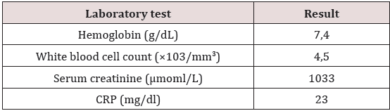
A 35 years old woman with a history of a right mastectomy in December 2014, for ductal carcinoma in situ with immediate breast reconstruction with silicone prosthesis. The assessment of extension is normal. She was hospitalized fifteen days after surgery for urinary infection with discovery of renal failure with a creatinine level of 500μmol/L; she was put under antibiotic treatment and is then output with a creatinine of 200mol/L. The patient was transferred to our department in late January 2015 for investigation of the renal failure (creatinine level 717μmol/l). Her physical examination was normal excluding significant edema of the lower members and normalarterial high blood pressure. Additional testing revealed microscopic hematuria, renal failure, elevated CRP and anaemia (Table 1).

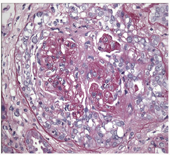
Based on acute progressive renal failure with proteinuria and hematuria, kidney biopsy done revealed focal segmental necrotizing and pauci-immune crescentic glomerulonephritis, with moderate activity and minimal chronicity, minimal tubular atrophy, and interstitial fibrosis (Figure 1). Immunofluorescence microscopy was negative for any significant immunoglobulins and complement deposition. All immunological tests were negative: Anti Nuclear Antibody (ANA), cryoglobulins,cytoplasmic antibodies neutophilespolynuclear (ANCA), Anti Membranes Basal Glomerular (anti MBG). The thoracic scanner did not show a diffuse alveolar hemorrhage. Anechocardiogram showed severe left ventricular dysfunction with ejection fraction of 32%. The silicone prosthesis was criminalized. Breast implant was removed. The patient was treated with a combination of pulses of methylprednisolone and cyclophosphamide. The evolution was unfavourable without improved renal function. The patient chose hemodialysis; immunosuppression was decreased and stopped.

Figure 1: Glomerulus showing cellular crescent formation (H and E stain ×100).

lupinepublishers-openaccess-journal-urology-nephrology

Table 1: Laboratory tests during hospital admission.

lupinepublishers-openaccess-journal-urology-nephrology

Discussion

The relationship between silicone breast implants and autoimmune or connective-tissue diseases has been the focus of considerable medical and legal discussion throughout the past decade. Although evidence of a causal relationship between the implantation and the development of connective tissue disease is circumstantial, removal of the silicone prosthesis has been reported to result in subsequent remission [1]. Silicones are not biologically inert. Injectable as well as implantable silicones have proven capable of eliciting inflammatory and fibro proliferative responses [2]. Silicone exposure is associated with different systemic autoimmune diseases such as systemic lupus erythematosus, rheumatoid arthritis, progressive systemic sclerosis, and vasculitis [3]. We postulate that silicone in silicone-filled breast implants may increase the risk of developing (auto) immune diseases and immune deficiencies [3].

Silicone implants may cause local complications (such as capsular contracture, rupture, closed capsulotomy, gel “bleed”, nodular foreign body granulomas in the capsular tissue and lymph nodes) or general symptoms. An adverse immune reaction with signs and symptoms of rheumatoid disorders is also possible [4]. The development of disease related to silicone implants would depend on genetic factors, so that only a very few women are potentially at risk. HLA-DR53 may be a marker of predisposed subjects [4]. From a public health perspective, breast implants appear to have a minimal effect on the number of women in whom connective-tissue diseases develop, and the elimination of implants would not be likely to reduce the incidence of connectivetissue diseases [5]. The mechanism of silica exposure in the development of small vessel vasculitis is not well understood but several potential mechanisms have been proposed [6]. One theory suggests that silica particles stimulate production of lymphocytes, including T cells and B cells, and that in certain clinical and genetic settings causes autoimmune disease as well as the production of auto antibodies, including ANCA [6]. A second theory suggests that silica particles activate monocytes and macrophages, resulting in the release of IL- 1 or tumour necrosis factor-𝛼, oxygen-derived free radicals, and lysosomal enzymes such as PR3 and MPO [6].

Conclusion

Silicone implantation is associated with scleroderma, systemic lupus erythematosus, and rheumatoid arthritis. This case report indicates the possibility of the development of small-vessel vasculitis after silicone breast implantation.

Read More Lupine Publishers Journal of Urology Please Click on Below Link:
https://lupine-publishers-urology-nephrology.blogspot.com/

Thursday, 15 April 2021

Lupine Publishers | How Might the UK Government Engage with the Public on Compulsory Vaccination Debate in the United Kingdom?

 Lupine Publishers | Journal of Clinical & Community Medicine


Abstract

This essay suggests three ways in which compulsory vaccination can be taken forward should the UK Government decide to do so. It addresses the question of; how can we ensure that public concerns about compulsory vaccination are considered if compulsory vaccination is to be introduced by the UK Government? Arguments for and against compulsory vaccination from the literature ate provided. However, the interest here is not whether compulsory vaccination should be adopted or not but how best to take this forward should the UK Government decide to take this forward.

Background

On the 29th of September 2019, the United Kingdom (UK) Government suggested that it was “looking very seriously” into the introduction of compulsory vaccination for school children [1]. This follows the recommendation of four London senior National Health Service (NHS) GP’s including Sir Sam Everington, Dr Mohini Parmar, Dr Andrew Parson and Dr Josephine Sauvage [1]. The UK Health Secretary, Matt Hancock, speaking at the Tory conference, said: “I’m very worried about falling rates of vaccinations, especially measles” ... “For measles, the falling vaccination rates are a serious problem, and it is unbelievable that Britain has lost its measles-free status.” [2]. Earlier this year, the UK lost it ‘measles eradicated’ status with the World Health Organisation (WHO) [3]. Worldwide, figures show a rise of 300% reported cases of measles in the first three months of 2018 when compared the same time period of the previous [4]. MMR vaccine rates in England has been in the decline since the last five years [5] and according to figures from the Childhood Vaccination Coverage Statistics England 2018-2019 – there was a decline from 91.2% of vaccinated children to 90.3% when data was compared with the previous year.

Compulsory vaccination has previously been introduced in other countries such as the United States and Australia [6]. The main argument is that compulsory vaccination protects other children who cannot be vaccinated for other medical reasons. Compulsory vaccination is neither new in the UK, for example, when compulsory smallpox vaccination was introduced for all children born after 1853 [7]. Childhood vaccination programmes have seen successful eradication of diseases such as smallpox in the UK and brought under control other diseases such polio, diphtheria, whooping cough, and meningitis, and they extend back to the nineteenth century. Nevertheless, vaccination remains voluntary in the UK, and as such, public communication, education and trust have been relied upon [8] as a means to maintain high uptake to ensure herd immunity [9].

Other concerns for health authorities, is the rise in unproven theories linking vaccines to diseases such as autism, multiple sclerosis and diabetics; and the rise of anti-vaccination movement on social media on the other hand. For example, Wakefield et al. [10] published in ‘The Lancet’ described twelve children aged between three and ten, suffering from developmental regression and gastrointestinal problems. Andrew Wakefield in a press conference prior to this publication suggested a possible link between measle, mumps and rubella (MMR) vaccine and autism. However, the study could not be replicated elsewhere. The Lancet paper was retracted and Andrew Wakefield was struck off the British medical register by the General Medical Council for serious professional misconduct in 2010 [11]. Nevertheless, this false hypothesis continues to shape the MMR vaccine debate especially with the rise of social media anti-vaccine movement. According to the UK Health Secretary, “If you don’t vaccinate your child and you can, then the person you are putting at risk is not only your own child, but it is also the child who can’t be vaccinated for medical reasons”. He called those who spread anti-vaccine messages as having “blood on their hands” asking social media platforms to do more to curb the ant—vaccination movement on social media [12].

Compulsory vaccination to a wide range of childhood diseases such as polio, measles, mumps, and rubella and pertussis have in the past met with fierce legal and legislative challenges justified by ideological, scientific, religious and political philosophies. Thus, it is expected that moves to make the vaccine compulsory in the UK would generate a new round of emotionally charged debates. In response to Government suggestion of the possibility of introducing compulsory vaccination, the British Society for Immunology chief executive Dr Doug Brown said: “to make this compulsory in the UK, there are concerns that it could increase current health inequities and alienate parents with questions on vaccination” [13]. Dr Brown noted that more can still be done in terms of information campaigns and the delivery of local immunisation in communities. The Royal Society for Public Health noted that “compulsory vaccination should be a last resort.

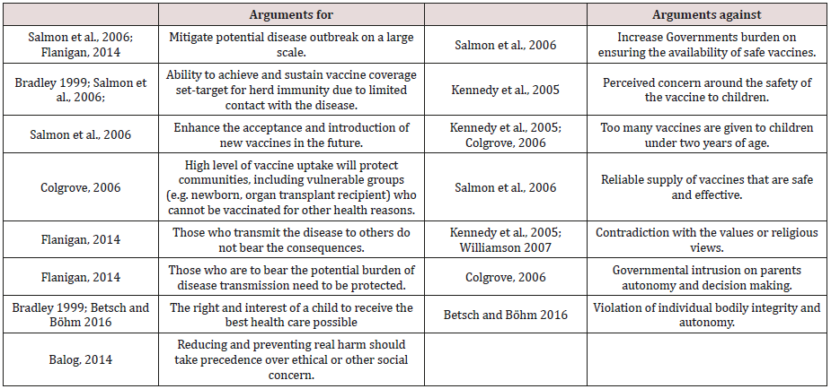
Table 1 below set out arguments for and against compulsory vaccination. This review is not meant to be exhaustive, but rather, it is a selection of literature that illustrates some of the core arguments for or against compulsory vaccination debate. The focus of this opinion essay is not whether compulsory vaccination should be adopted or not but how best to take this forward should the UK Government decides to take this forward.

Table 1: Arguments for or against compulsory vaccination.

Lupinepublishers-clinical-community-medicine

Compulsory Vaccination and Public Engagement

An interesting question therefore is; how can we ensure that public concerns about compulsory vaccination are considered if compulsory vaccination is to be introduced by the UK Government? Research on risk communication has shown that simply understanding the rationale and the science behind such policy intervention does not necessarily translate to trust and acceptance of public health policy interventions. Often times, the public would have concerns and questions which they would want to be answered even if the science is settled, see for example. This essay, therefore, suggests three ways in which compulsory vaccination can be taken forward should the UK Government decide to do so.

Firstly, debates about new public health policy interventions are never just about science and evidence. This is especially the case where science conflict with group beliefs, values or religious convictions as proven by psychometric and social theories of risk. People will interpret public health policy interventions in a range of different contexts including their situational context. Therefore, the Government must be willing to understand the variety of political, ethical, scientific, cultural, religious and ideological context that exist and how this would affect the interpretation that will be brought to bear on the compulsory vaccination intervention by the Government. This way, the Government could set out exceptions and boundaries, in a way that would effectively ease any backlash or resistance that could arise if the policy is implemented.

Secondly, there is a need for Government to understand what people’s hopes and concerns about compulsory vaccination are. This is important as the public, who are adopters of policy intervention, should have a say in how, where and who is affected by mandatory vaccination. Important questions that could potentially arise here are - would the public value their autonomy than been compelled? This sits within the libertarian or nannystate arguments where some critic would see this as a step too far into family matters. Other questions are: who decides who is compelled or not and in what conditions? What evidence decides this parameter? Can parents delay child vaccination should they have concerns? It would also be good for the Government to set out how public concern raised will be considered and addressed. This will ease the burden and pressure around public acceptability of any compulsory vaccination policies.

An example of a recent public engagement on public health interventions can be seen in the case of the electronic cigarette (EC) debate; even if the public engagement tended to be limited to the acceptability debate and not necessarily the technical debate [14]. The EC debate was triggered by the sharp rise in the use of EC in the UK, and a call by the WHO in 2008 who raised concerns that ECs were being marketed as a safer alternative to tobacco cigarettes despite a lack of, or insufficient, scientific understanding of the safety and efficacy of ECs at the time (WHO, 2008). EC is currently being regulated under the EU Tobacco Products Directive (TPD) following public consultation carried out in 2010 on whether to bring nicotine-containing products (NCPs), including ECs, within the medicines licensing regime. This regulation came into effect in May of 2016 and according to the directive, ECs containing up to 20mg/ml of nicotine will be regulated by the TPD. However, whether this regulation will change in light of recent EC health concern (vaping illness) in the US [2] and following Britain’s exit from the European Union, remains unknown as at the time of writing this article.

Finally, like much other public engagement, the Government will need to be clear and honest if, why and when they want to engage with the public if there are to take compulsory vaccination forward. Risk communication research has shown that public engagement can open up areas of agreement and conflict that can have implications for trust and risk acceptability. However, they must be used in ways that challenge, rather than reinforce powerful incumbent societal structures [15,16]. If compulsory vaccination is to be introduced, then public engagement will be beneficial to empower and give voice to concerned public groups. Then, the objective of this engagement should guide when and how the public debate should take place. In the case of the electronic cigarette debate, for example, the MHRA-led public consultation was influential in shaping the questions and arguments that were brought to bear on the vaping debate [17-24].

Read More About Lupine Publishers Clinical and Community Medicine Please Click on Below Link:
https://journalofclinicalcommunitymedicine.blogspot.com/

Wednesday, 14 April 2021

Lupine Publishers| Internet of Things and Privacy

 Lupine Publishers| Current Trends in Computer Sciences & Applications


Opinion

The Internet of Things is a concept that has been heard quite a lot in recent years, a concept that slowly emerged, but over time it has experienced a booming growth that was then adopted not only by giant IT companies, offering according to the directions of each company that adopts the corresponding services and applications to the end user. Undoubtedly this technology has come to stay for a long time, it has come to improve our living conditions and to simplify habits and functions that required time and many times difficulties. The Internet of Things is almost everywhere around us from the super market to the cars we drive, our everyday life is going easy and we are happy to live with, but there are two important points that we should consider:

a) Uncontrolled product design based on the Internet of Things.

b) Access to data managed by Internet of Things are inaccessible by the users.

According to the above, important questions arise, such as:

a) How and where these devices store and manage the data now?

b) How and where these devices store and manage data in the future?

c) What personal data are collected and for whom?

d) How protected they are from hacking attacks?

e) How capable is an Internet of Thing to take full control of an information system?

Surely for all of us who are involved in the security of information systems, we have a lot of work to do. The biggest challenges we have to face are:

a) No one knows an Internet of Thing how it collects, how it uses, and where stores personal data. One could construct an Internet of Thing that behaved properly according to the purpose it was created, but it could also act as intelligence espionage product to transmit personal sensitive data and information related to the online traffic and services of an organization, and the worst scenario is that this device can work for years without being noticed.

b) Depending on the complexity of the smart device, the amount of data it sends is increasing, for example a complex smart device can send up to 5 times the volume of unidentified data.

c) The Internet of Things, as I mentioned above, was designed to make our lives easier because technology evolves and these devices evolve, so many unknowingly use them in a computer room or in critical security infrastructures for which the devices have not been made for this purpose, and of course with unknown security implications for the company’s network since these devices do not have an management interface for the user, so it is impossible to access them.

d) Depending on the type of the device, third-party information is internally embeded, for example GPS maps, geostrategic data, human habits information, transaction information, which will then have to work all together for the Internet of Thing.

e) We do not know exactly what kind of data and metadata are collected, it is enough to ponder that an IoT card containing basic medical data of a patient sends about 200MB of unknown data per year.

f) The Internet of Things are not safe enough in external attacks, a denial of service attack on an Internet of thing could be the entry for violating an information system and collect sensitive information from it.

g) The construction of these devices varies, there are not enough standards to build and protect personal data, no one can guarantee that an Internet of Thing will protect its owner against an external threat, which for example could get the control of a car with unpleasant consequences for the driver.

Considering the above, we should consider whether we really need an Internet of Thing and if this is necessary, how much we can parameterize and access to it [1,2].

Read More About Lupine Publishers Current Trends in Computer Sciences & Applications Please Click on Below Link https://computer-sciences-lupine-publishers.blogspot.com/


Friday, 9 April 2021

Lupine publishers | Risk Stratification at Patients with the Defect Vessels and High Blood Pressure: New Mathematical Model

 Lupine Publishers | Journal of Complementary & Alternative Medicine


Short Communication

It is noted, that today the population of Uzbekistan unprecedentedly increases consumption of salt and have low physical activity in general. Besides for the last 25 years, the population of Uzbekistan was enlarged twice. Today such tendency of population increase is followed by its aging. The positive aspect of this phenomenon is augmentation of average life expectancy. However, a negative side that the augmentation of life expectancy doesn’t correspond to its quality. Deterioration first of all is bound to augmentation of prevalence of age chronic diseases, such as a hypertension. For example, today in Uzbekistan, one of three adults has raised by the ABP, and excess weight occurs at every second (WHO/STEPS 2014). As a result it caused double increasing of visits of the doctor and total number of patients in general. The purpose of our research was in developing the prognostic model of the turnpike arteries biological age at hypertensive patients. To study the arterial blood pressure daily profile at sick with the arterial hypertension (AH) with the metabolic syndrome (MS) [1].

Material and Methods

The study included 96 healthy volunteers and 96 men with arterial hypertension AH in average age of 56.06 ± 7.86 years, diagnosed abdominal obesity (BMI 34.54 ± 3.83 kg/m2) and metabolic disorders. In order to estimate the clinical status, the following risk factors were studied: elevated arterial BP, smoking; clinical and biochemical parameters: 12-lead ECG; 24-hour Holter ECG monitoring; exercise stress test; echocardiography (EchoCG); carotid artery intima-media thickness (IMT) [2,3].

Prognostic Model

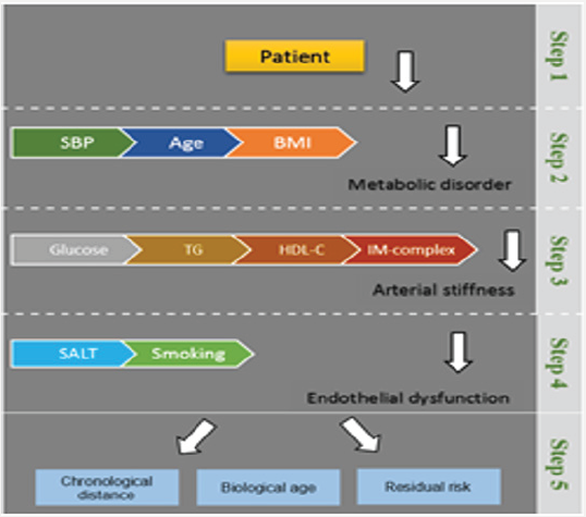
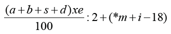
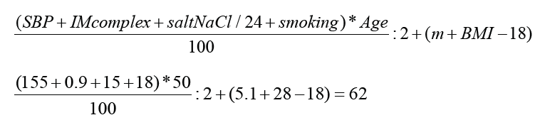
We selected clinical signs for creation of the prognostic table by means of the method of the consecutive diagnostic procedure based on a technique of the sequential analysis offered by A. Wald. For each informative sign gradation of this or that indicator were selected to equal the diagnostic value of each of indicators (Figure 1). On the basis of local research grant analysis the calculator of risk was developed to provide patients with AH I-II -degrees the important prognostic information. We also considered that population of modern Uzbekistan have tendencies of negative impact at vessels damage such as: salt and the increased body weight. As a result, nine best factors for stratification of risk at patients with the defect vessels and raised SBP, including easy available, clinical parameters were included in this calculator (Figure 1). We applied multistage stratification of risk, based on nine parameters which were included in the developed equation of nine factors [4]:

Figure 1: Prognostic model.

Lupinepublishers-openaccess-complementary-alternative-medicine-journal

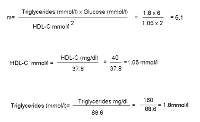
(a-SBP; b- IMT intima-media thickness; s- Salt; d-smoking; e-age; *m- metabolic index *m= t x g / h2; i-BMI; t- Triglycerides, g- Glucose; h- HDL-C).

Lupinepublishers-openaccess-complementary-alternative-medicine-journal

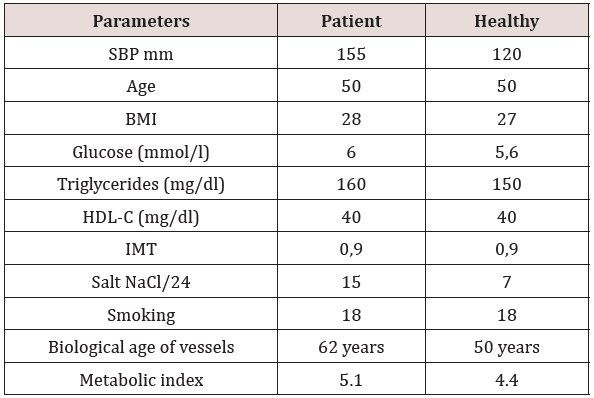
During this experiment, patients noted informational content of the calculator for the objective comprehension of their cardiovascular status (Sensitivity–85 %, Specificity–63%) (Table 1). However long-term preventive effect, in prevention of the recurrence was not confirmed [5-7].

Table 1: Example Clinical data.

Lupinepublishers-openaccess-complementary-alternative-medicine-journal

Conclusion

On completion of the therapy clinical tests indicated the high sensitivity at average specificity model that was acceptable in an opportunity to estimate medical effect. Unfortunately, the calculator could show the rate of damage vessels in the cases of actual disease, but not in cases of its prediction or probable emergence.

Read More About Lupine Publishers Complementary Medicine Journal Please Click on Below Link
https://lupine-journal-complementary-medicine.blogspot.com/

Thursday, 8 April 2021

Lupine Publishers | Approach to Neurodegenerative Disease in Children: A Short Review

 Lupine Publishers | Journal of Pediatrics and Neonatology


Abstract

Neurodegenerative disorders of childhood are complicated diseases with wide range of systematic involvement. These diseases often pose great challenge to clinicians in terms of diagnosis and management. The purpose of this article is to outline a systematic approach to a child presenting with suspected neurodevelopmental regression. Many inherited metabolic disorders present with neural regression. The clinical approach depends upon the age of presentation, site of involvement in brain. Sound clinical knowledge and better approach leads to early diagnosis, better management and above all genetic counselling. As the medical science is in the track of rapid progression several treatment modalities are in the pipeline for neurodegenerative syndromes, early diagnosis and referral to higher centres can bring a better future to the child.

Keywords: Neurodegenerative diseases; Hepatomegaly; White Matter; Grey Matter

Introduction

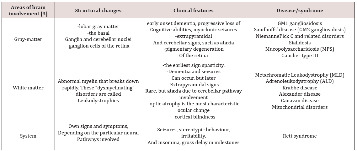
Neurodegenerative disorders of childhood include large, heterogeneous group of diseases that result from specific genetic and biochemical defects, chronic viral infections, and varied unknown causes. The hallmark of a neurodegenerative disease is regression and progressive deterioration of neurologic function with loss of speech, vision, hearing, or locomotion, often associated with seizures, feeding difficulties, and impairment of intellect [1]. The acquisition of new developmental milestones does not exclude the existence of a degenerative disorder. Most degenerative CNS disorders can be divided clinically into three groups: gray-matter diseases, white-matter diseases, and system diseases [1,2] (Tables 1-3).

Table 1:Area of brain involvement.

lupinepublishers-openaccess-journal-pediatrics-neonatology

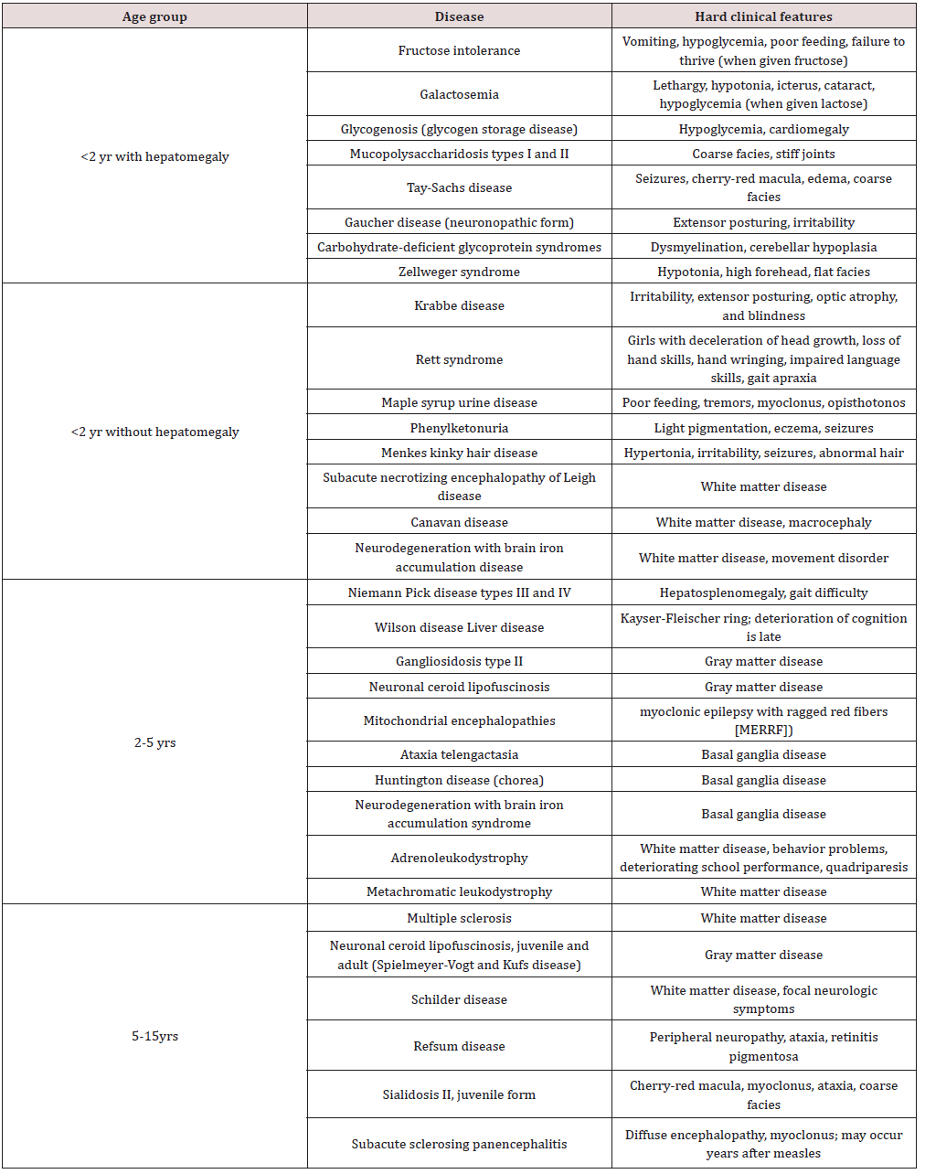
Table 2:Approach according to age of presentation [4].

lupinepublishers-openaccess-journal-pediatrics-neonatology

Table 3:Treatable conditions with neural regression [5] loss of social skills.

lupinepublishers-openaccess-journal-pediatrics-neonatology

Classification

Approach to Child with Marked Regression

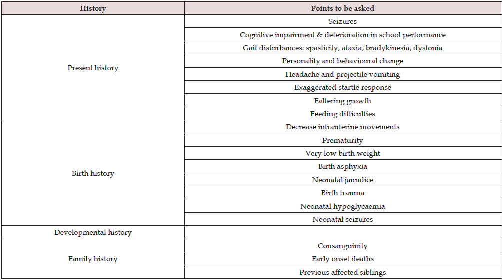
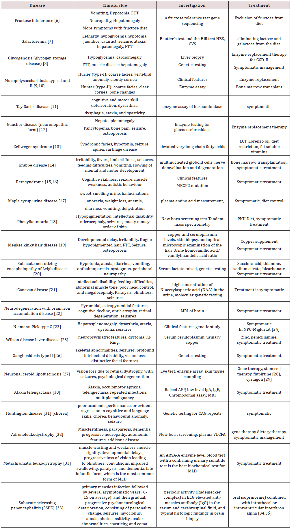
a) History: In a patient with developmental regression the history is very vital. Clues for diagnosis lie in the history. First step would be to ascertain the age of onset of regression and the acquisition of various milestones prior to that. Was the child completely normal before regression or was there a concern regarding development even prior to regression? Did the child attained milestones before losing it. Which milestones the child lost? In autism and other pervasive developmental disorders regression of language skills is noted first followed by loss of social skills (Tables 4 & 5).

Table 4:Specific pointers in history.

lupinepublishers-openaccess-journal-pediatrics-neonatology

Table 5:Individual disease and clues for diagnosis.

lupinepublishers-openaccess-journal-pediatrics-neonatology

Wednesday, 7 April 2021

Lupine Publishers| Nano Toxicity: Due to Drug Delivery and Environmental Exposure

 Lupine Publishers| Archives of Nanomedicine: Open Access Journal


Abstract

Nanotechnology is undergoing a vast expansion in materials science, Research and Development. Nano scientists are focusing on synthesis and development of nanoparticles, nanomaterials, and bio nano composite materials. The drug delivery is also a recent development where in bio nano materials are being used for diagnosis of the various diseases. The synthesis of nanomaterials at large scale causes health risk due to the exposure via inhalation, skin contacts and ingestion; based on the characterisation of bio nano materials. The use of bio nanomaterials in drug delivery as well as the environment exposure during the large-scale synthesis of nanomaterials, the bio nanomaterials into human body.The exact mechanisms, chemical reactivity and enzymatic reaction is not well understood, documented, and studied. Therefore, the intake bio nanomaterials via drug delivery or environment exposure amounts to health risk and need to be studied in detail.

Introduction

Nanosciences and Nanotechnology is the study and use of nanomaterials falls in the range of 0.1nm to 100nm which corresponds to 0.2nm- water molecule, 7nm-haemoglobin, 10-100nm - virus, -1μm - microbial cells and >2μm - protozoa. The synthesized and developed nanoparticles, nanomaterials, and Bio nanomaterials are being used in various fields. The recent advances in the field of material sciences include the synthesis of Bio nano material for use in drug delivery. Bio nanotechnology companies are designing drugs for various diseases such as heart disease, kidney stones, and cancer cosmetic generic products using a short fragment of DNA as a new type of drugs. These drugs are assembled in nano chips and as nanoparticles for delivering into human body and are effective in using the sick/diseased and healing the injuries. Bio nano products are diverged as bio chip and Nano medicine, bio nanotechnology products which include Nano medicine, nano material, micro detectors, Nano sensors and herbal medicine [1].

Drug nano crystals are particles made from 100% drug; typically, surfactants or polymeric steric stabilizers stabilize them. These particles possess a 100% drug loading in contrast to matrix nanoparticles consisting of a polymeric matrix (polymeric nanoparticles or a lipidic matrix i.e. Nano emulsions, liposomes | or lipid nanoparticles. Thus, the high loading makes them very efficient in transporting drug to or into cells, reaching a sufficiently high therapeutic concentration for the pharmacological effect [4-8].

Health Risk

The scientific evidence demonstrates the potential for nano material to be toxic to the humans or the environment; therefore, synthesis of nanoparticles and bio nano composites and their use causes health risk due to intake – drug delivery and environment exposure that need to be studied before making the wider application of bio nanomaterials. The smaller a particle, the greater it’s surface area to volume ratio and the higher its chemical reactivity and biological activity. The extremely small size of nanomaterials also means that they are more rapidly taken up by the human body than larger sized particles. Nanomaterials can enter into the body through inhalation, ingestion or skin contacts. Nanomaterials are able to cross biological membranes and access cell tissues and organs. The greater chemical reactivity of nanomaterials results in increased production of reactive oxygen species, including free radicals. Reactive oxygen species and free radical product is one of the primary mechanisms of nanoparticles toxicity. Other properties of nanomaterials that influence toxicity include chemical composition, shape, surface structure, surface charge, aggregation and solubility and the presence or absence of functional groups of other chemicals [9-11].

Mode of entry of Nano particle:

The Nano particle ranges between 1nm to 100nm which can enter into the body through inhalation, skin contact and ingestion. The synthesis of nano particle at large scale will cause exposure through these routes.

a. Inhalation:

Inhalation is the most important route for the intake of airborne nano particle. Depending on the size, particles are trapped in mucous layer and alveoli. For nano particle the position is more complex. Particles of 1 micron diameter or more tend to be deposited, but only those less than 7.0 microns, deposit deep inside the lungs. Those more than 7.0 micron deposit in the conductive airways. Particles in size less than 0.1 micron deposit in the alveolus. Most of the particles between 0.1 and 0 micron size are exhaled. The pattern and depth of breathing and irritant effects of inhaled material may alter the deposition of particles and may remain permanently within the lung tissue.

b. Skin contact:

The large scale synthesis of nano particles in industry for wider application will cause exposure of nano particle through skin absorption; the penetration of nano particle through skin occurs via lipids and dissolved material. Lipid solubility and molecular size are the most important factors, so that higher lipid solubility and small molecular size enhance penetration through skin. Abrasion and irritation also encourage penetration. This route is particularly important for organic solvents and can occur in a number of ways.

(i) Direct absorption through wounds or abrasions.

(ii) Degreasing of the skin followed by absorption of the degreasing agents.

(iii) Degreasing of the skin allowing absorption of other chemicals.

(iv) Sensitisation, local and general.

b. Skin contact:

Ingestion of nano materials during the process of synthesis may result from the contaminated object into the mouth. Ingestion of toxic substance along with food in the workroom occurs where housekeeping is not good, or where workers are careless to nano particles in their clothes, or wash their hands with soap. If the toxic nano dust swallowed with food or saliva is not soluble in body fluids, it is eliminated directly through the intestinal tract. Toxic materials that are readily soluble in body fluids are absorbed in the digestive system and circulated by the blood. Compared with inhalation and skin absorption, ingestion, plays a minor role in the absorption of toxic materials in industries [2-3].

Toxicity of Nanomaterials

The intake of bio nanomaterials in human body undergoes biochemical mechanism and enzymatic interaction and height cause. Toxicity of nano particles depending on nature of chemical used for the synthesis, type of precursor, concentration of precursor, duration of exposure, personal susceptibility, and mode of entry, size of nano particle, environmental factors, and threshold limit value.

Conclusion

The drug delivery is one of the routes for treating diagnosis using the bio nano material. The exact fate of bio chemical reactivity, enzymatic interaction is not well understood and studied and might lead to toxicity similar to that of exposure of nano material through inhalation, skin contact and ingestion. Therefore, synthesis of nano particle, bio nano composite, their use and environmental exposure need to be studied before making the wider application for the diagnosis of disease using bio nano materials. The detail of physico chemical characteristics, stability of nanomaterials and their specification to target organs as human body system need data base scientific research.

Read More Lupine Publishers Archives of Nanomedicine Open Access Journal Please Click on Below Link https://lupine-publishers-nano-science.blogspot.com/


Tuesday, 6 April 2021

Lupine Publishers| Reversal of Rocuronium-Induced Neuromuscular Block with Neostigmine in the Libyan Patients

 Lupine Publishers| LOJ Pharmacology & Clinical Research


Abstract

Background: Reversal of the enduring effect of rocuronium by neostigmine is a common procedure performed in the Libyan hospitals. The reversal of the continuing effect of rocuronium by neostigmine was also evaluated.

Methods: eighty adult surgical patients were included in the study using neostigmine 2.5mg (0.05-0.07mg/kg) to reversal the block induced by rocuronium 0.6 mg/kg. Anaesthesia was induced and maintained using i.v. propofol (2.5mg/kg) and fentanyl (1.5μg/kg). Reversal neuromuscular function was monitored using clinical signs includes patient responsiveness, subjective measurements of muscle strength (5 second head lift, hand grasp), eye opening, and tongue extrusion.

Results: Reversal of block was sustained in all patients from the enduring effect of rocuronium by neostigmine. Ninety-six patients were had a similar time of recovery but eleventh were not. There were no serious adverse effects from neostigmine and no significant changes in any measure of safety.

Conclusions: neostigmine is capable of reversing rocuronium-induced blockade in the Libyan patients by monitoring the muscle strength, eye opening, and tongue extrusion.

Keywords: Acetylcholinesterase; Neostigmine; Propofol; Fentanyl; Anaesthesia; Rocuronium

Introduction

Acetylcholinesterase (AChE) is catalyzing the quick hydrolysis of acetylcholine (ACh) to acetate and choline. The main biological function is being annihilation of impulse transmission at cholinergic synapses. In addition, AChE is believed to have nonclassical roles in nerve and muscle growth and in hematopoiesis [1,2]. Additionally, AChE has been concerned in Alzheimer’s disease [3,4], hypersensitivity to pesticides and Gulf War syndrome [5]. Rocuronium is frequently used non-depolarizing neuromuscular blocking agents (NMBAs) to facilitate tracheal intubation and affording muscle relaxation throughout surgery. Patients receiving these agents are at danger of outstanding curarization, which is a cause of postoperative pulmonary complications and might augment postoperative mortality [6,7]. Anticholinesterases drugs act mainly by inhibiting acetylcholinesterase and butyryl cholinesterase, prolonging the existence of acetylcholine at the motor end-plate [8]. Additionally, anticholinesterases may have a direct agonistic effect by increasing the release of acetylcholine from presynaptic nerve terminals [9]. For, edrophonium the maximum effective dose is 1.0-1.5mg/kg and for neostigmine, is 60-80μg/kg [8]. The use of two antagonists together is avoided as they are not additive and insufficient reversal can occur. It is not sensible to administer extra anticholinesterase if maximal doses of edrophonium (1.5mg/ kg), neostigmine (70μg/kg), or pyridostigmine (350μg/kg) fail to antagonize the residual blockade and might in turn increase the weakness [9]. They are combined with atropine or glycopyrrolate in order to counteract the muscarinic side-effects of these drugs. Neostigmine is the most potent and the preferred drug [9]. Highdose neostigmine or unnecessary use of neostigmine could interpret to increased post-operative respiratory morbidity [10,11]. Current rules identify the use of reversal with neostigmine based on the train of four (TOF) monitoring with the neuromuscular monitor. Neostigmine can be given for reversal in patients: who were receiving drugs which enhance the action of NMBAs (inhalational agents). The reversal should not be given in case of TOF counts four in patients receiving anaesthetic drugs which do not boost the blockade by NMBAs (intravenous anaesthetics) [10,11]. Moreover, the rules also identify that if the TOF counts less than 2 reversal should not be delayed. A lower dose of neostigmine (20μg/kg) must be considered if TOF counts four and no fade is apparent or if TOF ratio is 0.4:0.9 on qualitative neuromuscular monitoring [12]. Neostigmine is a cholinesterase inhibitors and it is wildly used in Libya as indicated in the literature as reversal agents for NMBAs [13,14]. Sugammadex, which is a novel agent for the reversal of neuromuscular blockade, is adapted gamma-cyclodextrin. Sugammadex is able to form a complex with rocuronium, eradicates it from the circulation and terminates neuromuscular blockade [15]. Sugammadex is a very safe agent with a little risk of serious side effects [16]. The high cost of sugammadex, which is one of the costliest drugs in anesthesia practice, prevents it from being used in Libya as a standard neuromuscular reversal drug. Even though it has been suggested by many doctors in Libya that the cost of sugammadex use in anesthesia could be reduced by shortening the duration of recovery [17,18], further clinical studies on sugammadex in Libya are needed to introduce it to the governmental and private hospitals.

It has been reported that the co-administration of neostigmine with some Nonsteroidal Anti-inflammatory Drugs (NSAIDs, e.g. aspirin) resulted in a synergistic interaction, which may provide evidence of supraspinal antinociception modulation by the increased acetylcholine concentration in the synaptic cleft of cholinergic interneurons. The interaction obtained between neostigmine and the NSAIDs could carry important clinical implications [19,20]. Acute toxicity from using cholinesterase inhibitors is related to the inhibition of acetylcholinesterase activity at the neuromuscular junction and in the brain, resulting in depression of circulatory centers in the medulla, weakness of the muscles of respiration, and pulmonary edema [21]. Because of the toxicity relates with inhibition of acetylcholinesterase activity rather than butyryl cholinesterase activity, it would seem reasonable to spotlight on genetic variants of acetylcholinesterase. Though genetic variants of human acetylcholinesterase exist, harmful mutations are uncommon and occur only in the heterozygous state [22]. The aim of this study is to investigate the effects of neostigmine’s introduction on the incidence of residual neuromuscular paralysis and postoperative Libyan patient outcome at Al-Shyfaa Private Clinic, Tripoli, Libya.

Materials and Methods

Qualitative and Quantitative Determination of Neostigmine: The Neostigmine methyl sulphate was obtained from four different companies which are: Neostigmine methyl sulphate (Rotexmedica, Germany); Plantigmine (Polifarma, Turkey); Flagstig (Thexopharma, UK) and Neostigmine (Adeka, Turkey). The reference standard was obtained from Sigma-Aldrich (cat. no. 2126). The identification test for the neostigmin was done using absorbance spectra which were measured on a Jenway UVvisible spectrophotometer, model 6505 (London, UK) using quartz cells of 1.00 cm path length. The UV-Vis absorbance spectra were recorded in the 200-500 nm range, and spectral bandwidth of 3.0 nm. It was performed the baseline subtraction of the water for the final spectrum of each solution analyzed. An equivalent to 5 mg of neostigmine methylsulfate Injection according to the labeled amount was taken and completed to 10 mL with distilled water, and the absorption spectrum of this solution was measured by Ultraviolet-visible Spectrphotometry which should exhibit a principle band at 261nm. In addition the identity of neostigmine was also determined using HPLC technique by determining the retention time of the major peak in the chromatogram of the four Neostigmine methylsulphate brands preparations compared to the retention time of the major peak in the chromatogram of the standard preparation. The quantitative assay of the neostigmine (Neostigmine Methylsulfate Injection) was perofromed by Waters HPLC using the Japanese Pharmacopoeia (Sixteenth Edition), page 1168.

Molecular Docking: The starting geometry of neostigmine was constructed using chem3D Ultra (version 8.0, Cambridgesoft Com., USA). The optimized geometry of neostigmine with the lowest energy was used in the molecular docking. The crystal structures of human acetylcholinesterase in a complex with a transition-state analogue were downloaded from the Protein Data Bank. The molecular dockings of neostigmine with human acetylcholinesterase was accomplished by AutoDock 4.2software from the Scripps Research Institute (TSRI). Firstly, the polar hydrogen atoms were added into human acetylcholinesterase and neostigmine molecules. Then, the partial atomic charges of the human acetylcholinesterase and neostigmine molecule were calculated using Kollman methods [23]. In the process of molecular docking, the grid maps of dimensions (62Å X 62Å X 62Å) with a grid-point spacing of 0.376Å and the grid boxes is centered. The number of genetic algorithm runs, and the number of evaluations was set to 100. All other parameters were default settings. Cluster analysis was performed on the results of docking by using a root mean square (RMS) tolerance of 2.0Å, and this was dependent on the binding free energy. Lastly, the dominating configuration of the binding complex of neostigmine and human acetylcholinesterase with minimum energy of binding can be determined.

Study Design and Patient Selection: This study was conducted at APC (Al-Shyfaa Private Clinic, Tripoli, Libya) during July/August 2018. The protocol was accepted by the Independent Ethics Committee at the clinic and conducted in fulfillment with the recent amendment of the Declaration of Libyan Guidelines, present regulatory requirements and Good Clinical Practice. Eighty patients were included in the study and they were aged 20-60 years and experiencing surgery in the supine position under general anaesthesia (Laparoscopy surgery) which are requiring muscle relaxation. Patients were excluded if they were anticipated to have a neuromuscular disorder, difficult intubation for anatomical reasons; family history of malignant hyperthermia, significant renal dysfunction; or a known allergy to NMBAs, narcotics, or other medication used throughout general anaesthesia. Patients receiving anticonvulsants, antibiotics, or magnesium at a time possible to hinder with neuromuscular block effect were also excluded. Female patients who were breastfeeding, pregnant, childbearing potential were also excluded. All patients have presented written consent. Subject numbers were allocated to patients in chronological order of their participation into this study. Anaesthesia was induced with i.v. propofol (2.5mg/kg) and fentanyl (1.5 μg/kg) and maintained using a continuous infusion of propofol and additional increments or infusions of analgesic as needed. After the establishment of neuromuscular scrutinizing, rocuronium 0.6mg/kg was given as an i.v. bolus over 10 seconds into a quick running i.v. infusion. Tracheal intubation was performed once the maximum neuromuscular block effect is achieved and intermittent positive pressure ventilation started to achieve a standard end-tidal CO2 concentration (4.5-5.5 kPa). Additional doses of rocuronium 0.1-0.2mg/kg up to a highest of two dosages were administered if required. Once the last dose of NMBA was administered and reappearance of T2 then neostigmine 0.05-0.07mg/kg (maximum of 2.5mg) was administered within 10 seconds into a quick-running i.v. infusion.

Monitoring: Using clinical signs comprises patient responsiveness, individual measurements of eye opening, muscle strength (5 second head lift, hand grasp), and tongue extrusion. After extubation (removal of the endotracheal tube (ETT) and it is the final step in liberating a patient from mechanical ventilation), clinical evaluation of level of consciousness and neuromuscular recovery (5 s head lift and general muscle weakness on a scale of 1-9) were carried out each 15 min until the first continued head lift for 5 second was attained. Oxygen saturation using breathing frequency and pulse oximetry were watched during anesthesia and in the recovery room for at least one hour after operation. Heart rate and arterial blood pressure were traced at the screening, before administration of rocuronium, before and two, five, ten, and thirty minutes after administration of neostigmine, and during the post-anaesthetic visit, which was carried out within the first twenty-four postoperative hours. Diastolic pressure of ≤45 or ≥95 mm Hg, systolic arterial pressure of ≤90 or ≥160 mm Hg, and heart rates of ≤50 or ≥120 beats per minutes were admitted as clinically noteworthy. The ECG was monitored constantly in the operating theater and the recovery ward in a way consistent with classical anaesthetic practice APC (Al-Shyfaa Private Clinic, Tripoli, Libya). Physical examination was done prior to the surgery and at the postanaesthetic visit. Ten milliliters of blood samples were withdrawn from every patient for haematology and biochemistry evaluations prior administration of rocuronium, from four to six hours after administration of neostigmine, and at the post-anaesthetic visit. Urine samples were gathered for analysis on the day prior surgery or on the same day of surgery before anaesthesia and at the postanaesthetic visit.

Anesthesia and Neuromuscular Block: To evaluate the efficacy of neostigmine, the time from the start of administration of neostigmine to recovery was determined. In addition, the clinical signs of recovery after extubation, but before transfer to the recovery room and before discharge from the recovery room were also recorded. The time from administration of the intubating dose of rocuronium to event of highest block was also studied (i.e. onset time).

Statistics: Results were expressed as mean ± standard error (SE). Statistical differences between the three parameters (muscle strength, eye opening, and tongue extrusion) were evaluated by one-way analysis of variance (ANOVA). All data were analyzed with SPSS 10.0 software. P < 0.05 was considered statistically significant.

Results and Discussion

Qualitative and Quantitative Determination of Neostigmine: The qualitative determination of neostigmine was confirmed by the presence of the principle band at 261 nm of the UVvisible absorption spectrum of the four Neostigmine methlsulphate brands which is corresponding to the spectrum of the neostigmine reference standard (cat. no. 2126, Sigma-Aldrich) and all spectra exhibit identical absorption spectrum. In addition, retention time (9 min) of HPLC chromatogram is also confirming the identity of the neostigmine. Regarding to the quantitative assay; the HPLC results have shown that the content profiles of the four brands are within the pharmacopeia limit which is not less than 93% and not more than 107% of the labeled amount of Neostigmine methylsulphate C13H22N2O6S:334.39.

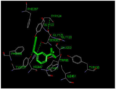
Molecular Docking Analysis: The modeling study was performed in this paper showed great interactions between neostigmine and human acetylcholinesterase. The binding energies of neostigmine and acetylcholine human acetylcholinesterase were -6.52 and -4.83 kcal/mole, respectively. The geometry of docking obtained with neostigmine with human acetylcholinesterase as shown in Figure 1. Neostigmine was able to form hydrogen bonds (HBs) with the residues Try337 of the enzyme, pi-pi stacking with the residue Trp86 and Pi-alkyl interaction with the residue Trp86. In addition, the molecular docking results showed that other amino acids residues are involved in the interactions with the neostigmine.

Figure 1: Three-dimensional representation of neostigmine interacting with active site of target macromolecule human acetylcholinesterase.

lupinepublishers-openaccess-journal-pharmacology-clinical-research-journal

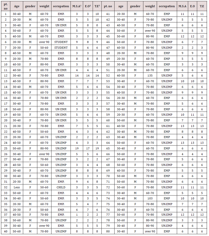
Clinical Elements: Eighty patients were randomized to treatment (rocuronium–neostigmine, n=80). All treated patients had three efficacy parameters that assessment carried out as shown in Table 1. The time from the start of administration of the reversal agent (neostigmine) to recovery, 11 patients used neostigmine had very late recovery time (≥ 9 min). These data were not excluded from the analysis. Mean systolic and diastolic arterial pressures and heart rates were not very similar in all patients. Systolic arterial pressures of ≥150 or ≤90 mm Hg, diastolic pressures of ≥98 or ≤45 mm Hg, and heart rates of ≥121 or ≤55 beats min−1 were observed in six patients used neostigmine. None of these was believed clinically important. Full recovery from neuromuscular block postoperative is crucial since residual neuromuscular block may augment morbidity (hypoxia, dyspnea, airway obstruction,). The use and monitoring of classical reversal agent is important to avoid residual neuromuscular blockage [24,25]. The usual neostigmine therapeutic dose is 0.04-0.07mg/kg whereas a toxic dose is 0.08mg/kg. Neostigmine uses has restrictions such as recurarization, muscarinic effect, weakness of respiratory muscle and ceiling effect [26,27]. Some adverse effects of neostigmine are dependent on the dose. Recent studies were performed to estimate the lowest neostigmine dose to obtain recovery from neuromuscular block (TOF ratio, TOFR 90%, the lower TOFR value, the deeper the neuromuscular blockade) lacking adverse effects [28]. The allocation of essential characteristic in this study is including age, gender, body weight and occupation for all patients. The duration of surgery in this study was limited to 30–90 minutes owing to the pharmacokinetics of rocuronium. Rocuronium has a moderate duration of action between 35-75 minutes [29,30]. However, Debane et al. has reported that the pharmacological effect of rocuronium is existing until 120 minutes after single dose administration [31]. All patients do not need to use additional neuromuscular blocking agents dose during operation in this study. Table 1 showed that a dose of neostigmine 2.5mg was as efficient as attuned dose based on the TOFR value for reversal of single dose rocuronium 0.6mg/kg. Table 1 showed that the total patients who reached TOFR ≥ 90% at 0-9 minutes after reversal administration were 69 and who reached TOFR ≥ 90% at 10-20 minutes after reversal administration were 11 (underline numbers). However, the differences were not statistically significant (p=0.718, F=0.331, F critical 3.034). All patients (except one patients had 19 minutes) had already reached TOFR ≥ 90% in interval of 0-15 minutes after reversal administration, the data obtained in this study in which the mean of the time required to reach TOFR ≥ 90% after reversal was 0-15 minutes were consistent with the data reported by Fuchs- Buder et. al which showed that the time required was 10 or more minutes [32]. In addition to that our results are consistent with other study by Cappellini et. al who reported that the recovery time of the patient was 10 minutes after neostigmine 0.01-0.03mg/kg with a shallow degree of neuromuscular block at the end surgery [33].

Table 1 also showed that the recovery time of the patient number 25 (who is on Aspirin tablets) was 17-20 minutes after neostigmine administration and this delayed action could be related to the fact that co-administration of neostigmine with some aspirin or other NSAIDs like diclofenac resulted in a synergistic interaction, which may provide evidence of supraspinal antinociception modulation by the increased acetylcholine availability in the synaptic cleft of cholinergic interneurons. The interaction obtained in this study between neostigmine and the NSAIDs have important clinical implications and it is consistent with the literature as reported by Hugo et.al [20]. For the present study, patients No. 12, 41, 45, 53, 59, 61, 64, 72, 74, and 77 had delayed action could be related to the mutational structure perturbation approach to expose longrange communication and the presence of conformational switches in the interior of the acetylcholinesterase enzyme. It is known that mutations are changes to the base sequence. The base sequence determines the amino acid sequence. A different base sequence therefore codes for a different amino acid sequence. Amino acids interact with each other by H-bonds, ionic bonds, and disulphide bridges. When the amino acids sequence of the acetylcholinesterase is changing then these interactions and bonds will be affected and the 3D shape of the acetylcholinesterase changes (tertiary structure). acetylcholinesterase work by interacting with substrates through their ‘active site’ as shown in Figure 1. A change in acetylcholinesterase shape will change the shape of the active site. The neostigmine cannot bind to the new shape of the active site so no acetylcholinesterase-neostigmine complexes are formed easily so the acetylcholinesterase cannot be inhibited or inhibited very slowly. Hasin et. al. has reported a total of 13 acetylcholinesterase single nucleotide polymorphisms (SNPs) and they might have deleterious adverse drug responses to acetylcholinesterase inhibitors [34]. In addition, Johnson and Moore have reported that the homo sapiens acetylcholinesterase mutations and they study the active site residues and they found the mutations could lead to differences in both steric and electrostatic properties of the active site [35]. The delayed onset of action of neostigmine as shown in Table 1 could be attributed to SNPs and mutation in Libyan patients. Our findings in addition to the published record suggest that the genetic variation could effect on neostigmine pharmacological action. Further studies need on Libyan people to be done in the future in order to confirm these results.

Table 1: Time (min) from start of administration of neostigmine to recovery.

lupinepublishers-openaccess-journal-environmental-soil-sciences

1M.S.A = Muscle Strength Assessment (5-second head lift, hand grasp),

2E.O = eye opening

3T.E = tongue extrusion

Conclusion

This Libyan study was the first comparative study between the three parameters (measurements of muscle strength, eye opening, and tongue extrusion) that are used to reversal of the enduring effect of rocuronium by neostigmine. It is found that rocuroniuminduced neuromuscular block and can be reversed by neostigmine. It increased the amount of safety data available about neostigmine, although this was not the main reason for the study. Clinical signs of recovery were similar with the three parameters, which is to be expected. This indicates that the results presented are realistic in the Libyan patients. The faster time to recovery with most of patients (n= 69) compared with eleventh patients (n=11) in this study is consistent with that previously reported. The rapid time to recovery with neostigmine observed in Libyan patients is similar to that reported in previous studies, demonstrating a consistency in observed efficacy.

Read More Lupine Publishers LOJ Pharmacology & Clinical Research Please Click on Below Link https://lupine-publishers-pharmacology.blogspot.com/


Monday, 5 April 2021

Resurrection Day!!!


Resurrection Day!!! Easter is the time to rejoice and be thankful for the gift of life , love and joy. Have a Blessed Day from our Lupine Family

Lupine Publishers| Pregnancy after Liver Transplantation

 Lupine Publishers|  Current Trends in Gastroenterology and Hepatology


Introduction

In 1978, Walcott and coworkers documented the first pregnancy in a liver transplant recipient [1]. The recipient’s immunosuppressive regimen included prednisone and azathioprine. Her pregnancy was uncomplicated, and she delivered a healthy male, weighing 2400 g, at term. Since Walcott’s report, there have been hundreds of pregnancies reported worldwide in liver transplant recipients. Whether the transplant could take in an adverse effect of the pregnancy or whether the pregnancy could potentiate liver transplant it is not settled [2].

Liver Transplant and Timing of Pregnancy

a) Women should wait at least 1 year after receiving a solidorgan transplant before attempting to get significant, and then should do so but when earned by the transplant team and obstetrician, with close monitoring [3].

b) The best contraceptive method, at least early after liver transplantation, is a barrier method. Later, when the patient is stabilized, hormonal methods may be considered. Sterilization is an option for patients who have completed their family [4].

c) Liver transplant recipients considering pregnancy should have preconception counseling with a maternal–fetal medicine specialist and their transplant physician. There are no specific guidelines for liver transplant recipients to decide whether an allograft is functioning sufficiently well for pregnancy as there is for kidney transplant recipients. Nevertheless, a general assessment of graft function and the status of comorbidities, such as high blood pressure, should be done before conception. The preconceptional visit also provides an opportunity to look back with the patient outcomes of pregnancies with liver transplants, potential effects of immunosuppressive agents on the pregnancy and fetus, and the plan of management for the gestation.

Pregnancy outcome after liver transplantation

Despite advances in immunosuppressive therapy, pregnancies in liver transplant recipients are still more risky than in the general population for both the mother and the fetus. Pregnancies in liver transplant recipients should be followed up more cautiously by a multidisciplinary team because [5,6]:

a) Overall, 78% of pregnancies were successful.

b) Many recipients of solid-organ transplants, including liver transplants, develop high blood pressure and renal dysfunction. It is easily grounded in the obstetrical literature that these conditions are independently linked with adverse pregnancy events. The most common obstetric complications were high blood pressure (28%), preeclampsia (12%), and reversible liver dysfunction (27%) [7].

c) The rate of acute rejection was 10% [8].

d) The incidence of perinatal and neonatal death was about 4%.

e) Thither was a 3% incidence of congenital malformations, after exclusion of an infant with the presumed fetal alcohol syndrome and an infant with an autosomal dominant genetic disorder inherited from the female parent.

f) The maternal death rate was 5.5%, over a 2-year period following delivery [9].

g) Additionally, it seems that the danger of infection during gestation is also significantly increased after liver transplant; this finding may be a consequence of chronic immunosuppressive therapy [1].

h) Liver transplantation does not significantly modify pretransplant diabetes mellitus. Diabetes mellitus frequently develops de novo after liver transplantation, although this complication is usually short-lived and likely linked to immunosuppressive drug administration. The prognosis of patients with post-transplant diabetes mellitus is worse than that of those without this complication [10].

i) The incidence of preterm delivery is 24% [8].

j) The rate of cesarean delivery for liver transplant recipients is higher (20% -100%) than the national average [11].

k) The rate of newborn complications was also high, at 29%.

l) Subjective hearing impairment is frequent in patients after OLT and contributes to post-OLT morbidity. Calcineurin inhibitor-related neurotoxicity appears as a potential mechanism. Further prospective investigations with objective hearing tests are necessary to substantiate these results and to assess the use of immunosuppression [12].

Management Recommendations

a) Patients with liver transplants considering pregnancy should receive preconception counseling. The preconceptional evaluation will require an appraisal of the current graft status and episodes of rejection.

b) Preconceptional counseling should be provided to both the patient and her spouse. For patients who bear a genetic disorder underlying their liver disease, genetic counseling should also be provided. These will include patients with alpha-1antitrypsin deficiency and hemochromatosis, as well as less common disorders such as Alagille syndrome and Caroli syndrome. Prenatal testing may be usable for certain disorders.

c) Ideally, immunosuppressive medications are at stable doses, and no episodes of rejection have occurred in the old yr. An assessment of renal, cardiovascular, and pulmonary status should be performed.

d) Comorbidities such as diabetes and hypertension should be addressed and ideally should be easily-controlled before conception.

e) The status of hepatitis B and C infections as easily as the CMV status should be known and the possible impact on pregnancy discussed. Patients should have up-to-date vaccinations for influenza, pneumococcus, tetanus, and hepatitis B if indicated. In summation, a discussion of pregnancy results in transplant recipients should take place, concentrating especially on the potential for and long-term outcomes of preterm parturition.

f) Pregnancies in patients with liver transplants should be done in an interdisciplinary fashion between a maternal–fetal medicine specialist, a transplant physician, and other specialists as indicated.

g) Transplant recipients will require frequent prenatal visits throughout the class of their pregnancies.

h) Close monitoring of line pressure, proteinuria, and laboratory studies throughout the pregnancy are all-important.

i) Patients on chronic prednisone should have early screening for gestational diabetes. Although the frequency of congenital birth defects does not appear to be increased with immunosuppressive agents in studies thus far, a genetic sonogram is indicated for transplant patients to boot out the presence of congenital abnormalities.

j) Serial growth scans should be done throughout pregnancy to monitor for fetal growth restriction.

k) In the third trimester, antenatal testing may be done, although the benefit of testing in this population has not been established.

l) Cesarean delivery should be performed for standard obstetrical indications.

m) Stress-dose steroids are shown at the time of delivery for patients who have been taking corticosteroids during pregnancy.

n) An increase in baseline liver function needs an aggressive evaluation in these pregnancies. On that point is no contraindication to liver biopsy in these patients to evaluate for graft rejection.

o) Several other complications of pregnancy, such as preeclampsia, HELLP syndrome, and cholestasis of pregnancy, may result in elevations of liver enzymes. Some small reports have demonstrated an increase in the rate of cholestasis of pregnancy in liver transplant recipients, but this has not been measured in larger works. It may be rather challenging to identify these complications of pregnancy from graft rejection or exacerbation of the underlying liver disease, such as hepatitis C.

p) The goal of managing these pregnancies is to optimize maternal health, including graft function and co morbidities, to find and manage complications of pregnancy such as preeclampsia and infection, and to downplay the complications of preterm birth whenever possible.

q) From the literature, it appears that many liver transplant recipients can look at safe gestation and neonatal outcomes. However, at that place is a big deal of research needed to clarify the optimal management of these pregnancies.

Read More  Lupine Publishers Current Trends in Gastroenterology and Hepatology Please Click on Below Link https://currenttrendsingastroenterology.blogspot.com/