Journal of orthopaedics | Lupine Publishers
Van Neck-Odelberg disease is considered a benign skeletal
developmental abnormality seen in children, comprising hyperostosis
of the ischiopubic synchondrosis (IPS). During skeletal development,
typically there is unilateral and asymmetric enlargement of
the IPS of no pathological relevance. Nevertheless, in spite of being
considered a physiological phenomenon, some children can be
clinically symptomatic and experience unspecific groin pain and limping.
Importantly, the diagnosis of IPS opsteochondritis cannot
be established by radiographic findings solely and needs to be supported
by the appropriate clinical scenario and by the exclusion
of other causes such as neoplasia, stress fracture, osteomyelitis, and
post-traumatic osteolysis. The role of imaging is essentially to
exclude these causes.
Keywords:Musculoskeletal; Radiography; MRI; Osteochondrosis
Clinical History
A 10-year-old boy presented with right groin pain and a limping
leg aggravated after football practice.
Imaging Findings
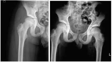
In the hip radiographs (Figure 1), there was no evidence of an
acute fracture, but a sharp asymmetric shadow was seen at the right
ischiopubic synchondrosis with no associated periosteal reaction
or soft tissue thickening.The triradiate cartilage and hip joint spaces
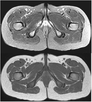
were symmetric and unremarkable. The MRI (Magnetic Resonance
Imaging) depicted a prominent asymmetric synchondrosis of the
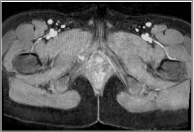
right inferior pubic ramus with mild associated edema (Figure 2)
and minimal enhancement (Figure 3). There was no evidence of
fracture or stress response. These imaging findings in the current
clinical setting lead to a diagnosis of VND.
Figure 1: AP radiographs of the Hip: A sharphly demarcated assymetric shadow was seen at the right ischiopubic synchondrosis
and extending into the obturator foramen, with no associated periosteal reaction or soft tissue thickening.
Figure 2: Bony pelvis MRI (Upper image T2FS ; Bottom image T1)- Assymetric prominent right isquiopubic synchondrosis
with hyperintense signal and mild surrounding sof tissue edema on fluid sensitive sequence and hypointense signal on T1.
Figure 3: Bony pelvis MRI, Axial T1 post intravenous paramagnetic contrast administration: There is mild enhancement of the
assymetric prominent right isquiopubic synchondrosis.
VND is a benign skeletal developmental abnormality seen in
children, comprising hyperostosis of the ischiopubic synchondrosis
(IPS), and it was first described by Odelberg and Van Neck in
radiographs of prepubescents [1,2]. The IPS consists of cartilaginous
tissue lying between the two ossification centers of the ischiopubic
region: the superomedial pubic center and posterolateral ischial
center [3,4]. IPS acts as a temporary joint between the ischium
and pubis, and its ossification begins early in childhood completes
before puberty [3,4]. In the latter stage of this process there is
unilateral asymmetric enlargement, of no pathological relevance
[4,5]. IPS are commonly asymmetric in asymptomatic children,
with 50% of asymptomatic children of ages between 4-5 years old
exhibiting asymmetric closure of this site, which can persist for
several years [6].
In spite of being considered a physiological phenomenon which
some authors relate to mechanical stress conditioned by iliopsoas,
adductors, and gemellus, leading to delayed union of the cartilage
and ossification centers [4,5,7], some children can be clinically
symptomatic and experience groin pain and limping, regarded
by some authors as a consequence of the inflammatory reaction
conditioned by the constant movement of the IPS [4,7].
Clinical Perspective
Patients may complain of an unspecific groin or buttock pain [8]. The differential diagnosis includes stress fractures, osteomyelitis, post-traumatic osteolysis or neoplasia [3,7,8]. Ostechondritis of the IPS is often a diagnosis of exclusion and laboratory test are generally unremarkable [7].
Imaging Perspective
An enlarged and assymetric IPS is generally consided as an
anatomical developmental variant [6], furthermore, the diagnosis
of VND cannot be established by radiographic findings solely and
needs to be supported by the appropriate clinical scenario and
the primary role of imaging is to exclude other causes [3,5]. The
typical finding of VNP in radiographs is of an asymmetric sharply
circumscribed shadow located in the ischiopubic region extending
towards the obturator foramen without periosteal reaction or soft
tissue thickening [1,2,8]. CT (computed tomography) and MRI
usually show asymmetric enlargement of the IPS, furthermore, on
MRI there is hyperintense signal on T2 with fat saturation and STIR
sequences and hypointense signal on T1, with fibrous bridging and
fusiform swelling of the IPS [3,5,8]. There can be mild associated
soft tissue edema and minimal post contrast enhancement [5].
Radiologically it can extend for several months to a year period,
although when the ossification completes, no signs will subsist [7].
Take Home Message, Teaching Points
a) The Diagnosis of VND is clinically challenging and other
entities such as stress fracture, osteomyelitis, post-traumatic
osteolysis, should be excluded.
b) Clinical symptoms are a must condition to identify a
delayed unilateral ischiopubic synchondrosis as VND.
c) Imaging value is in excluding other entities.
https://lupinepublishersgroup.com/
For more Journal of orthopaedics Please Click Here: https://lupinepublishers.com/orthopedics-sportsmedicine-journal/
To Know more Open Access Publishers Click on Lupine Publishers


No comments:
Post a Comment
Note: only a member of this blog may post a comment.